. 2018 Sep 21;50(9):1-10.
doi: 10.1038/s12276-018-0151-9.

Notch ligand Jagged1 promotes mesenchymal stromal cell-based cartilage repair

Affiliations

Notch ligand Jagged1 promotes mesenchymal stromal cell-based cartilage repair

Junkui Sun et al. Exp Mol Med. .

Abstract

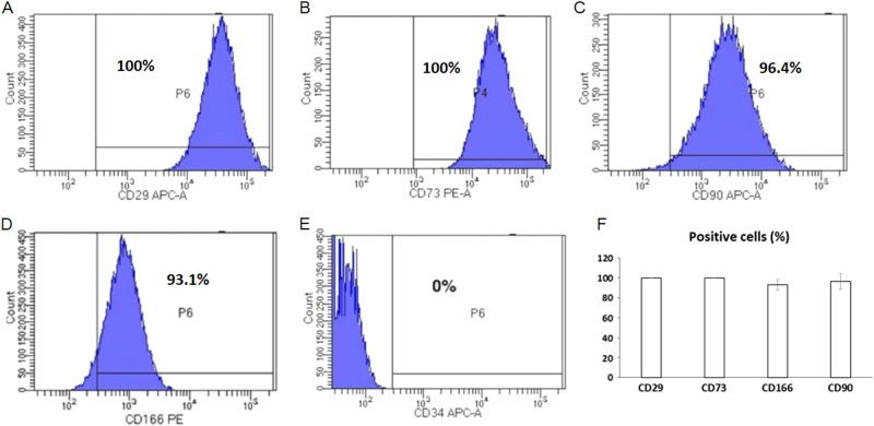
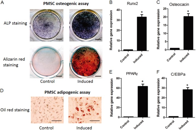
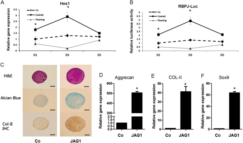
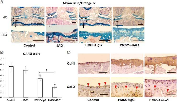
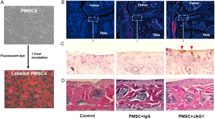
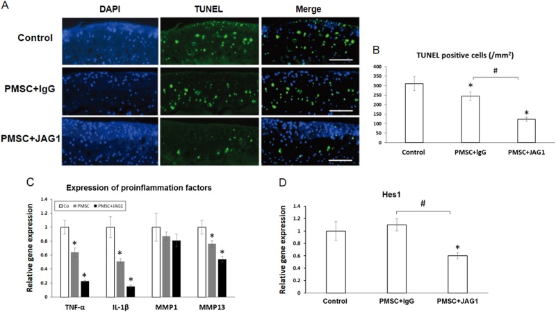
Placenta-derived mesenchymal stromal cells (PMSCs) provide a promising cell source for tissue regeneration. However, rapid induction of PMSC chondrogenic differentiation during therapeutic transplantation remains extremely challenging. Here we undertook a study to determine if Notch inhibition by soluble Jagged1 (JAG1) peptides could be utilized to accelerate PMSC-induced cartilage regeneration in a mouse post-traumatic osteoarthritis (PTOA) model. Our results showed that treatment of PMSCs with soluble JAG1 significantly enhanced chondrogenesis in culture as shown by increased alcian blue staining and decreased Notch target Hes1 expression when compared to those in lgG-treated control cells. Importantly, significantly enhanced cartilage formation and decreased joint inflammation were observed when JAG1-treated PMSCs were injected into mouse PTOA knee joints. Finally, in vivo cell tracing showed that more JAG1-treated PMSCs remained in knee joint tissues and that JAG1-treated PMSCs exhibited greater PMSC chondrogenic differentiation than lgG-treated control PMSCs at 4 weeks after injection. These data indicate that transient Notch inhibition by soluble JAG1 could be used to enhance PMSC survival and chondrogenic differentiation, thereby increasing the therapeutic potential of PMSCs for cartilage regeneration.


Conflict of interest statement

The authors declare that they have no conflict of interest.

Figures

Fig. 1
Fig. 1. Characterization of human PMSCs.
ae Representative flow cytometry histograms showing CD29, CD73, CD90 and CD166-positive PMSCs and CD34-negative PMSCs in passage 4 cultures, respectively. f Quantified percentage of CD29, CD73, CD90 and CD166-positive MSCs in passage 4 cultures. Data are the means ± SD of three independent experiments
Fig. 2
Fig. 2. Analysis of PMSC osteogenic and adipogenic differentiation.
a Osteogenic differentiation was examined by alkaline phosphatase staining and alizarin red staining without (Control) induction or with induction (Induced). Bar indicates 100 µm. b, c Osteogenic differentiation markers Runx2 and osteocalcin were further examined by real-time PCR. d Adipogenic differentiation was studied by the detection of lipid vacuoles by oil red O staining with or without induction. Bar indicates 50 µm. e, f Adipogenic differentiation markers PPARr and C/EBPa were inspected by real-time PCR. The data are the means ± SD of three independent experiments and all the results were normalized to the internal control (*p < 0.05 compared with control PMSCs without induction)
Fig. 3
Fig. 3. Inhibition of Notch signaling by unbound JAG1 enhances inducted PMSC chondrogenic differentiation.
a Quantification of gene expression of the Notch target gene Hes1 indicates a significant increase (p < 0.05) in cells with surface-bound (coated) JAG1 (10 µg/ml) treatment, and a reduced expression was observed in cells treated with a single treatment of unbound (Floating) JAG1 (10 µg/ml) for up to 5 days. b Luciferase assays showed a significant decrease in Notch-responsive reporter activity in bound JAG1-treated MSCs and reduced activity in day 1 and day 3 with unbound JAG1 treatment. The data are the means ± SD of three independent experiments performed in duplicate, and the gene expression level in control cells (Co) was set at 1 (*p < 0.05 compared with control). c An increase in chondrogenesis was observed in unbound JAG1-treated PMSCs at day 14, with stronger staining of alcian blue and type II collagen (Col-II). Scale bars, 100 µm. df Quantification of gene expression of cartilage matrix aggrecan and Col-II, as well as transcription factor Sox9, using RNA from day 14 culture pellets. Data are the means ± SD of three independent experiments performed in triplicate, and all the results were normalized to internal control (*p < 0.05 compared with control PMSCs (Co)
Fig. 4
Fig. 4. Mice develop more severe osteoarthritis without PMSC injection.
At week 4 post-intra-articular injection of PBS, JAG1 (10 µg/ml) in PBS, PMSCs with lgG (10 µg/ml) or JAG1 (10 µg/ml), right knee joints were harvested for histological assessment. a Representative images of medial compartment of knee sections from MLI mice with injection of PBS (Control), JAG1, PMSC + lgG and PMSC + JAG1 stained with alcian blue/orange G. b Osteoarthritic changes in knee joints (n = 12) as quantified with Osteoarthritis Research Society International (OARSI) score. Data are the means ± SD (*p < 0.05 compared with control; #P < 0.05 between two groups). c Immunohistochemistry staining (IHC) of chondrogenic marker type II collagen (Col-ll) and type X collagen (Col-X) in tibia articular cartilage from 8-week MLI mice with or without PMSC injection. Increased Col-X expression was detected, and more Col-X-positive cells were located toward the articular surface in control and PMSC + lgG mice, not in PMSC + JAG1 mice 8-weeks after MLI surgery (Col-X positive cells: red arrowheads)
Fig. 5
Fig. 5. Tracing of PMSCs in the mouse knee joint.
a Qtracker® reagent-labeled PMSCs showed strong red fluorescence after 1 h of incubation in the culture. b Mouse knee joints injected with 0.2 million red fluorescent-labeled JAG1-treated PMSCs showed more live PMSCs invading synovial tissue surrounding the artificial cartilage than joints injected with untreated PMSCs at 4 weeks after cell injection. c Immunohistochemistry staining (IHC) of human PMSCs using the anti-human nuclear antigen antibody in tibia articular cartilage from 8-week MLI mice (human nuclear antigen positive cells: red arrowheads). d H&E staining of subchondral bone in the mouse knee joint from control, PMSC and JAG1/PMSC groups at 4 weeks after cell injection
Fig. 6
Fig. 6. JAG1 treatment inhibits chondrocyte apoptosis and inflammation in the mouse OA joint.
a Top panel: The representative images of TUNEL staining of the tibia plateau from a control OA mouse injected with 8 µl PBS. Middle panel: The stained tibia plateau from a control OA mouse injected with 8 µl PBS containing 0.2 million PMSCs and lgG (10 µg/ml). Low panel: The stained tibia plateau from a control OA mouse injected with 8 µl PBS containing 0.2 million PMSCs and JAG1 (10 µg/ml). b Quantification of TUNEL-positive cells in areas of the stained tibia plateau (n = 12). Data are the means ± SD (*p < 0.05 compared with control; #P < 0.05 between two groups). c Relative mRNA expression of proinflammation cytokines was analyzed by quantitative real-time PCR after synovial tissue obtained from OA mice was treated with PMSCs with or without JAG1 for 4 weeks. The data are the means ± SD (*p < 0.05 compared with control). d Relative mRNA expression of the Notch target gene Hes1 in synovial tissue was analyzed by quantitative real-time PCR. Data are the means ± SD (*p < 0.05 compared with control; #P < 0.05 between two groups)

References

    1. Parolini O, et al. Concise review: isolation and characterization of cells from human term placenta: outcome of the first international Workshop on Placenta-Derived Stem Cells. Stem Cells. 2008;26:300–311. doi: 10.1634/stemcells.2007-0594. - DOI - PubMed
    1. Jeon YJ, Kim J, Cho JH, Chung HM, Chae JI. Comparative analysis of human mesenchymal stem cells derived from bone marrow, placenta, and adipose tissue as sources of cell therapy. J. Cell Biochem. 2016;117:1112–1125. doi: 10.1002/jcb.25395. - DOI - PubMed
    1. Fan ZX, et al. Placenta- versus bone-marrow-derived mesenchymal cells for the repair of segmental bone defects in a rabbit model. FEBS J. 2012;279:2455–2465. doi: 10.1111/j.1742-4658.2012.08625.x. - DOI - PubMed
    1. Mokbel AN, et al. Homing and reparative effect of intra-articular injection of autologus mesenchymal stem cells in osteoarthritic animal model. BMC Musculoskelet. Disord. 2011;12:259. doi: 10.1186/1471-2474-12-259. - DOI - PMC - PubMed
    1. Dong Y, et al. RBPjkappa-dependent Notch signaling regulates mesenchymal progenitor cell proliferation and differentiation during skeletal development. Development. 2010;137:1461–1471. doi: 10.1242/dev.042911. - DOI - PMC - PubMed

Publication types

LinkOut - more resources