. 2018 Sep 21;8(1):14223.
doi: 10.1038/s41598-018-31773-z.

Misfolded SOD1 pathology in sporadic Amyotrophic Lateral Sclerosis

Affiliations

Misfolded SOD1 pathology in sporadic Amyotrophic Lateral Sclerosis

Bastien Paré et al. Sci Rep. .

Abstract

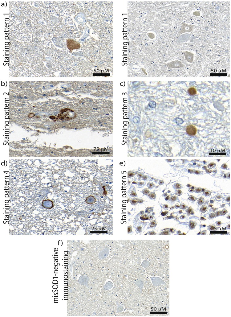
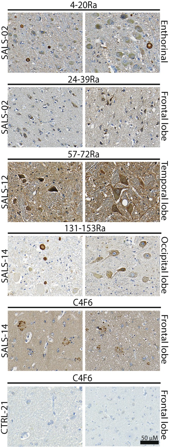
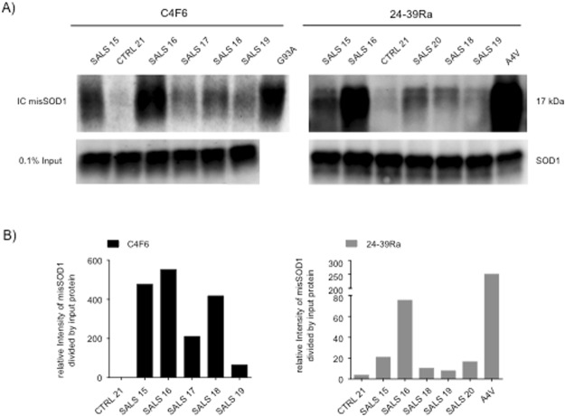
Aggregation of mutant superoxide dismutase 1 (SOD1) is a pathological hallmark of a subset of familial ALS patients. However, the possible role of misfolded wild type SOD1 in human ALS is highly debated. To ascertain whether or not misfolded SOD1 is a common pathological feature in non-SOD1 ALS, we performed a blinded histological and biochemical analysis of post mortem brain and spinal cord tissues from 19 sporadic ALS, compared with a SOD1 A4V patient as well as Alzheimer's disease (AD) and non-neurological controls. Multiple conformation- or misfolded-specific antibodies for human SOD1 were compared. These were generated independently by different research groups and were compared using standardized conditions. Five different misSOD1 staining patterns were found consistently in tissue sections from SALS cases and the SOD1 A4V patient, but were essentially absent in AD and non-neurological controls. We have established clear experimental protocols and provide specific guidelines for working, with conformational/misfolded SOD1-specific antibodies. Adherence to these guidelines will aid in the comparison of the results of future studies and better interpretation of staining patterns. This blinded, standardized and unbiased approach provides further support for a possible pathological role of misSOD1 in SALS.


Conflict of interest statement

The authors declare no competing interests.

Figures

Figure 1
Figure 1
Human SOD1 protein schematic representation with highlighted misSOD1 epitope mapping regions (a) Linear representation of the 5 tested misfolded/conformational SOD1 specific antibody’s binding regions. (b) 3D protein structure representation with highlighted misfolded/conformational SOD1 antibody’s bindging regions. Ra: Rabbit polyclonal. C4F6: mouse monoclonal.
Figure 2
Figure 2
Immunodetection of misSOD1 accumulation in spinal cord of A4V SOD1 FALS patient Lumbar spinal cord sections from A4V-SOD1 FALS autopsied patient (positive control) and non-neurological control individual (negative control) immunostained with 5 misSOD1 conformational antibodies (4–20Ra, 24–39Ra, 57–72Ra, C4F6 and 131–153 R). Characteristic misSOD1-positive cytoplasmic accumulation in spinal motor neurons can be observed with all tested antibodies in the A4V-SOD1 FALS patient, whereas no immunostaining is detected in the non-neurological control. Scale bar: 50 mm.
Figure 3
Figure 3
Detected misSOD1 immunostaining patterns MisSOD1 immunostaining patterns detected in SALS individuals were (a) Diffuse misSOD1 staining in the cytoplasm of motor neurons (immunostaining pattern 1). Note that different misSOD1 immunosignal intensity levels were detected. As misSOD1-positive motor neurons can be detected beside misSOD1-negative motor neurons on the same tissue section, it is unlikely that the observed misSOD1 accumulation represents a false-positive signal (b) misSOD1-positive deposits detecetd in the cytoplasm of motor neurons (immunostaining pattern 2) (c) Dendritic and axonal misSOD1 accumulation (immunostaining pattern 3) (d) MisSOD1-positive perivacuolar ring-like structures (immunostaining pattern 4) (e) MisSOD1 nuclear accumulation (immunostaining pattern 5) and (f) misSDO1-negative immunostaining observed in non-neurological control. Scale bars are presented in the lower right corner for each detected immunostaining patterns.
Figure 4
Figure 4
Detection of misSOD1-positive ring-like structure in the SALS and control individuals Representative images of misSOD1-positive ring-like structures in the extracellular space. (AD) misSOD1-positive serpiginous structures detected in SALS cases detected in spinal horn grey matter. (EG) misSOD1-negative CA-like structures detected in SALS cases located in the spinal cord tissue section periphery. (H) Spinal ventral horn grey matter accumulation of misSOD1-positive CA-like structures and, peripheral white matter negative misSOD1 CA-like deposits within the spinal cord.
Figure 5
Figure 5
Immunodetection of misfolded SOD1 aggregates detected in brain tissue sections of SALS patients by immunohistochimestry using the misfolded SOD1/conformational-specific 4–20Ra, 24–39Ra, 57–72Ra, C4F6 and 131–153Ra antibodies Brain tissue sections from SALS patients and non-ALS control individuals immunostained using 5 different misSOD1/conformational-specific antibodies. Two representative pictures per SALS patients and control are showed. Different brain areas were analysed and misSOD1-positive accumulation can be observed.
Figure 6
Figure 6
Immunocapture of misSOD1 in SALS spinal cord tissue samples using the C4F6 and 24–39Ra misSDO1 specific antibodies (A) Immunocapture of misSOD1 in total spinal cord protein extracts, obtained from SALS patients, A4V-SOD1 FALS patient, non-ALS control individual and transgenic mice over-expressing G93ASOD1 mutant protein is illustrated. (B) Quantification data of the immunocaptured misSOD1 protein shown in panel A. Immunodetection of the transferred immunucaptured products, using the 24–39Ra and the C4F6 misSOD1/conformational specific antibodies, revealed that both antibodies yielded clear and positive immunoreactivity signals and a high degree of specificity whilst only very low levels were detected in spinal cord extracts collected from the non-ALS control individual. A fraction of the total amount of proteins used (0.1%) was also run on a SDS–PAGE to validate that an equal amount of protein was loaded from each sample. Note that, due to the availability of patient materials, SALS17 and SALS20 spinal cord extracts were only immunocaptured using either the C4F6 or 24–39Ra antibodies.

References

    1. Mulder DW, Kurland LT, Offord KP, Beard CM. Familial adult motor neuron disease: amyotrophic lateral sclerosis. Neurology. 1986;36:511–517. doi: 10.1212/WNL.36.4.511. - DOI - PubMed
    1. Robberecht W, Philips T. The changing scene of amyotrophic lateral sclerosis. Nat. Rev. Neurosci. 2013;14:248–64. doi: 10.1038/nrn3430. - DOI - PubMed
    1. Chen S, Sayana P, Zhang X, Le W. Genetics of amyotrophic lateral sclerosis: an update. Mol. Neurodegener. 2013;8:28. doi: 10.1186/1750-1326-8-28. - DOI - PMC - PubMed
    1. Gros-Louis F, Gaspar C, Rouleau GA. Genetics of familial and sporadic amyotrophic lateral sclerosis. Biochim. Biophys. Acta. 2006;1762:956–72. doi: 10.1016/j.bbadis.2006.01.004. - DOI - PubMed
    1. Andersen PM, Al-Chalabi A. Clinical genetics of amyotrophic lateral sclerosis: what do we really know? Nat. Rev. Neurol. 2011;7:603–615. doi: 10.1038/nrneurol.2011.150. - DOI - PubMed

Publication types