. 2018 Sep 21;8(1):14174.
doi: 10.1038/s41598-018-32640-7.

Two-photon microscopy of Paneth cells in the small intestine of live mice

Affiliations

Two-photon microscopy of Paneth cells in the small intestine of live mice

Won Hyuk Jang et al. Sci Rep. .

Abstract

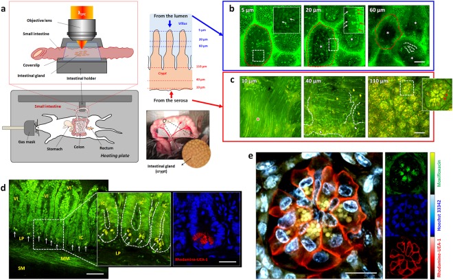
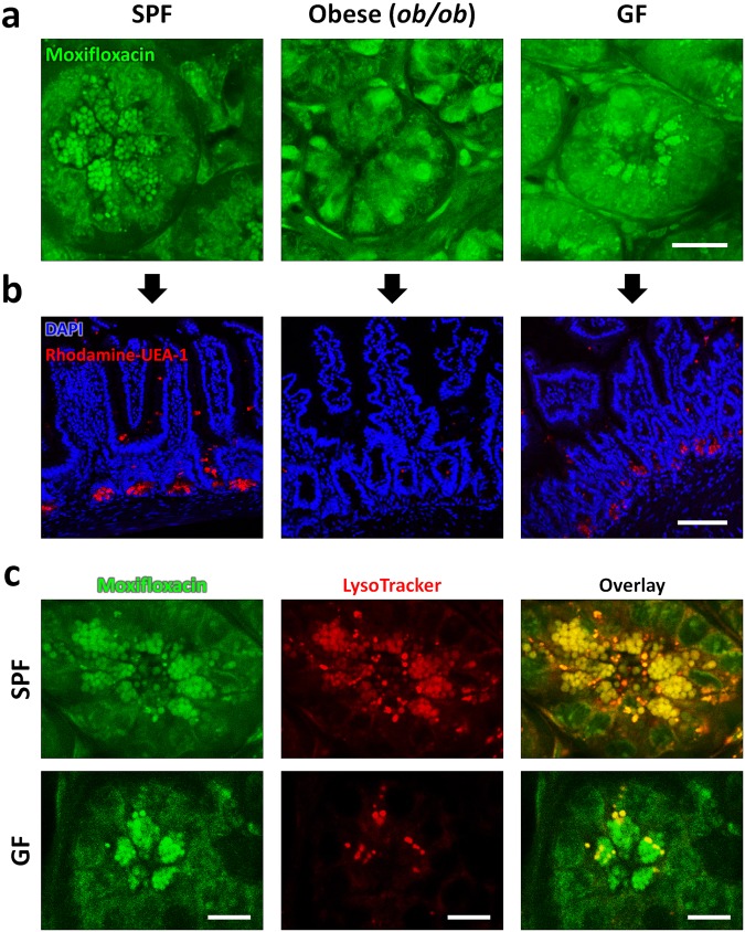
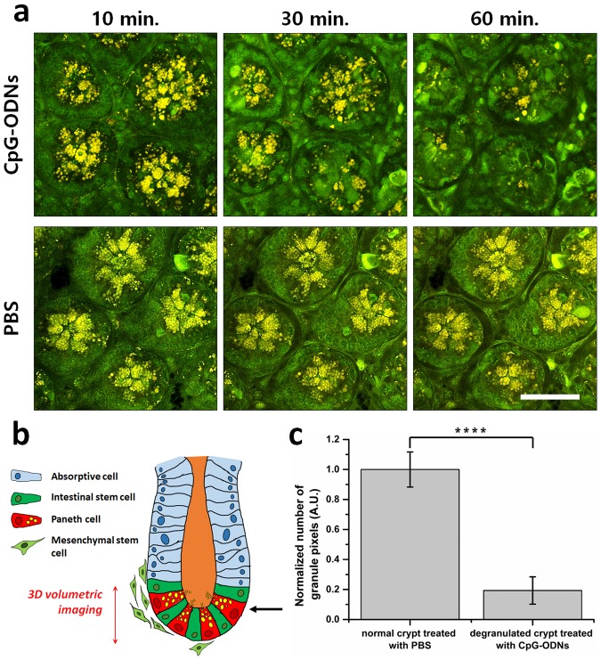
Paneth cells are one of the principal epithelial cell types in the small intestine, located at the base of intestinal crypts. Paneth cells play key roles in intestinal host-microbe homeostasis via granule secretion, and their dysfunction is implicated in pathogenesis of several diseases including Crohn's disease. Despite their physiological importance, study of Paneth cells has been hampered by the limited accessibility and lack of labeling methods. In this study, we developed a simple in vivo imaging method of Paneth cells in the intact mouse small intestine by using moxifloxacin and two-photon microscopy (TPM). Moxifloxacin, an FDA-approved antibiotic, was used for labeling cells and its fluorescence was strongly observed in Paneth cell granules by TPM. Moxifloxacin labeling of Paneth cell granules was confirmed by molecular counterstaining. Comparison of Paneth cells in wild type, genetically obese (ob/ob), and germ-free (GF) mice showed different granule distribution. Furthermore, Paneth cell degranulation was observed in vivo. Our study suggests that TPM with moxifloxacin labeling can serve as a useful tool for studying Paneth cell biology and related diseases.


Conflict of interest statement

The authors declare no competing interests.

Figures

Figure 1
Figure 1
In vivo TP images of the mouse small intestine with moxifloxacin labeling. (a) TPM setup for in vivo imaging of the anesthetized mouse small intestine. A portion of the small intestine was gently pulled out from the abdominal cavity, held on the temperature-controlled moisture-maintained intestinal holder, and sealed with a coverslip. (b) Moxifloxacin-based 3D TPM images taken from the luminal side of the small intestine in a wild type mouse (Supplementary Video 1). 3D TPM images revealed well-organized enterocytes (white-arrows) on the surface of the villi (red-dashed-circles), some cells inside the villi (white-asterisks), and vascular structures inside the villi (white-dashed-line), and strong moxifloxacin fluorescence expressing microstructures (red arrows in inset), which are likely lysosomes in enterocytes. (c) Moxifloxacin-based 3D TPM images taken from the serosal side of the small intestine in a wild type mouse (Supplementary Video 2). 3D TPM images showed the outer muscle (red-asterisks), myenteric plexus (white-dashed-lines), fibrous structures, (yellow-arrow), and glial cells (white-arrows) at the bottom of intestinal crypts. In the relatively deep region from the serosal surface, Paneth cells with multiple, clustered granules (red arrowheads in inset) were detected at the base of intestinal crypts. (d) Moxifloxacin-based cross-sectional TP images of the in vivo small intestine and ex vivo cross-sectional fluorescence image of the sectioned small intestine tissue of wild type mice (Supplementary Video 3). TP images showed cross-sectional structures of the small intestine including intestinal villi (VI), intestinal crypts (IC), lamina propria (LP), muscularis mucosa (MM), and submucosa (SM). Bright granular structures (white-arrows) were visible at the base of intestinal crypts owing to the stronger expression of moxifloxacin fluorescence compared with other subcellular structures. Corresponding fluorescence image of the sectioned small intestinal tissue showed the location and the morphologies of the Paneth cell granules at the epithelial lining of the small intestine. DAPI (blue) and rohdamine-UEA-1 (red) stained nuclei and Paneth cell granules, respectively, in the fixed and permeabilized small intestinal section. (e) Counterstained TPM images of the small intestinal crypt base of a live mouse (Supplementary Video 4). Cells containing spherical-shaped granules, highly labeled with moxifloxacin fluorescence (green), were confirmed to be Paneth cells by counterstaining the cell membrane with rhodamine-UEA-1 (red). Cell nuclei were labeled with Hoechst 33342 (blue). Individual fluorescence signal from each fluorescence contrast agents are displayed in small subsets (right panel, moxifloxacin: green, Hoechst 33342: blue, rhodamine-UEA-1: red). In this experiment, the number of mice used are as follows; wild type C57BL/6 mice bred in a SPF condition (n = 5). Scale bars, (b,c): 50 µm; (d), 100 µm and 15 µm; (e), 15 µm.
Figure 2
Figure 2
In vivo Moxifloxacin-based TPM images and ex-vivo fluorescently labeled images of small intestinal crypts in mice under different environmental and metabolic conditions. (a) In vivo moxifloxacin-based TPM images at the base of intestinal crypts in SPF, obese (ob/ob), and GF mice. In wild type SPF mice, Paneth cell granules are clearly visible via moxifloxacin labeling. In obese (ob/ob) mice, Paneth cell granules did not appear at the base of intestinal crypts, but there were some cells expressing moxifloxacin fluorescence in the cytoplasm. In wild type GF mice, Paneth cell granules appeared similarly to those of SPF mice in moxifloxacin-based TPM. (b) Ex vivo fluorescence images of the small intestine sections from SPF, obese (ob/ob), and GF mice. Cell nuclei and Paneth cell granules in the fixed and permeablilized small intestinal sections were stained with DAPI (blue) and rhodamine-UEA-1 (red), respectively. Fluorescence images of the sectioned small intestine tissues showed epithelial linings and Paneth cell granules at the bottom. Paneth cell granules were visible in SPF and GF mice only, and not detected in obese (ob/ob) mice. These fluorescence image results of the sectioned intestinal crypts were consistent with in vivo moxifloxacin-based TPM results. (c) In vivo TPM images of Paneth cell granules in SPF and GF mice with the counterstaining of both moxifloxacin and LytoTracker. TPM images of SPF mice showed that both moxifloxacin and LysoTracker similarly labeled Paneth cell granules. On the other hand, TPM images of GF mice showed that LysoTracker labeled only a portion of moxifloxacin-labeled Paneth cell granules. In this experiment, the number of mice used are as follows; wild type C57BL/6 mice bred in a SPF condition (n = 5), germfree (GF) condition (n = 9), obese (ob/ob) mice (n = 8). Scale bars in (a,b), and (c) are 20 μm, 100 μm, and 10 μm, respectively.
Figure 3
Figure 3
In vivo observation of Paneth cell degranulation in SPF mice using moxifloxacin-based TPM. (a) Longitudinal moxifloxacin-based TPM images of intestinal crypts at the base after intraluminal injection of CpG-ODNs (top panel, 10 µM), and PBS (lower panel) at different time points. Longitudinal TPM images are presented at single representative depth planes in 10 min, 30 min, and 60 min post-injection. (b) A schematic of the small intestine showing the depth range of 3D TPM (red-arrow), and single representative depth plane (black-arrow). (c) Quantitative analysis of total granule pixel counts in the moxifloxacin-based 3D TPM images of degranulated intestinal crypts in SPF mice in 5 hours after intraperitoneal injection of CpG-ODNs. Statistical analysis was performed by Welch’s t-test; ****p < 0.0001. In this experiment, the number of mice used are as follows; wild type C57BL/6 mice bred in a SPF condition (n = 5). Scale bars in (a) indicate 100 µm.

References

    1. Peterson LW, Artis D. Intestinal epithelial cells: regulators of barrier function and immune homeostasis. Nat. Rev. Immunol. 2014;14:141. doi: 10.1038/nri3608. - DOI - PubMed
    1. Abreu MT. Toll-like receptor signalling in the intestinal epithelium: how bacterial recognition shapes intestinal function. Nat. Rev. Immunol. 2010;10:131–144. doi: 10.1038/nri2707. - DOI - PubMed
    1. Wilson CL, et al. Regulation of intestinal α-defensin activation by the metalloproteinase matrilysin in innate host defense. Science. 1999;286:113–117. doi: 10.1126/science.286.5437.113. - DOI - PubMed
    1. Clevers HC, Bevins CL. Paneth cells: maestros of the small intestinal crypts. Annu. Rev. Physiol. 2013;75:289–311. doi: 10.1146/annurev-physiol-030212-183744. - DOI - PubMed
    1. Sato T, et al. Paneth cells constitute the niche for Lgr5 stem cells in intestinal crypts. Nature. 2011;469:415. doi: 10.1038/nature09637. - DOI - PMC - PubMed

Publication types

LinkOut - more resources