Review
. 2018 Oct;10(5):1443-1463.
doi: 10.1007/s12551-018-0456-x. Epub 2018 Sep 22.

Coherent phenomena in photosynthetic light harvesting: part two-observations in biological systems

Affiliations
Review

Coherent phenomena in photosynthetic light harvesting: part two-observations in biological systems

Harry W Rathbone et al. Biophys Rev. 2018 Oct.

Abstract

Considerable debate surrounds the question of whether or not quantum mechanics plays a significant, non-trivial role in photosynthetic light harvesting. Many have proposed that quantum superpositions and/or quantum transport phenomena may be responsible for the efficiency and robustness of energy transport present in biological systems. The critical experimental observations comprise the observation of coherent oscillations or "quantum beats" via femtosecond laser spectroscopy, which have been observed in many different light harvesting systems. Part Two of this review aims to provide an overview of experimental observations of energy transfer in the most studied light harvesting systems. Length scales, derived from crystallographic studies, are combined with energy and time scales of the beats observed via spectroscopy. A consensus is emerging that most long-lived (hundreds of femtoseconds) coherent phenomena are of vibrational or vibronic origin, where the latter may result in coherent excitation transport within a protein complex. In contrast, energy transport between proteins is likely to be incoherent in nature. The question of whether evolution has selected for these non-trivial quantum phenomena may be an unanswerable question, as dense packings of chromophores will lead to strong coupling and hence non-trivial quantum phenomena. As such, one cannot discern whether evolution has optimised light harvesting systems for high chromophore density or for the ensuing quantum effects as these are inextricably linked and cannot be switched off.

Keywords: Light harvesting; Photosynthesis; Protein; Quantum biology; Quantum coherence.


Conflict of interest statement

Conflict of interest

Harry W. Rathbone declares that he has no conflict of interest. Jeffery A. Davis declares that he has no conflict of interest. Katharine A. Michie declares that she has no conflict of interest. Sophia C. Goodchild declares that she has no conflict of interest. Neil O. Robertson declares that he has no conflict of interest. Paul M.G. Curmi declares that he has no conflict of interest.

Ethical approval

This article does not contain any studies with human participants or animals performed by any of the authors.

Figures
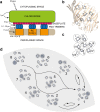
Fig. 1
Fig. 1
a Morphology of the green sulphur bacteria light harvesting apparatus. In green non-sulphur bacteria, the RC type I is replaced with RC type II and the FMO complex is missing. Transfer and charge separation times are given. b Structure of the FMO monomer (PDB 3eni). c Chlorophyll arrangement and chromophore labels for the FMO monomer. d Exciton states and energy flow downhill between them from state 8 to state 1. Acutely studied transfer rates and coherence times are labelled as arrows and ellipses respectively. Exciton states were calculated by finding eigenvectors of the FMO Hamiltonian (Adolphs and Renger 2006) in Mathematica. Exciton states are calculated from the Hamiltonian given by. Chlorophyll tails are removed for clarity. Structures were rendered in PyMol
Fig. 2
Fig. 2
a Representative relaxation and energy transfer times between different states and rings and morphology of proteins embedded in the chromatophore membrane. b Crystal structure of LH I (PDB: 3wmm) with the chlorophyll ring labelled. The inset highlights the alternating chlorophyll orientations. c Crystal structure of LH II (PDB: 2fkw) with the chlorophyll rings labelled. The inset highlights the alternating chlorophyll orientations of B850. d A selection of important exciton states of the B850 ring; two isoenergetic first excited states at the top (with their dipole moments) and the ground state at the bottom. Exciton states were calculated by finding eigenvectors of the B850 Hamiltonian (Strümpfer and Schulten 2009) in Mathematica. Chlorophyll tails have been removed for clarity and structures were rendered in PyMol
Fig. 3
Fig. 3
a Morphology of the phycobilisome light harvesting apparatus. Also highlighted is the flow of energy down the phycobilisome. Note that the relative amounts of each PE/PC are light dependent. b Crystal structure of R-PE (PDB: 1b8d) with and without protein. c Crystal structure of APC in two orientations (PDB: 1kn1) with and without protein. Structures rendered in PyMol
Fig. 4
Fig. 4
a Morphology of the cryptophyte light harvesting apparatus and suggested energy transfer pathway. b PC645 energy transfer rates as illustrative of other PBPs. Energy transfer and relaxation are shown on one half of the dimer to avoid crowding the figure. Coherences are shown as dashed ellipses (PDB: 4lms). c Crystal structure of the closed form PE545 with and without protein. The two dimer halves are shaded and the central pair is circled (PDB: 1xg0). d Crystal structure of the closed form PE555 with and without protein. The two dimer halves are shaded with the central pair circled and arrows highlighting their increased separation. (PDB: 4lmx). Phycocyanin, green; mesobiliverdin, blue; dihydrobiliverdin, orange; and phycoerythrin, red. Structures rendered in PyMol
Fig. 5
Fig. 5
a Morphology of the dinoflagellate light harvesting apparatus with putative energy transfer path. b Crystal structure of PCP monomer (PDB: 1ppr) in two orientations and with protein and lipid stripped away highlighting chlorophyll (in green) surrounded by 4 peridinins each (in magenta). Structures rendered in PyMol
Fig. 6
Fig. 6
Structure of the reaction centres (types I and II) with labelled chromophores and electron transport between them. Reaction centres have been stripped of accessory peptide chains and other electron transport chain proteins (PDB: 3ZKI - type II and PDB: 1JB0 - type I). Structures rendered in PyMol

Similar articles

Cited by

References

    1. Adolphs J, Renger T. How proteins trigger excitation energy transfer in the FMO complex of green sulfur bacteria. Biophys J. 2006;91:2778–2797. - PMC - PubMed
    1. Arpin PC, Turner DB, McClure SD, et al. Spectroscopic studies of cryptophyte light harvesting proteins: vibrations and coherent oscillations. J Phys Chem B. 2015;119:10025–10034. - PubMed
    1. Baghbanzadeh S, Kassal I. Geometry, supertransfer, and optimality in the light harvesting of purple bacteria. J Phys Chem Lett. 2016;7:3804–3811. - PubMed
    1. Book LD, Ostafin AE, Ponomarenko N, et al. Exciton delocalization and initial dephasing dynamics of purple bacterial LH2. J Phys Chem B. 2000;104:8295–8307.
    1. Bricker WP, Lo CS. Efficient pathways of excitation energy transfer from delocalized S2 excitons in the peridinin-chlorophyll a-protein complex. J Phys Chem B. 2015;119:5755–5764. - PubMed

LinkOut - more resources