

3D Brain Imaging in Vascular Segmentation of Cerebral Venous Sinuses
- PMID: 30242780
- PMCID: PMC6456638
- DOI: 10.1007/s10278-018-0125-4
3D Brain Imaging in Vascular Segmentation of Cerebral Venous Sinuses
Abstract
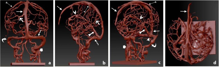
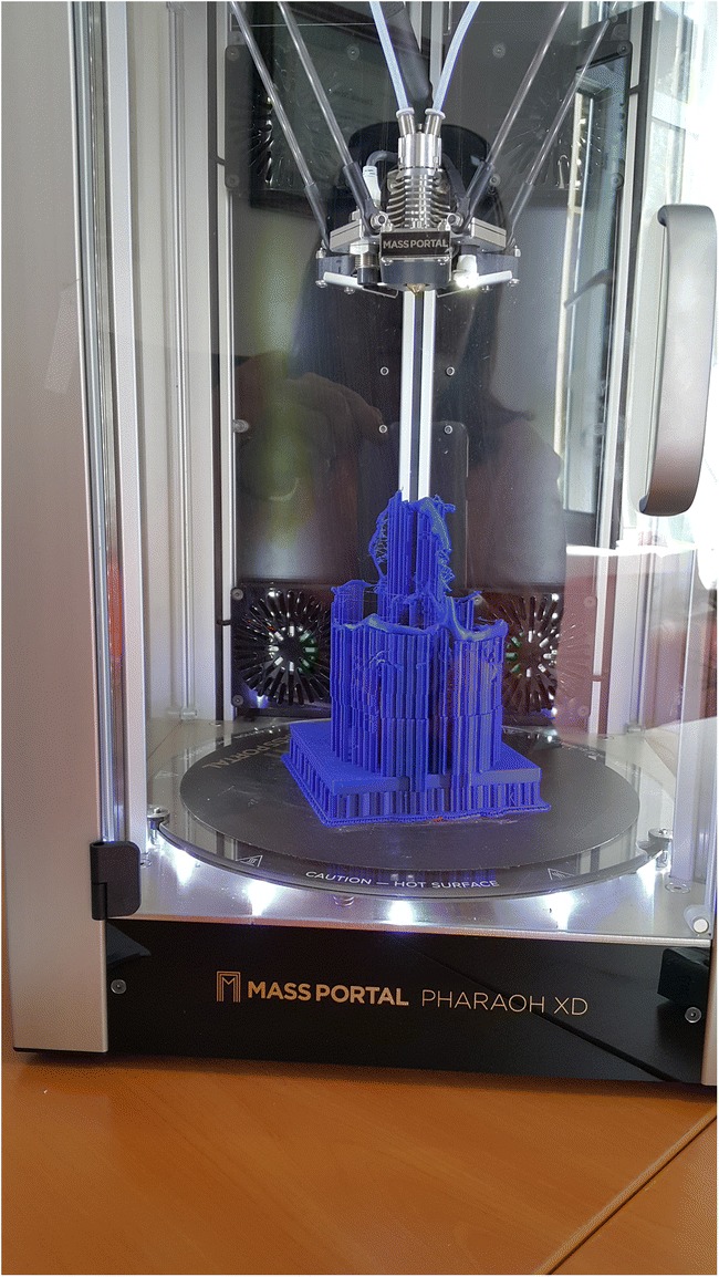
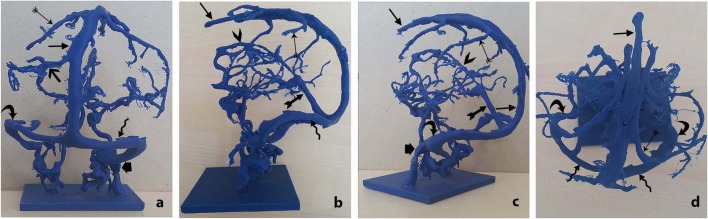
The three-dimensional (3D) visualization of dural venous sinuses (DVS) networks is desired by surgical trainers to create a clear mental picture of the neuroanatomical orientation of the complex cerebral anatomy. Our purpose is to document those identified during routine 3D venography created through 3D models using two-dimensional axial images for teaching and learning neuroanatomy. Anatomical data were segmented and extracted from imaging of the DVS of healthy people. The digital data of the extracted anatomical surfaces was then edited and smoothed, resulting in a set of digital 3D models of the superior sagittal, inferior sagittal, transverse, and sigmoid, rectus sinuses, and internal jugular veins. A combination of 3D printing technology and casting processes led to the creation of realistic neuroanatomical models that include high-fidelity reproductions of the neuroanatomical features of DVS. The life-size DVS training models were provided good detail and representation of the spatial distances. Geometrical details between the neighboring of DVS could be easily manipulated and explored from different angles. A graspable, patient-specific, 3D-printed model of DVS geometry could provide an improved understanding of the complex brain anatomy. These models have various benefits such as the ability to adjust properties, to convert two-dimension images of the patient into three-dimension images, to have different color options, and to be economical. Neuroanatomy experts can model such as the reliability and validity of the designed models, enhance patient satisfaction with improved clinical examination, and demonstrate clinical interventions by simulation; thus, they teach neuroanatomy training with effective teaching styles.
Keywords: 3D neuroanatomical models; Brain imaging; Clinical skills; Dural venous sinuses; Minimally invasive neurosurgery; Neurosurgical education; Surgical trainers.
Conflict of interest statement
The authors declare that they have no conflict of interest.
This article has not been submitted or published elsewhere in part or in whole and that it is original work.
Figures
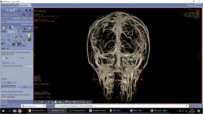
 superior sagittal sinus,
  superior anastomotic vein,
  inferior anastomotic vein,
  transverse sinus,
  straight sinus,
  sigmoid sinus,
  inferior sagittal sinus,
  occipital sinus,
  internal jugular vein
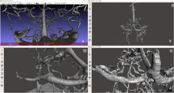
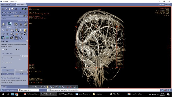
 superior sagittal sinus,
  superior anastomotic vein,
  inferior anastomotic vein,
  transverse sinus,
  straight sinus,
  sigmoid sinus,
  inferior sagittal sinus,
  occipital sinus,
  internal jugular vein
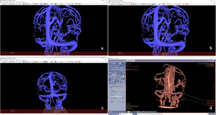
References
MeSH terms
LinkOut - more resources
Full Text Sources
Other Literature Sources
Medical
