Review
. 2018:81:385-455.
doi: 10.1016/bs.ctm.2018.07.006. Epub 2018 Aug 27.

Cell Volume Control in Healthy Brain and Neuropathologies

Affiliations
Review

Cell Volume Control in Healthy Brain and Neuropathologies

Corinne S Wilson et al. Curr Top Membr. 2018.

Abstract

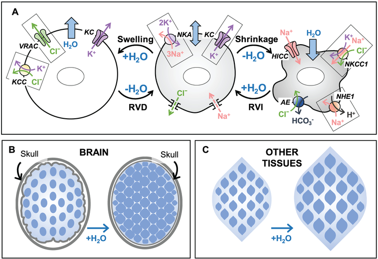
Regulation of cellular volume is a critical homeostatic process that is intimately linked to ionic and osmotic balance in the brain tissue. Because the brain is encased in the rigid skull and has a very complex cellular architecture, even minute changes in the volume of extracellular and intracellular compartments have a very strong impact on tissue excitability and function. The failure of cell volume control is a major feature of several neuropathologies, such as hyponatremia, stroke, epilepsy, hyperammonemia, and others. There is strong evidence that such dysregulation, especially uncontrolled cell swelling, plays a major role in adverse pathological outcomes. To protect themselves, brain cells utilize a variety of mechanisms to maintain their optimal volume, primarily by releasing or taking in ions and small organic molecules through diverse volume-sensitive ion channels and transporters. In principle, the mechanisms of cell volume regulation are not unique to the brain and share many commonalities with other tissues. However, because ions and some organic osmolytes (e.g., major amino acid neurotransmitters) have a strong impact on neuronal excitability, cell volume regulation in the brain is a surprisingly treacherous process, which may cause more harm than good. This topical review covers the established and emerging information in this rapidly developing area of physiology.

Keywords: Brain; Cell swelling; Cell volume regulation; Central nervous system; Epilepsy; Excitotoxicity; Glutamate release; Hyponatremia; Hyrerammonemia; Stroke; Volume-regulated anion channel.


Figures

Figure 12.1
Figure 12.1
Basic principles of cell volume regulation in mammalian cells and major differences in the osmotic homeostasis within the brain and in other tissues. (A) Ion transport mechanisms responsible for cell volume regulation. Center panel: Under isosmotic conditions, the work of the Na+,K+-ATPase (NKA) and the dominant activity of K+ channels (KC), set the transmembrane ionic gradients and negative membrane potential, as well as compensate for the persistent Na+ uptake via a variety of mechanisms (Na+ leak). Importantly, the negative membrane potential drives out intracellular Cl, offsetting the presence of the negatively charged impermeant macromolecules and metabolites in the cytosol. Left panel: Hypoosmotic cell swelling triggers activation of the volume-regulated Cl/anion channel (VRAC) and several K+,Cl cotransporters (KCC). Cooperative activity of VRAC, KC, and KCC mediates the loss of cytosolic KCl and powers regulatory volume decrease (RVD). Right panel: Cell shrinkage upon exposure to hyperosmotic media stimulates the ubiquitous Na+,K+,Cl cotransporter 1 (NKCC1) and/or the Na+/H+ exchanger 1 (NHE1). NHE works in cooperation with the volume-insensitive Cl/HCO3anion exchangers (AE). In some cell types, cell shrinkage also opens hypertonicity induced non-selective cation channels (HICC). The combined activity of these transporters and channels leads to cytosolic accumulation of NaCl and KCl and mediates regulatory volume increase (RVI). (B and C) The major differences in cell volume control between the brain and other tissues are due to the fixed volume of (extracellular + intracellular) space in the CNS. (B) In the brain, due to spatial limitations imposed by the rigid skull, cell swelling under hypoosmotic conditions or in pathologies occurs at the expense of the interstitial volume and may also compress blood vessels and cause ischemia. Also, due to the restricted ion transport across the blood-brain barrier, there is a “fixed” total pool of extracellular and intracellular ions. Therefore, during cell volume regulation, the electrochemical driving forces for ionic fluxes dissipate very quickly. (C) In contrast to the brain, the majority of peripheral tissues are not restricted in terms of their osmotic expansion or shrinkage, and allow for the relatively rapid exchange of electrolytes between the interstitial space and the blood.
Figure 12.2
Figure 12.2
Swelling-activated Cl currents and amino acid release through VRAC in vitro and in vivo. (A) Representative whole-cell recordings of Cl currents in primary rat astrocytes exposed to hypoosmotic medium (−60 mOsm). Activity of Cl channels was measured by holding cells at 0 mV and alternately administering ± 40 mV voltage pulses. Currents were inhibited by treatment with the VRAC blocker DCPIB (20 μm). (B) Swelling-activated Cl currents in astrocytes in response to 20 mV step pulses from −100 to +100 mV, displaying the characteristic outward rectification and time-dependent inactivation at positive potentials. (C) Effect of DCPIB on swelling-activated glutamate release in primary astrocytes, traced with the non-metabolizable glutamate analog D-[3H]aspartate. ***p < 0.001, effect of DCPIB. (D) Effect of the non-specific VRAC blocker DNDS on swelling-activated glutamate release in the rat cortex after stimulation with hypoosmotic artificial cerebrospinal fluid, measured by microdialysis approach and analyzed with HPLC. *p <0.05, effect of DNDS. (A-C) adapted from I.F. Abdullaev et al. (2006), with permission. (D) Reproduced from R.E. Haskew-Layton et al., 2008, under the Creative Commons Attribution (CC BY) license.
Figure 12.3
Figure 12.3
Astrocytic properties that may be responsible for preferential propensity of this cell type to cell swelling in neuropathologies. (A) Astrocyte membranes are highly permeable to water due to expression of the water channel aquaporin-4 (AQP4); (B) Astrocytes take up neurotransmitters from the extracellular space in order to maintain normal neuronal activity. Excitatory amino acid transporters (EAAT) take one glutamate in together with 3 Na+ and in exchange of 1 K+, and promote the accumulation of osmotically obligated water; (C) Buffering of [K+]o through potassium channels (KC) can be associated with the concomitant influx of Cl through VRAC and promote Donnan cell swelling; (D) In normal brain and neuropathologies, astrocytes accumulate extracellular ammonia (NH3) via passive transmembrane diffusion and ammonium ions (NH4+) through potassium channels (KCs) and NKCC1. Inside the cell, NH3/NH4+ is then assimilated to produce glutamine from glutamate. (E) Upregulation and activation of the nonselective SUR1-TRPM4 channels and their assembly with AQP4 promotes Na+ uptake and water accumulation; (F) Astrocytic NKCC1 cotransporter contributes to electrolyte and water accumulation in response to high [K+]o. Cell swelling may paradoxically activate NKCC1 via the low [Cl]i-sensing WNK/SPAK/OSR1 cascade and in such a way further amplify the persistent astrocytic swelling.
Figure 12.4
Figure 12.4
Cell swelling in status epilepticus revealed by electron microscopy (EM). (A) Structural analysis of the control rat neocortex reveals neuronal cell bodies [N], capillaries [Cap], and pericytes [P]. (B and C) Two hours following injection of 4-aminopyridine, EM shows swollen perivascular astrocytic endfeet [asterisks in (B)], which surround an endothelial cell [E]. Swollen astrocytic processes [asterisks in (C)] also border an apparently shrunken pyramidal neuronal body [N]. (D) A higher magnification image in the CA3 layer of the hippocampus displays a swollen dendrite [D] with apparently normal mitochondrion [M], and adjacent electron-transparent swollen astrocytic processes [asterisks]. Additionally labeled are: an axon terminal [A] and dendritic spines [1,2]. The scale bars in the fields (A-C) = 5 μm, and in (D) = 1 μm. Reproduced from P.F. Fabene et al., 2006, with permission.
Figure 12.5
Figure 12.5
MRI imaging modes which are used in clinical settings to visualize changes in water mobility (cell swelling) following ischemic stroke. Left panel: At early times after the onset of focal ischemic stroke, fast spin-echo T2-weighted MRI imaging has yet to show brain damage. Center panel: In contrast to the T2 imaging, diffusion weighted imaging (DWI) shows a large hyperintense area in the territories of the prefrontal artery (arrowhead) and a smaller signal in the territory of the anterior cerebral artery (arrow). Hyperintensity reflects a decrease in water mobility upon its shift from the extracellular to the intracellular space. Right panel: Apparent diffusion coefficient (ADC) of water identifies the regions of low water mobility as hypointense signals. In 2–3 days, the DWI/ADC positive areas will likely develop full infarction that will be apparent in T2 imaging (not shown). Reproduced from M.G. Lansberg et al., 1999, with permission.
Figure 12.6
Figure 12.6
Intracerebral treatment with the VRAC blocker DCPIB potently protects against ischemic brain damage in a rat model of stroke induced by 2-hour occlusion of the middle cerebral artery (MCAo). (A) Representative images of brain sections prepared from MCAo animals after intracerebral injection of vehicle (DMSO) or 20 μg/kg DCPIB. (B) Brain sections from MCAo animal which were treated systemically with intravenous injection of vehicle or 10 mg/kg DCPIB. Animals were euthanized 3 days after ischemia and the extent of brain infarction was revealed by staining brain sections with 2,3,5-triphenyltetrazolium chloride (viable tissue is red). Modified from Y. Zhang et al., 2008, with permission.
Figure 12.7
Figure 12.7
RNAi analysis reveals that astrocytes express two types of heteromeric LRRC8-containing VRAC channels with differential permeability to charged and uncharged organic osmolytes. (A–D) Primary rat astrocytes were transfected with negative control siRNA (Control), or the gene-specific siRNA targeting the expression of one or more members of the proteins belonging to the family of leucine-rich repeat-containing 8 (LRRC8) proteins. After a 4-day incubation with the gene specific siRNA constructs, astrocytes were simultaneously loaded with [3H]taurine and the glutamate analog D-[14C]aspartate, and superfused with isosmotic or hypoosmotic (HYPO) media to activate VRAC and quantify the amino acid release. Taurine and glutamate fluxes have been measured simultaneously but, for clarity, are presented in separate panels (A-B) and (C-D). Knockdown of LRRC8A reduced release of all amino acids. Knockdown of LRRC8D reduced [3H]taurine release by ~50% (A), but had no effect on the release of D-[14C]aspartate (B). Conversely, the combined knockdown of LRRC8C + LRRC8E inhibited the efflux of D-[14C]aspartate (D), but not [3H]taurine (C). Finally, and surprisingly, the combined knockdown of LRRC8C + LRRC8D inhibited the release of all amino acids to the same extent as the knockdown of LRRC8A. (E) The proposed composition of the two populations of LRRC8-containing VRAC heteromers which explains the data in (A-D). The LRRC8A+D combination is responsible for the movement of uncharged molecules such as taurine and myo-inositol (Ino) (data not shown). Another channel composed of LRRC8A+C+D+E is permeable to negatively charged molecules, including glutamate. Because zwitterionic taurine is partially negatively charged, it shares both pathways. **p<0.01, ***p<0.001, amino acid release rates vs. cells treated with the negative control siRNA. Adapted from A.L. Schober et al. (2017), with permission.

References

    1. Abdullaev IF, Rudkouskaya A, Schools GP, Kimelberg HK, & Mongin AA (2006). Pharmacological comparison of swelling-activated excitatory amino acid release and Cl- currents in rat cultured astrocytes. J.Physiol, 572, 677–689. - PMC - PubMed
    1. Adrogue HJ & Madias NE (2000). Hyponatremia. N.Engl.J.Med, 342, 1581–1589. - PubMed
    1. Aitken PG, Borgdorff AJ, Juta AJ, Kiehart DP, Somjen GG, & Wadman WJ (1998). Volume changes induced by osmotic stress in freshly isolated rat hippocampal neurons. Pflugers Arch., 436, 991–998. - PubMed
    1. Aitken PG, Fayuk D, Somjen GG, & Turner DA (1999). Use of intrinsic optical signals to monitor physiological changes in brain tissue slices. Methods, 18, 91–103. - PubMed
    1. Akita T, Fedorovich SV, & Okada Y (2011). Ca2+ nanodomain-mediated component of swelling-induced volume-sensitive outwardly rectifying anion current triggered by autocrine action of ATP in mouse astrocytes. Cell Physiol Biochem., 28, 1181–1190. - PubMed

Publication types

LinkOut - more resources