RF-induced heating in tissue near bilateral DBS implants during MRI at 1.5 T and 3T: The role of surgical lead management
- PMID: 30243973
- PMCID: PMC6475594
- DOI: 10.1016/j.neuroimage.2018.09.034
RF-induced heating in tissue near bilateral DBS implants during MRI at 1.5 T and 3T: The role of surgical lead management
Abstract
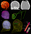
Access to MRI is limited for patients with deep brain stimulation (DBS) implants due to safety hazards, including radiofrequency (RF) heating of tissue surrounding the leads. Computational models provide an exquisite tool to explore the multi-variate problem of RF heating and help better understand the interaction of electromagnetic fields and biological tissues. This paper presents a computational approach to assess RF-induced heating, in terms of specific absorption rate (SAR) in the tissue, around the tip of bilateral DBS leads during MRI at 64MHz/1.5 T and 127 MHz/3T. Patient-specific realistic lead models were constructed from post-operative CT images of nine patients operated for sub-thalamic nucleus DBS. Finite element method was applied to calculate the SAR at the tip of left and right DBS contact electrodes. Both transmit head coils and transmit body coils were analyzed. We found a substantial difference between the SAR and temperature rise at the tip of right and left DBS leads, with the lead contralateral to the implanted pulse generator (IPG) exhibiting up to 7 times higher SAR in simulations, and up to 10 times higher temperature rise during measurements. The orientation of incident electric field with respect to lead trajectories was explored and a metric to predict local SAR amplification was introduced. Modification of the lead trajectory was shown to substantially reduce the heating in phantom experiments using both conductive wires and commercially available DBS leads. Finally, the surgical feasibility of implementing the modified trajectories was demonstrated in a patient operated for bilateral DBS.
Keywords: Computational modeling and simulations; Deep brain stimulation (DBS); Finite element method (FEM); MRI safety; Magnetic resonance imaging (MRI); Medical implants; Neuromodulation; Neurostimulation; Specific absorption rate (SAR).
Copyright © 2018 Elsevier Inc. All rights reserved.
Figures


References
-
- Limousin P et al. , “Effect on parkinsonian signs and symptoms of bilateral subthalamic nucleus stimulation,” The Lancet, vol. 345, no. 8942, pp. 91–95, 1995 - PubMed
-
- Benabid AL, “Deep brain stimulation for Parkinson’s disease,” Current opinion in neurobiology, vol. 13, no. 6, pp. 696–706, 2003. - PubMed
-
- Benabid AL, Chabardes S, Mitrofanis J, and Pollak P, “Deep brain stimulation of the subthalamic nucleus for the treatment of Parkinson’s disease,” The Lancet Neurology, vol. 8, no. 1, pp. 67–81, 2009. - PubMed
-
- Kumar R, Dagher A, Hutchison W, Lang A, and Lozano A, “Globus pallidus deep brain stimulation for generalized dystonia: clinical and PET investigation,” Neurology, vol. 53, no. 4, pp. 871-871, 1999. - PubMed
Publication types
MeSH terms
Grants and funding
LinkOut - more resources
Full Text Sources
Other Literature Sources
Medical
Miscellaneous

