

Hypersensitivity Pneumonitis: Radiologic Phenotypes Are Associated With Distinct Survival Time and Pulmonary Function Trajectory
- PMID: 30243979
- PMCID: PMC6514431
- DOI: 10.1016/j.chest.2018.08.1076
Hypersensitivity Pneumonitis: Radiologic Phenotypes Are Associated With Distinct Survival Time and Pulmonary Function Trajectory
Abstract
Background: Hypersensitivity pneumonitis (HP) is an interstitial lung disease with a better prognosis, on average, than idiopathic pulmonary fibrosis (IPF). We compare survival time and pulmonary function trajectory in patients with HP and IPF by radiologic phenotype.
Methods: HP (n = 117) was diagnosed if surgical/transbronchial lung biopsy, BAL, and exposure history results suggested this diagnosis. IPF (n = 152) was clinically and histopathologically diagnosed. All participants had a baseline high-resolution CT (HRCT) scan and FVC % predicted. Three thoracic radiologists documented radiologic features. Survival time is from HRCT scan to death or lung transplant. Cox proportional hazards models identify variables associated with survival time. Linear mixed models compare post-HRCT scan FVC % predicted trajectories.
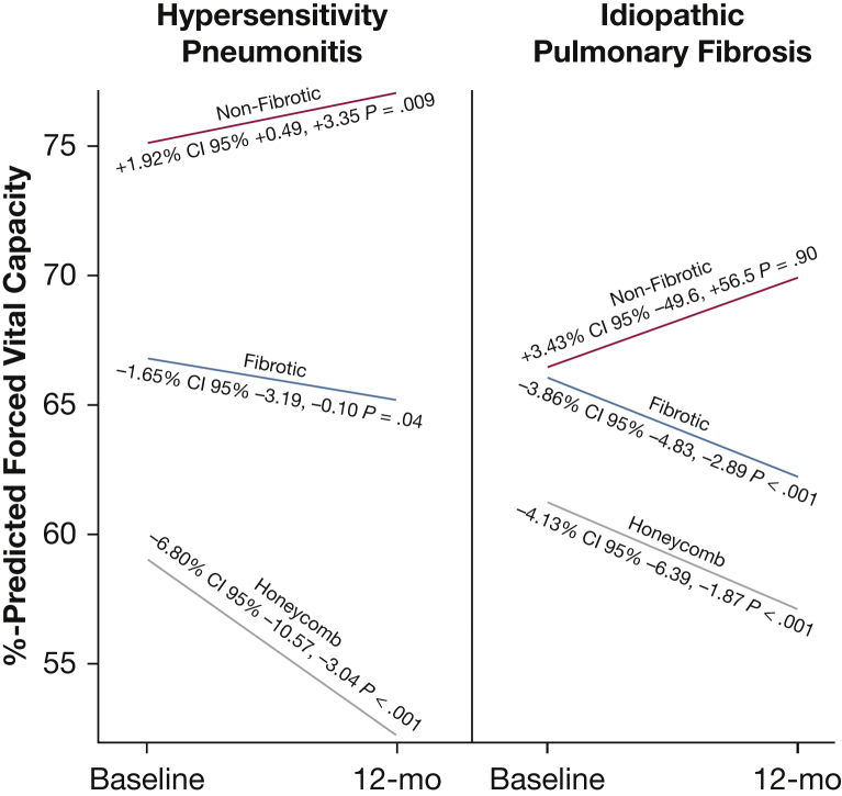
Results: Subjects were grouped by clinical diagnosis and three mutually exclusive radiologic phenotypes: honeycomb present, non-honeycomb fibrosis (traction bronchiectasis and reticulation) present, and nonfibrotic. Nonfibrotic HP had the longest event-free median survival (> 14.73 years) and improving FVC % predicted (1.92%; 95% CI, 0.49-3.35; P = .009). HP with non-honeycomb fibrosis had longer survival than IPF (> 7.95 vs 5.20 years), and both groups experienced a significant decline in FVC % predicted. Subjects with HP and IPF with honeycombing had poor survival (2.76 and 2.81 years, respectively) and significant decline in FVC % predicted.
Conclusions: Three prognostically distinct, radiologically defined phenotypes are identified among patients with HP. The importance of pursuing a specific diagnosis (eg, HP vs IPF) among patients with non-honeycomb fibrosis is highlighted. When radiologic honeycombing is present, invasive diagnostic testing directed at determining the diagnosis may be of limited value given a uniformly poor prognosis.
Keywords: interstitial lung disease; prognostic model; pulmonary fibrosis.
Copyright © 2018 American College of Chest Physicians. Published by Elsevier Inc. All rights reserved.
Figures


Comment in
-
CT Phenotypes in Hypersensitivity Pneumonitis.Chest. 2019 Apr;155(4):655-656. doi: 10.1016/j.chest.2018.10.048. Chest. 2019. PMID: 30955565 No abstract available.
References
-
- Churg A., Sin D.D., Everett D., Brown K., Cool C. Pathologic patterns and survival in chronic hypersensitivity pneumonitis. Am J Surg Pathol. 2009;33(12):1765–1770. - PubMed
-
- Hanak V., Golbin J.M., Hartman T.E., Ryu J.H. High-resolution CT findings of parenchymal fibrosis correlate with prognosis in hypersensitivity pneumonitis. Chest. 2008;134(1):133–138. - PubMed
-
- Mooney J.J., Elicker B.M., Urbania T.H. Radiographic fibrosis score predicts survival in hypersensitivity pneumonitis. Chest. 2013;144(2):586–592. - PubMed
Publication types
MeSH terms
Grants and funding
LinkOut - more resources
Full Text Sources
Other Literature Sources
Research Materials
Miscellaneous
