

Regulation of tumor cell - Microenvironment interaction by the autotaxin-lysophosphatidic acid receptor axis
- PMID: 30243984
- PMCID: PMC6433480
- DOI: 10.1016/j.jbior.2018.09.008
Regulation of tumor cell - Microenvironment interaction by the autotaxin-lysophosphatidic acid receptor axis
Abstract
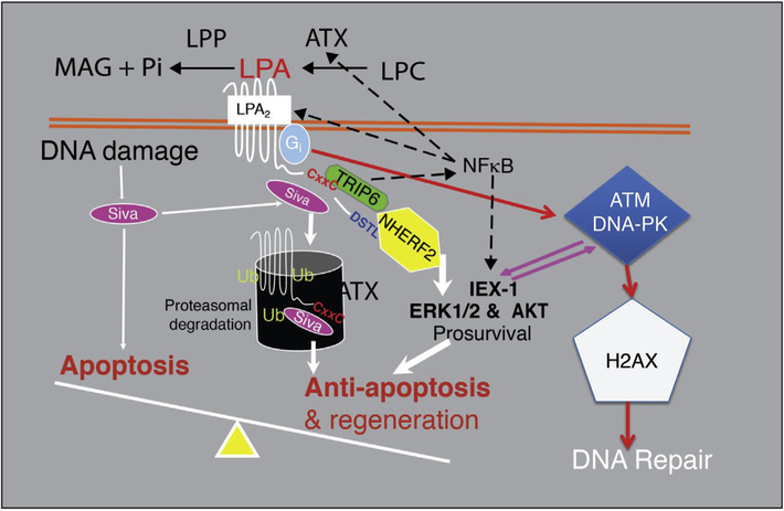
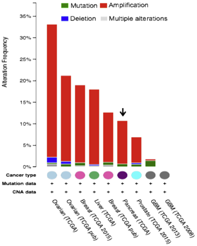
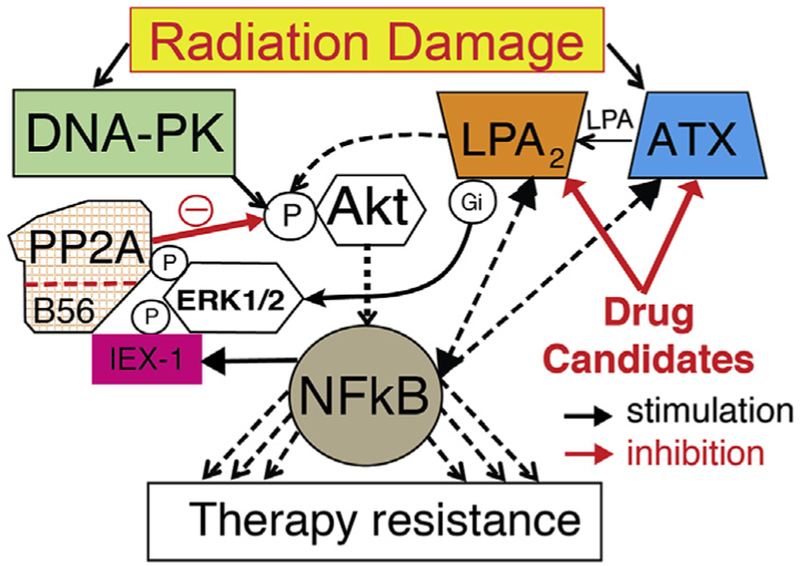
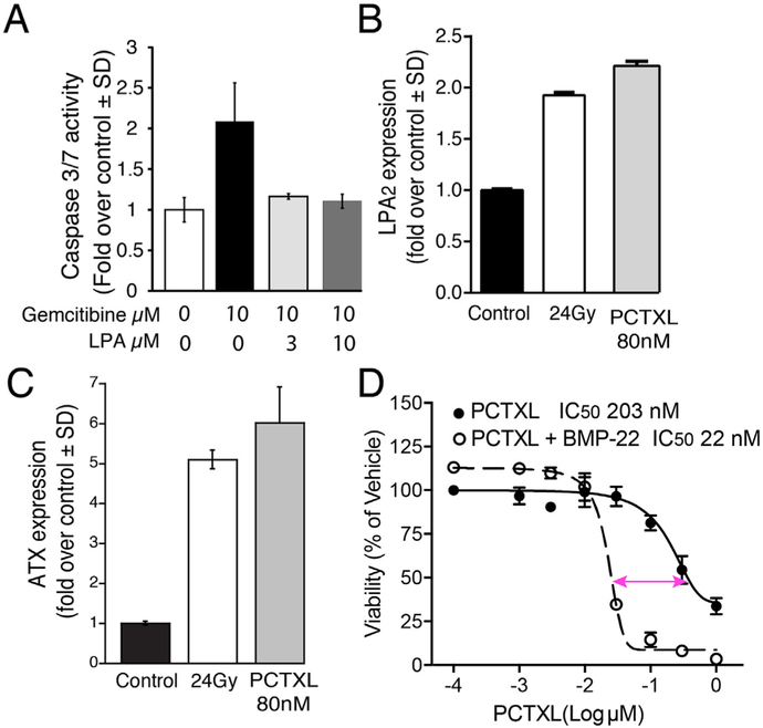
The lipid mediator lysophosphatidic acid (LPA) in biological fluids is primarily produced by cleavage of lysophospholipids by the lysophospholipase D enzyme Autotaxin (ATX). LPA has been identified and abundantly detected in the culture medium of various cancer cell types, tumor effusates, and ascites fluid of cancer patients. Our current understanding of the physiological role of LPA established its role in fundamental biological responses that include cell proliferation, metabolism, neuronal differentiation, angiogenesis, cell migration, hematopoiesis, inflammation, immunity, wound healing, regulation of cell excitability, and the promotion of cell survival by protecting against apoptotic death. These essential biological responses elicited by LPA are seemingly hijacked by cancer cells in many ways; transcriptional upregulation of ATX leading to increased LPA levels, enhanced expression of multiple LPA GPCR subtypes, and the downregulation of its metabolic breakdown. Recent studies have shown that overexpression of ATX and LPA GPCR can lead to malignant transformation, enhanced proliferation of cancer stem cells, increased invasion and metastasis, reprogramming of the tumor microenvironment and the metastatic niche, and development of resistance to chemo-, immuno-, and radiation-therapy of cancer. The fundamental role of LPA in cancer progression and the therapeutic inhibition of the ATX-LPA axis, although highly appealing, remains unexploited as drug development to these targets has not reached into the clinic yet. The purpose of this brief review is to highlight some unique signaling mechanisms engaged by the ATX-LPA axis and emphasize the therapeutic potential that lies in blocking the molecular targets of the LPA system.
Keywords: Cancer stem cell; ENPP2; Invasion; LPA; Metastasis; Therapy resistance.
Copyright © 2018. Published by Elsevier Ltd.
Figures

Similar articles
-
Autotaxin-Lysophosphatidate Axis: Promoter of Cancer Development and Possible Therapeutic Implications.Int J Mol Sci. 2024 Jul 15;25(14):7737. doi: 10.3390/ijms25147737. Int J Mol Sci. 2024. PMID: 39062979 Free PMC article. Review.
-
The autotaxin-lysophosphatidic acid-lysophosphatidic acid receptor cascade: proposal of a novel potential therapeutic target for treating glioblastoma multiforme.Lipids Health Dis. 2015 Jun 18;14:56. doi: 10.1186/s12944-015-0059-5. Lipids Health Dis. 2015. PMID: 26084470 Free PMC article. Review.
-
Autotaxin and LPA receptor signaling in cancer.Cancer Metastasis Rev. 2011 Dec;30(3-4):557-65. doi: 10.1007/s10555-011-9319-7. Cancer Metastasis Rev. 2011. PMID: 22002750 Review.
-
Controlling cancer through the autotaxin-lysophosphatidic acid receptor axis.Biochem Soc Trans. 2012 Feb;40(1):31-6. doi: 10.1042/BST20110608. Biochem Soc Trans. 2012. PMID: 22260662 Free PMC article. Review.
-
Role of the autotaxin-lysophosphatidate axis in the development of resistance to cancer therapy.Biochim Biophys Acta Mol Cell Biol Lipids. 2020 Aug;1865(8):158716. doi: 10.1016/j.bbalip.2020.158716. Epub 2020 Apr 16. Biochim Biophys Acta Mol Cell Biol Lipids. 2020. PMID: 32305571 Review.
Cited by
-
Novel Autotaxin Inhibitor ATX-1d Significantly Enhances Potency of Paclitaxel-An In Silico and In Vitro Study.Molecules. 2024 Sep 10;29(18):4285. doi: 10.3390/molecules29184285. Molecules. 2024. PMID: 39339280 Free PMC article.
-
Regulation of Tumor Immunity by Lysophosphatidic Acid.Cancers (Basel). 2020 May 10;12(5):1202. doi: 10.3390/cancers12051202. Cancers (Basel). 2020. PMID: 32397679 Free PMC article. Review.
-
An NMF-Based Methodology for Selecting Biomarkers in the Landscape of Genes of Heterogeneous Cancer-Associated Fibroblast Populations.Bioinform Biol Insights. 2020 May 8;14:1177932220906827. doi: 10.1177/1177932220906827. eCollection 2020. Bioinform Biol Insights. 2020. PMID: 32425511 Free PMC article.
-
Targeting Lysophosphatidic Acid in Cancer: The Issues in Moving from Bench to Bedside.Cancers (Basel). 2019 Oct 10;11(10):1523. doi: 10.3390/cancers11101523. Cancers (Basel). 2019. PMID: 31658655 Free PMC article. Review.
-
Anti-cancer strategies targeting the autotaxin-lysophosphatidic acid receptor axis: is there a path forward?Cancer Metastasis Rev. 2021 Mar;40(1):3-5. doi: 10.1007/s10555-021-09955-5. Cancer Metastasis Rev. 2021. PMID: 33454844 Free PMC article. No abstract available.
References
-
- Altman MK, Gopal V, Jia W, Yu S, Hall H, Mills GB, McGinnis AC, Bartlett MG, Jiang G, Madan D, Prestwich GD, Xu Y, Davies MA, Murph MM, 2010. Targeting melanoma growth and viability reveals dualistic functionality of the phosphonothionate analogue of carba cyclic phosphatidic acid. Mol. Canc 9, 140. - PMC - PubMed
-
- Baker DL, Morrison P, Miller B, Riely CA, Tolley B, Westermann AM, Bonfrer JM, Bais E, Moolenaar WH, Tigyi G, 2002. Plasma lysophosphatidic acid concentration and ovarian cancer. Jama 287 (23), 3081–3082. - PubMed
-
- Balogh A, Shimizu Y, Lee SC, Norman DD, Gangwar R, Bavaria M, Moon C, Shukla P, Rao R, Ray R, Naren AP, Banerje S, Miller DD, Balazs L, Pelus L, Tigyi G, 2015. The autotaxin-LPA2 GPCR axis is modulated by gamma-irradiation and facilitates DNA damage repair. Cell. Signal 27 (9), 1751–1762. - PMC - PubMed
-
- Bandoh K, Aoki J, Hosono H, Kobayashi S, Kobayashi T, Murakami-Murofushi K, Tsujimoto M, Arai H, Inoue K, 1999. Molecular cloning and characterization of a novel human G-protein- coupled receptor, EDG7, for lysophosphatidic acid. J. Biol. Chem 274 (39), 27776–27785. - PubMed
Publication types
MeSH terms
Substances
Grants and funding
LinkOut - more resources
Full Text Sources
Other Literature Sources
Miscellaneous
