. 2018 Nov 1;372(1):73-82.
doi: 10.1016/j.yexcr.2018.09.014. Epub 2018 Sep 21.

L-plastin phosphorylation regulates the early phase of sealing ring formation by actin bundling process in mouse osteoclasts

Affiliations

L-plastin phosphorylation regulates the early phase of sealing ring formation by actin bundling process in mouse osteoclasts

Meenakshi A Chellaiah et al. Exp Cell Res. .

Abstract

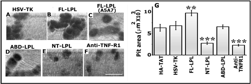
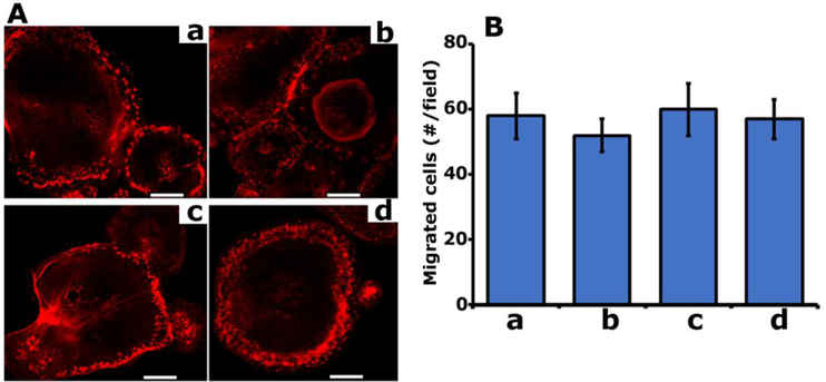
The process of sealing ring formation requires major actin filament reorganization. We previously demonstrated that an actin-bundling protein L-plastin has a role in the cross-linking of actin filaments into tight bundles and forms actin aggregates (denoted as nascent sealing zones). These nascent sealing zones mature into fully functional sealing rings. We have shown here that TNF-alpha signaling regulates the phosphorylation of serine-5 and -7 in L-plastin which increases the actin bundling capacity of L-plastin and hence the formation of nascent sealing zones in mouse osteoclasts. Using the TAT-mediated transduction method, we confirmed the role of L-plastin in nascent sealing zones formation at the early phase of the sealing ring assembly. Transduction of TAT-fused full-length L-plastin peptide significantly increases the number of nascent sealing zones and therefore sealing rings. But, transduction of amino-terminal L-plastin peptides consisting of the serine-5 and -7 reduces the formation of both nascent sealing zones and sealing rings. Therefore, bone resorption in vitro was reduced considerably. The decrease was associated with the selective inhibition of cellular L-plastin phosphorylation by the transduced peptides. Neither the formation of podosomes nor the migration was affected in these osteoclasts. Phosphorylation of L- plastin on serine 5 and -7 residues increases the F-actin bundling capacity. The significance of our studies stands on laying the groundwork for a better understanding of L-plastin as a potential regulator at the early phase of sealing ring formation and could be a new therapeutic target to treat bone loss.

Keywords: Bone resorption; Cytoskeleton; L-Plastin; Nascent sealing zones; Osteoclasts; Sealing rings.


Conflict of interest statement

Disclosures

All authors declare that they have no conflicts of interest.

Figures

Figure 1:
Figure 1:. Purification and analysis of dose and time-dependent uptake of TAT-fused LPL peptides.
Schematic diagram demonstrating various LPL constructs generated in a pTAT-HA expression vector is shown (Panel A). The domain organization of LPL is shown in full-length LPL (FL-LPL). The following are cloned separately into the pTAT-HA expression vector: FL-LPL, mutated FLLPL (A5A7), amino-terminal LPL containing S5 and S7 (NT-LPL), and actin-binding domains of LPL (ABD-LPL). The number within the parentheses indicates the first and last amino acid of the corresponding LPL peptide. The expression vector contains TAT protein transduction domain (10aa) and an HA- tag (20aa). SDS-PAGE analysis of purified TAT-fused LPL peptides is shown in Panel B. TAT-fused peptides were subjected to 8% (lanes 1 −3) and 15% (lanes 4 and 5) SDS-PAGE and stained with Coomassie blue. The purified proteins and their approximate molecular mass (kDa) are indicated below each lane. The standard molecular weight markers (kDa) are also indicated for 8% (left) and 15% (right) polyacrylamide gels (Panel B). Demonstration of a dose- and time-dependent uptake of TAT-fused FL-LPL in osteoclasts (Panels C and D). Immunoblotting analysis with an HA antibody was done to detect the transduced protein levels in osteoclast lysates. The blot was stripped and blotted with a GAPDH antibody for normalization (bottom panels of C and D). The results shown in B-D are representative of two different experiments from two different osteoclasts and TAT-protein preparations.
Figure 2:
Figure 2:. Immunoblotting analysis of phosphorylation of transduced and endogenous LPL protein in lysates made from osteoclasts
Osteoclasts treated with bone particles and TNF-α were also transduced with the following TATfused LPL and control (HSV-TK) peptides (150nM) for 3h as described in the Methods section: FL-LPL (Panel A; lanes 2 and 5); mutated FL-LPL (A5A7; lane 6); NT-LPL (lane 3), ABD-LPL (lane 4), and HSV-TK (lane 7). Lysates were immunoprecipitated with an antibody to LPL (lanes 1–7), HA (Panel D) or non-immune serum (NI; Panels A and D). The immunoprecipitates were subjected to 10% (Panel A) or 15% (Panel D) SDS-PAGE and immunoblotted with an antibody to phosphoserine (p-Serine; Panels A and D-lanes 1 and 2). Phosphorylated transduced LPL peptides and endogenous LPL protein are indicated in panels A and B. Stripping and reblotting of blot A with an antibody to LPL showed endogenous LPL and transduced FL-LPL (Panel B). Stripping and reblotting of blot D (left) with an antibody to HA shows the immunoprecipitated levels of transduced NT-LPL peptides (lane 4). Equal amount of total protein (Input) used for immunoprecipitation was assessed by direct immunoblotting of the lysates with a GAPDH antibody (Panels C and D-lanes 5 and 6). The results shown are representative of three different experiments from three different osteoclast preparations.
Figure 3:
Figure 3:. The effect of transduction of indicated TAT- LPL peptides on the formation of NSZs and total cellular F-actin content
Osteoclasts transduced with indicated LPL peptides were plated on dentine slices and incubated for 3–4h with TNF-α. Staining was performed with rhodamine-phalloidin for actin. Confocal images of osteoclasts are shown (Panels A-F). Arrows point to NSZs. Wavy arrows point to podosome-like structures. Scale bar-25μm. The number of NSZs were counted in ~ 75 osteoclasts and provided as a graph (Panel G). The data shown are the mean ± SEM of one of the three experiments performed with the same results. **p<0.01 versus HSV-TK transduced cells. F-actin content levels were determined by rhodamine-phalloidin binding to osteoclasts treated with anti-TNFR1 antibody as well as transduced or untransduced with indicated TATLPL peptides (Panel H). The F-actin content of the 0-min cells was assigned a value of 1.0 and all other values were expressed relative to the 0-mins values Values plotted are mean ± SD from three experiments *p<0.05 versus untreated (−) or HSV-TK transduced cells.
Figure 4:
Figure 4:. Analysis of the formation of sealing rings and resorption in osteoclasts
Osteoclasts transduced with indicated TAT-fused HSV-TK (A) and LPL (B-D) peptides were plated on dentine slices and incubated for 10–12h with TNF-α. Some cultures were treated with a neutralizing antibody to TNFR-1 (E). Confocal microscopy images of osteoclasts stained for actin (red) is shown. The reflected light in green is dentine. Overlay images show the distribution of actin protein (red) in osteoclasts plated on dentine slices (green). Resorption pits were found underneath where sealing rings were found in osteoclasts (indicated by arrows in red panels; AC). Resorption pits were outlined with white lines in green panels (A-C). Asterisks indicate punctate podosome-like structures in osteoclasts transduced with NT-LPL (panel D) or treated with a neutralizing antibody to TNFR-1 (E). Scale bar- 25μm. These results represent one of the three experiments performed with the similar results.. Sealing rings were counted in ~75 osteoclasts and provided as a graph in panel G; mean ±SEM. *** p<0.001; **p<0.01 versus HSV-TK transduced cells. Data provided are the representative of at least three independent experiments with comparable results.
Figure 5:
Figure 5:
Analysis of the effects of transduced TAT-LPL peptides on resorption by osteoclasts using dentine slices. Osteoclasts transduced with indicated TAT-fused peptides (Panels A-E) or treated with a neutralizing antibody to TNFR-1 (panel F) were cultured on dentine slices for 1012 h in the presence of TNF-α. Pits were scanned in a Bio-Rad confocal microscopy. Scale bar- 25μm. Resorbed area is seen as dark areas. Statistic measurements for the pit area are provided as a graph in G. ***p<0.001 and **p<0.05 versus HSV-TK transduced cells. The resorbed pit areas (20–25 pits/slice) were quantified and data were compiled from four slices per treatment. The data showed (G) is the mean±SEM of one experiment performed. Experiments were repeated three times with three different osteoclast preparations.
Figure 6:
Figure 6:
Analysis of the effect of TAT-fused LPL peptides on actin distribution in podosomes and migration. A. Actin staining with rhodamine phalloidin. TAT-fused FL-LPL (a), mutated FLLPL (b), ABD (c), and NT-LPL (d) were used. Confocal microscopy analysis of the actin stained cells is shown. Scale bar-25μm. B. Transwell migration assay. Data are presented as the number of cells per migrated field (mean ± SD) from one experiment of the three experiments performed
Figure 7:
Figure 7:
Schematic representation of the regulation of NSZ formation by TNF- α or RANKL signaling and maturation of NSZs to sealing rings by αvβ3 signaling.

Similar articles

Cited by

References

    1. Al TZ, Schaffner-Reckinger E, Halavatyi A, Hoffmann C, Moes M, Hadzic E, Catillon M, Yatskou M, Friederich E, Quantitative kinetic study of the actin-bundling protein L-plastin and of its impact on actin turn-over. PLoS.ONE. 5 (2010) e9210. - PMC - PubMed
    1. Arpin M, Friederich E, Algrain M, Vernel F, Louvard D, Functional differences between L- and T-plastin isoforms. J.Cell Biol 127 (1994) 1995–2008. - PMC - PubMed
    1. Babb SG, Matsudaira P, Sato M, Correia I, Lim S-S, Fimbrin in podosomes of monocyte-derived osteoclasts. Cell Motil.Cytosk. 37 (1997) 308–325. - PubMed
    1. Batsir S, Geiger B, Kam Z, Dynamics of the sealing zone in cultured osteoclasts. Cytoskeleton (Hoboken.) 74 (2017) 72–81. - PMC - PubMed
    1. Biswas R, Yuen D, Hruska K, MA. Chellaiah. Functional assessment of specific gelsolin peptide domains in osteoclast Actin Assembly and Motility. J Bone Miner Res 18 suppl1[Functional assessment of specific gelsolin peptide domains in osteoclast Actin Assembly and Motility], S279 10-10-2003.

Publication types

MeSH terms