. 2018 Dec;40(11-12):1507-1518.
doi: 10.1007/s10529-018-2605-5. Epub 2018 Sep 22.

Generation of Cas9 transgenic zebrafish and their application in establishing an ERV-deficient animal model

Affiliations

Generation of Cas9 transgenic zebrafish and their application in establishing an ERV-deficient animal model

Zhe Yang et al. Biotechnol Lett. 2018 Dec.

Abstract

Objectives: To investigate the effect of endogenous Cas9 on genome editing efficiency in transgenic zebrafish.

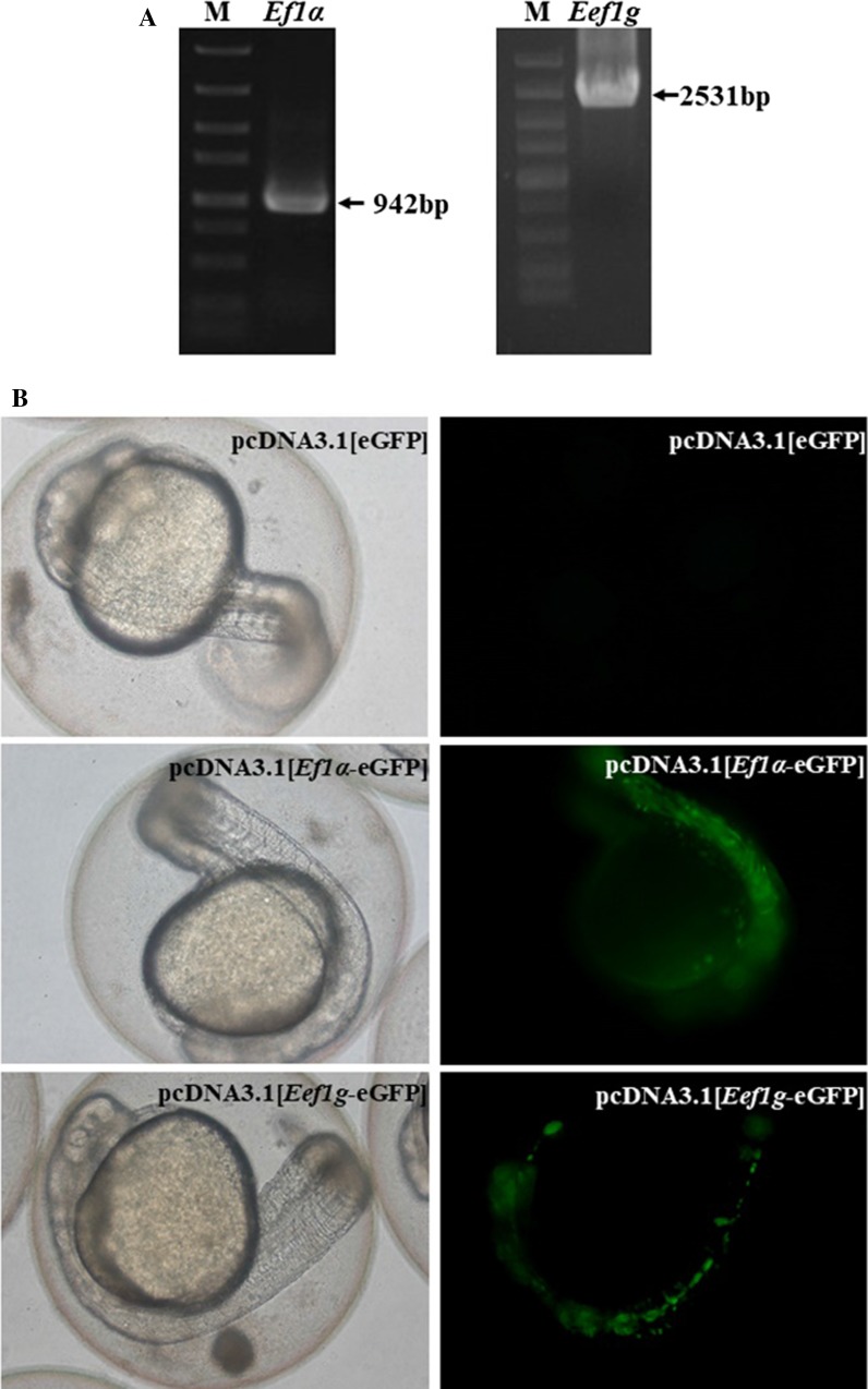
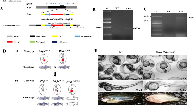
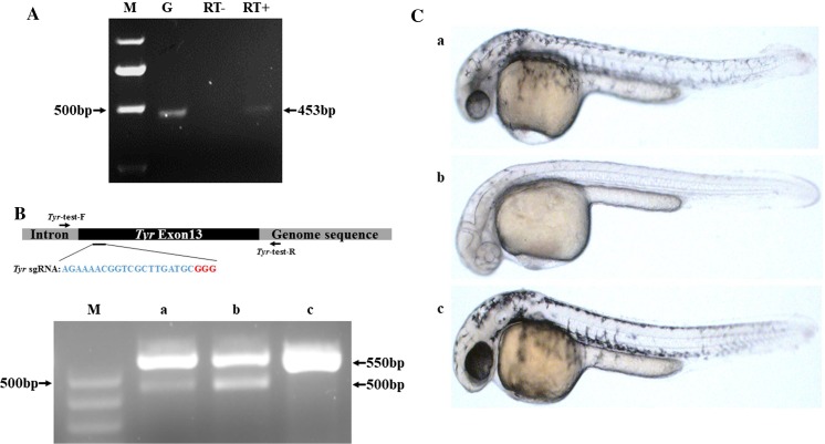
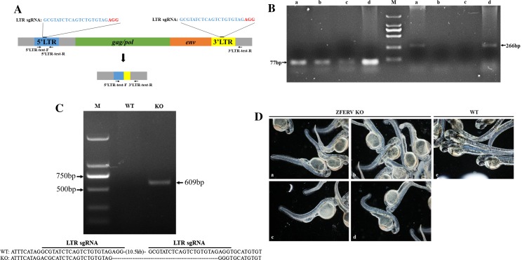
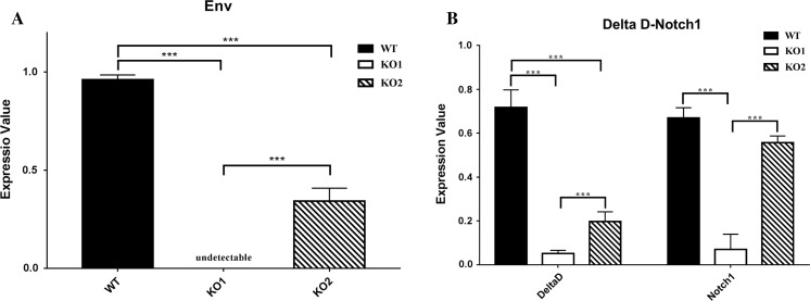
Results: Here we have constructed a transgenic zebrafish strain that can be screened by pigment deficiency. Compared with the traditional CRISPR injection method, the transgenic zebrafish can improve the efficiency of genome editing significantly. At the same time, we first observed that the phenotype of vertebral malformation in early embryonic development of zebrafish after ZFERV knockout.

Conclusions: The transgenic zebrafish with expressed Cas9, is more efficient in genome editing. And the results of ZFERV knockout indicated that ERV may affect the vertebral development by Notch1/Delta D signal pathway.

Keywords: CRISPR/Cas9; Embryonic development; Genomic editing; Spinal abnormality; Zebrafish.


Conflict of interest statement

The authors declare that they have no conflicts of interest with the contents of this article.

Figures

Fig. 1
Fig. 1
Selection of a promoter to drive transgenic Cas9 expression. a Amplification of Ef1α (942 bp) and Eef1g (2531 bp) promoter sequences. b Zebrafish embryos injected with pcDNA3.1 [eGFP], pcDNA3.1 [Ef1α-eGFP], and pcDNA3.1 [Eef1g-eGFP]. eGFP expression levels were indicated by fluorescent intensity
Fig. 2
Fig. 2
Generation of Cas9 transgenic zebrafish. a Schematic illustration of Mitfα locus-specific integration of the Ef1α-Cas9 expression cassette based on homologous recombination using Cas9 mRNA and sgRNA targeted to the zebrafish Mitfα gene. The indicated primers were used to confirm integration. HR denotes homologous recombination arm, and NLS denotes nuclear localization signal. b Confirmation of Cas9 gene insertion into the zebrafish genome, indicated by amplification of Cas9 by PCR with Cas9-specific primers. The expected size of the Cas9 amplicons is 453 bp. TU denotes genomic DNA isolated from wild-type zebrafish, while Cas9 denotes genomic DNA isolated from Cas9 transgenic zebrafish. M denotes the DNA ladder. c Confirmation of Mitfα-locus-specific integration of the Cas9 gene into the zebrafish genome, indicated by amplification of Cas9 by PCR with both Mitfα- and Cas9-specific primers. The expected size of the Cas9 amplicons is 1383 bp. TU denotes genomic DNA isolated from wild-type zebrafish, while Cas9 denotes genomic DNA isolated from Cas9 transgenic zebrafish. M denotes the DNA ladder. d Selection of zebrafish with biallelic transgenic Cas9 gene based on the nacre phenotype (pigment-deficient mutant), including genotyping and breeding of F0 zebrafish and selection for nacre phenotype-carrying zebrafish in F1. Based on phenotype, 23% (260/1130) of F1 zebrafish carry the nacre phenotype. The red box denotes the transgenic Cas9 gene. e Comparison of phenotypes of TU wild-type zebrafish with those of Mitfα-locus-specific transgenic Cas9 zebrafish at different developmental stages. Hpf and mpf indicate hours post-fertilization and months post-fertilization, respectively
Fig. 3
Fig. 3
Validation of Cas9 activity in F2 transgenic zebrafish. a Transgenic Cas9 expression was detected by reverse transcription (RT)-PCR in F2 transgenic zebrafish. Lane G: genomic DNA from F2 transgenic zebrafish as the positive control. Lane RT-: RT with no reverse transcriptase as the negative control. Lane RT + : cDNA synthesized by RT with total RNA isolated from F2 transgenic zebrafish. Lane M: DNA ladder. The expected size of the amplicon is 453 bp. b Validation of transgenic Cas9 activity by Tyr mutation assay and comparison of the editing efficiency of exogenous Cas9 mRNA and transgenic Cas9. The top panel shows the targeting site of the Tyr sgRNA in the Tyr gene, as well as the binding sites of the PCR primers used to detect the Tyr mutation in F2 zebrafish. The bottom panel shows the detection of the Tyr mutation via PCR and T7E1 digestion in fertilized eggs from TU wild-type zebrafish, which were injected with Tyr sgRNA plus Cas9 mRNA (a) vs. fertilized eggs from Cas9 transgenic zebrafish, which were injected with Tyr sgRNA (b), and TU wildtype zebrafish as control (c). The Tyr mutation was indicated by two fragments (approximately 550 bp and 500 bp, as indicated). c Different phenotypic changes in pigmentation fading in Tyr knockout zebrafish embryos produced via different gene editing methods. The representative images show a embryos injected with Cas9 mRNA and sgRNA, b Cas9 transgenic embryos injected with sgRNA and c wild-type embryos
Fig. 4
Fig. 4
Generation of ERV-deficient zebrafish. a Schematic overview of ZFERV gene knockout. 5′LTR-test-F and 5′LTR-test-R primers were used to PCR-amplify the 5′LTR sequence, while 5′LTR-test-F and 3′LTR-test-R primers were used to PCR-amplify the sequence between the 5′ and 3′LTR. b Gel images of PCR amplicons synthesized using GAPDH primers and the 5′LTR-test-F and 5′LTR-test-R primers for the ZFERV 5‘LTR used in the KO test. The primers 5′LTR-test-F and 5′LTR-test-R were used to amplify the ZFERV 5′LTR sequence. The expected sizes of the amplicons are indicated. Lane a: collected fertilized zebrafish eggs injected with LTR sgRNAs; Lane b and Lane c: zebrafish embryos with spinal deformity at 48 h and 120 h post-fertilization; Lane d: TU wild-type zebrafish embryos; M denotes the DNA ladder. c Detection of ZFERV gene deletion, which is indicated by the gel image of PCR amplicons synthesized from zebrafish genomic DNA using the primers 5′LTR-test-F and 3′LTR-test-R. The primers 5′LTR-test-F and 3′LTR-test-R were used to amplify the region between the 5′ and 3′LTR sequences. The expected size of the amplicons was 609 bp. The amplicon sequences were determined by sequencing analysis and are shown at the bottom. WT genomic DNA isolated from TU wild-type zebrafish, KO genomic DNA isolated from ERV-knockout zebrafish, M DNA ladder. (d) Spinal abnormality was caused by various sgRNAs in ZFERV-knockout (KO) zebrafish embryos. ZFERV knockout embryos produced by separately injecting LTR-sgRNA (a), sgRNA1 (b), sgRNA2 (c), or sgRNA3 (d) into the Cas9-transgenic zebrafish embryos. e Normal spinal morphology in wild-type embryos
Fig. 5
Fig. 5
Quantitative analysis of Env, Delta D, and notch1 gene expression. a Env gene expression in TU wild-type zebrafish and ERV-knockout zebrafish generated from Cas9 transgenic fish (KO1) or by microinjection of Cas9 mRNA and LTR sgRNAs into fertilized eggs (KO2). The expression level is normalized to that in TU wild-type zebrafish. ***p < 0.001. b Delta D and Notch1 gene expression in TU wild-type zebrafish and ERV-knockout zebrafish generated from Cas9-transgenic fish (KO1) or by microinjection of Cas9 mRNA and LTR sgRNAs into fertilized eggs (KO2). The expression level is normalized to that in TU wild-type zebrafish (Values are adjusted mean ± SEM, n = 3 for WT, n = 5 for ZFERV KO). ***p < 0.001

Similar articles

Cited by

References

    1. Burket CT, Montgomery JE, Thummel R, Kassen SC, LaFave MC, et al. Generation and characterization of transgenic zebrafish lines using different ubiquitous promoters. Transgenic Res. 2008;17:265–279. doi: 10.1007/s11248-007-9152-5. - DOI - PMC - PubMed
    1. Chen B, Gilbert LA, Cimini BA, Schnitzbauer J, Zhang W, et al. Dynamic imaging of genomic loci in living human cells by an optimized CRISPR/Cas system. Cell. 2013;155:1479–1491. doi: 10.1016/j.cell.2013.12.001. - DOI - PMC - PubMed
    1. Chen S, Lee B, Lee AY, Modzelewski AJ, He L. Highly efficient mouse genome editing by CRISPR ribonucleoprotein electroporation of zygotes. J Biol Chem. 2016;291:14457–14467. doi: 10.1074/jbc.M116.733154. - DOI - PMC - PubMed
    1. Chuong EB, Elde NC, Feschotte C. Regulatory evolution of innate immunity through co-option of endogenous retroviruses. Science. 2016;351:1083–1087. doi: 10.1126/science.aad5497. - DOI - PMC - PubMed
    1. Jao LE, Wente SR, Chen W. Efficient multiplex biallelic zebrafish genome editing using a CRISPR nuclease system. Proc Natl Acad Sci USA. 2013;110:13904–13909. doi: 10.1073/pnas.1308335110. - DOI - PMC - PubMed

LinkOut - more resources