. 2018 Dec;48(12):1676-1687.
doi: 10.1111/cea.13284. Epub 2018 Nov 5.

Excision release of 5?hydroxycytosine oxidatively induced DNA base lesions from the lung genome by cat dander extract challenge stimulates allergic airway inflammation

Affiliations

Excision release of 5?hydroxycytosine oxidatively induced DNA base lesions from the lung genome by cat dander extract challenge stimulates allergic airway inflammation

Koa Hosoki et al. Clin Exp Allergy. 2018 Dec.

Abstract

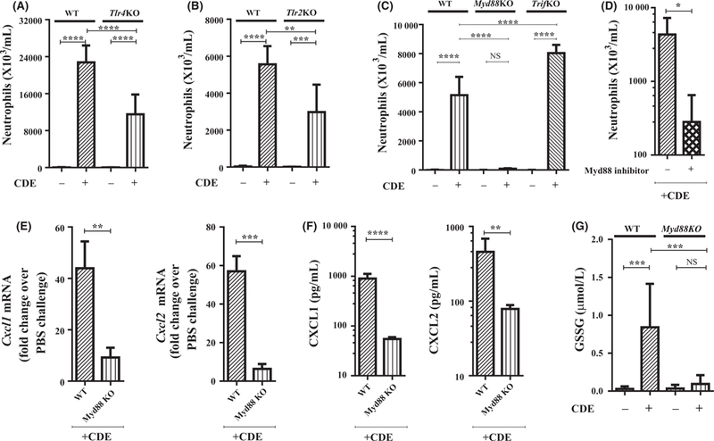
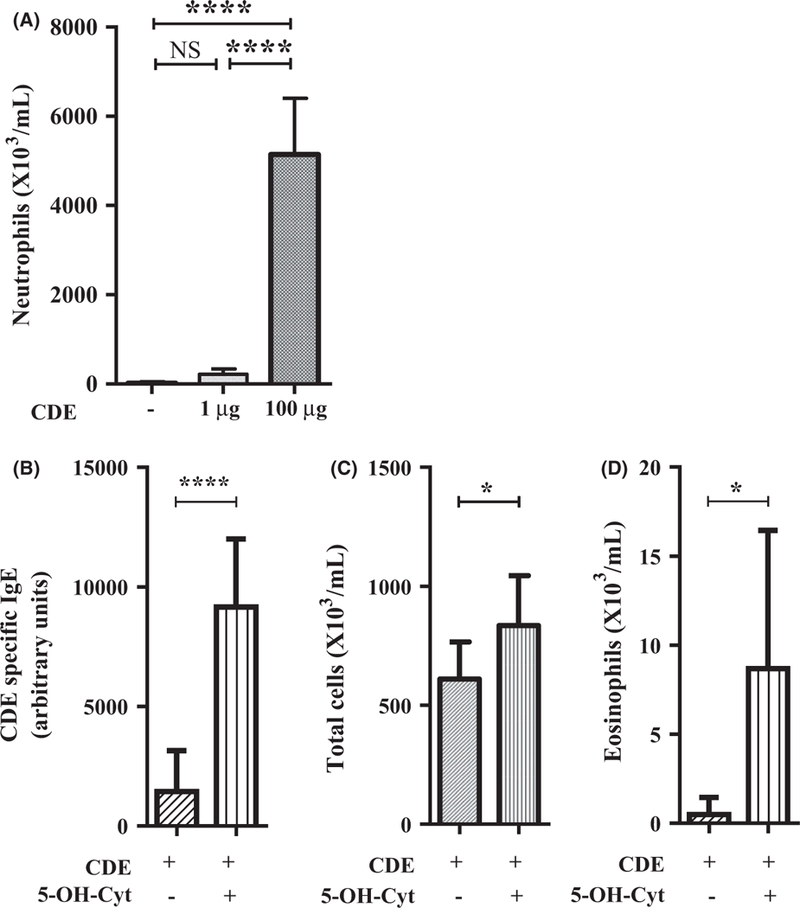
Background: Ragweed pollen extract (RWPE) induces TLR4-NFκB-CXCL-dependent recruitment of ROS-generating neutrophils to the airway and OGG1 DNA glycosylase-dependent excision of oxidatively induced 8-OH-Gua DNA base lesions from the airway epithelial cell genome. Administration of free 8-OH-Gua base stimulates RWPE-induced allergic lung inflammation. These studies suggest that stimulation of innate receptors and their adaptor by allergenic extracts initiates excision of a set of DNA base lesions that facilitate innate/allergic lung inflammation.

Objective: To test the hypothesis that stimulation of a conserved innate receptor/adaptor pathway by allergenic extracts induces excision of a set of pro-inflammatory oxidatively induced DNA base lesions from the lung genome that stimulate allergic airway inflammation.

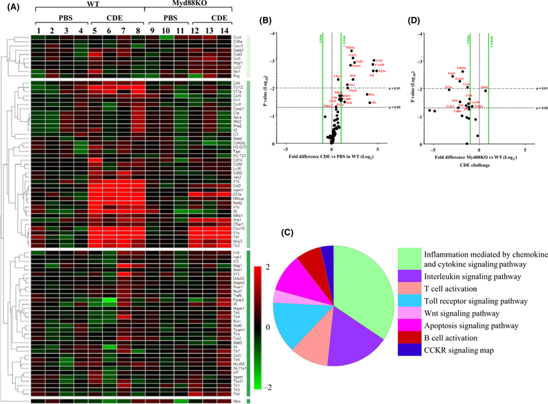
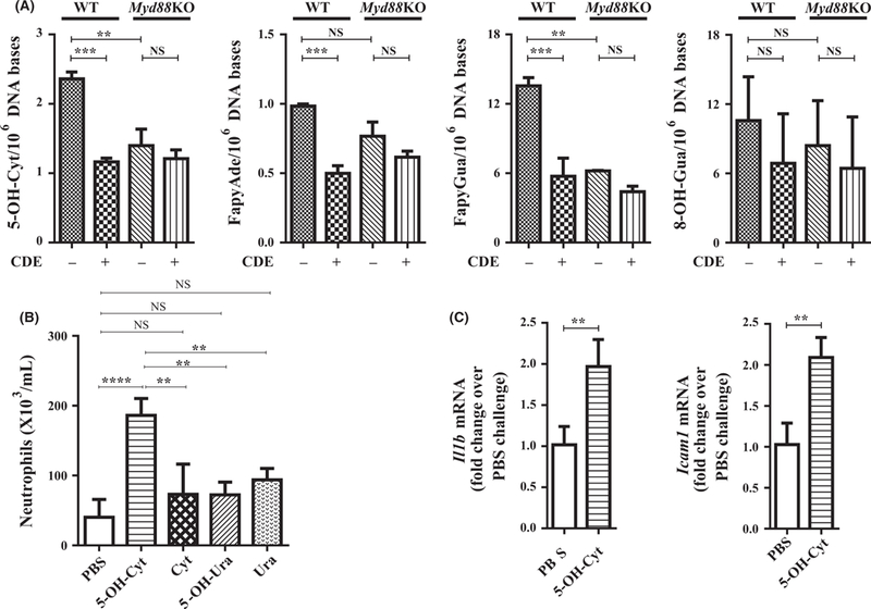
Methods: Wild-type (WT), Tlr4KO, Tlr2KO, Myd88KO, and TrifKO mice were intranasally challenged once or repeatedly with cat dander extract (CDE), and innate or allergic inflammation and gene expression were quantified. We utilized GC-MS/MS to quantify a set of oxidatively induced DNA base lesions after challenge of naïve mice with CDE.

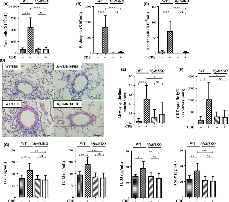
Results: A single CDE challenge stimulated innate neutrophil recruitment that was partially dependent on TLR4 and TLR2, and completely on Myd88, but not TRIF. A single CDE challenge stimulated MyD88-dependent excision of DNA base lesions 5-OH-Cyt, FapyAde, and FapyGua from the lung genome. A single challenge of naïve WT mice with 5-OH-Cyt stimulated neutrophilic lung inflammation. Multiple CDE instillations stimulated MyD88-dependent allergic airway inflammation. Multiple administrations of 5-OH-Cyt with CDE stimulated allergic sensitization and allergic airway inflammation.

Conclusions and clinical relevance: We show for the first time that CDE challenge stimulates MyD88-dependent excision of DNA base lesions. Our data suggest that the resultant-free base(s) contribute to CDE-induced innate/allergic lung inflammation. We suggest that blocking the MyD88 pathway in the airways with specific inhibitors may be a novel targeted strategy of inhibiting amplification of innate and adaptive immune inflammation in allergic diseases by oxidatively induced DNA base lesions.


Conflict of interest statement

CONFLICT OF INTEREST

The authors declares no conflict of interest.

Figures

FIGURE 1
FIGURE 1
MyD88 mediates CDE-induced innate lung inflammation. A-G, BALF innate immune response and lung mRNA expression in CDE-induced single-challenge model (Figure S1A). A-D, BALF neutrophil counts in (A) WT and Tlr4KO mice (n = 5 per group); (B) WT and Tlr2KO mice (n = 3-4 per group); (C) WT, TrifKO, and Myd88KO mice (n = 3-5 per group); (D) WT mice treated with a MyD88 inhibitor (Figure S1B) prior to CDE challenge (n = 4 per group) (E) CDE-induced Cxcl1 and Cxcl2 lung mRNA expression in WT and Myd88KO mice (n = 3-4 per group). (F) CDE-induced CXCL1 and CXCL2 secretion into the BALF in WT and Myd88 KO mice (n = 4-6 per group). (G) BALF level of oxidized glutathione (GSSG) in WT and Myd88 KO mice in single-challenge model (Figure S1A) (n = 4-5 per group). Data are expressed as means ± standard deviations, Std. *=P < 0.05, **=P < 0.01, ***=P < 0.001, and ****=P < 0.0001, analyzed by t test or ANOVA and posthoc test Bonferroni
FIGURE 2
FIGURE 2
CDE challenge induces MyD88-dependent innate gene expression. A-D, Innate gene expression in the lung after a PBS or CDE single-challenge model (Figure S1A) (n = 3-4 per group). A, Hierarchical clustering analysis showing single CDE challenge-induced fold change of mRNA over PBS control in WT mice (mouse #1-8) and Myd88KO mice (mouse #9-14) after 2 h post challenge. B,D, Volcano plot showing fold difference of mRNA in the lung and p value between 2 h post CDE challenge vs PBS control in WT mice (B), and between WT mice and Myd88KO mice (D) 2 h post CDE challenge. C, Pie chart showing predicted signalling pathways activated in WT mice after a single CDE challenge
FIGURE 3
FIGURE 3
CDE challenge induces MyD88-dependent excision of DNA base lesions. A, The content of 5-OH-Cyt, FapyAde, FapyGua, and 8-OH-Gua in the lung genomic DNA of WT or Myd88KO mice 16 h after a single CDE challenge (Figure S1A) (n = 3 per group). B, BALF neutrophil counts in WT mice 16 h after a single challenge (Figure S1A) with PBS, 60 μL of 1 μmol/L solution cytosine (Cyt), 5-OH-Cyt, uracil (Ura), or 5-OH-Ura (n = 3-5 per group). C, 5-OH-Cyt-induced Il1b and Icam1 lung mRNA expression in WT mice (n = 4 per group). Data are expressed as means ± standard deviations, Std. *=P < 0.05, **=P < 0.01, ***=P < 0.001, and ****=p < 0.0001, analyzed by t test or ANOVA and post-hoc test Bonferroni
FIGURE 4
FIGURE 4
MyD88 mediates CDE-induced allergic sensitization and inflammation. A-G, CDE multiple-challenge model in WT and Myd88 KO mice (Figure S1C) (n = 5-7 per group). Number of total inflammatory cells (A), eosinophils (B), and neutrophils (C), in BALF, and mucin secreting airway epithelial cells (D, E). Original magnifications, ×400. F, Serum CDE-specific IgE. G, BALF level of IL5, IL13, IL33, and TSLP. Data are expressed as means ± standard deviations, Std. *=P < 0.05, **=P < 0.01, ***=P < 0.001, and ****=P < 0.0001, analyzed by ANOVA and posthoc test Bonferroni
FIGURE 5
FIGURE 5
Lung mRNA expression in CDE multiple-challenge model. A-D, Gene expression and signalling pathways in CDE multiple-challenge model (Figure S1C) (n = 3 per group). Cluster 4 (A) of the hierarchical clustering analysis (Figure S2B) showed the greatest change of lung mRNA expression 2, 4, and 16 h after CDE challenge in WT mice. (B, D) Volcano plot showing fold difference of lung mRNA expression and p value 4 h post CDE challenge. B, Comparison between CDE vs. PBS in WT mice. D, Comparison between WT mice and Myd88KO mice. C, Pie chart showing predicted signalling pathways activated in CDE multiple-challenge model
FIGURE 6
FIGURE 6
5-OH-Cyt facilitates CDE-induced allergic airway inflammation. A, BALF neutrophil counts in WT mice 16 h after a single challenge (Figure S1A) with PBS or 1 μg CDE or 100 μg CDE (n = 4-5 per group). B-D, Allergic sensitization or allergic inflammation induced by multiple administration 1 μg CDE with or without 60 μL of 1 μmol/L solution 5-OH-Cyt in WT mice (Figure S1C) (n = 6-7 per group). B, Serum CDE-specific IgE. Number of total inflammatory cells (C) and eosinophils (D) in BALF. Data are expressed as means ± standard deviations, Std. *=P < .05, **=P < 0.01, ***=P < 0.001, and ****=P < 0.0001, analyzed by t test or ANOVA and post-hoc test Bonferroni

Similar articles

Cited by

References

    1. Boldogh I, Bacsi A, Choudhury BK, et al. ROS generated by pollen NADPH oxidase provide a signal that augments antigen-induced allergic airway inflammation. J Clin Invest. 2005;115(8):2169–2179. - PMC - PubMed
    1. Hazra TK, Das A, Das S, Choudhury S, Kow YW, Roy R. Oxidative DNA damage repair in mammalian cells: a new perspective. DNA Repair. 2007;6(4):470–480. - PMC - PubMed
    1. David SS, O’Shea VL, Kundu S. Base-excision repair of oxidative DNA damage. Nature. 2007;447(7147):941–950. - PMC - PubMed
    1. Bacsi A, Aguilera-Aguirre L, Szczesny B, et al. Down-regulation of 8-oxoguanine DNA glycosylase 1 expression in the airway epithelium ameliorates allergic lung inflammation. DNA Repair (Amst). 2013;12 (1):18–26. - PMC - PubMed
    1. Aguilera-Aguirre L, Bacsi A, Radak Z, et al. Innate inflammation induced by the 8-oxoguanine DNA glycosylase-1-KRAS-NF-kappaB pathway. J Immunol. 2014;193(9):4643–4653. - PMC - PubMed

Publication types

MeSH terms