. 2018 Sep 24;15(1):26.
doi: 10.1186/s12987-018-0112-7.

Blood-brain and blood-cerebrospinal fluid barrier permeability in spontaneously hypertensive rats

Affiliations

Blood-brain and blood-cerebrospinal fluid barrier permeability in spontaneously hypertensive rats

Daphne M P Naessens et al. Fluids Barriers CNS. .

Abstract

Background: Hypertension is an important risk factor for cerebrovascular disease, including stroke and dementia. Both in humans and animal models of hypertension, neuropathological features such as brain atrophy and oedema have been reported. We hypothesised that cerebrovascular damage resulting from chronic hypertension would manifest itself in a more permeable blood-brain barrier and blood-cerebrospinal fluid barrier. In addition, more leaky barriers could potentially contribute to an enhanced interstitial fluid and cerebrospinal fluid formation, which could, in turn, lead to an elevated intracranial pressure.

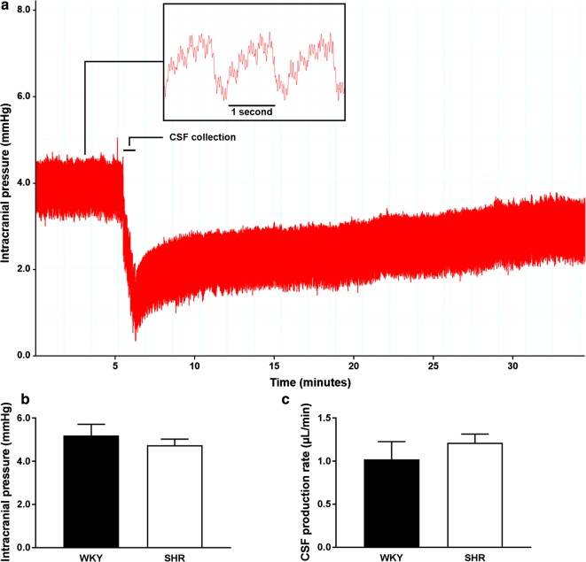
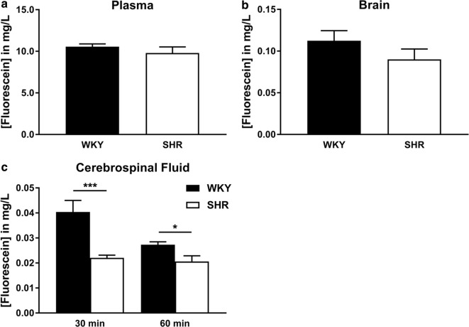
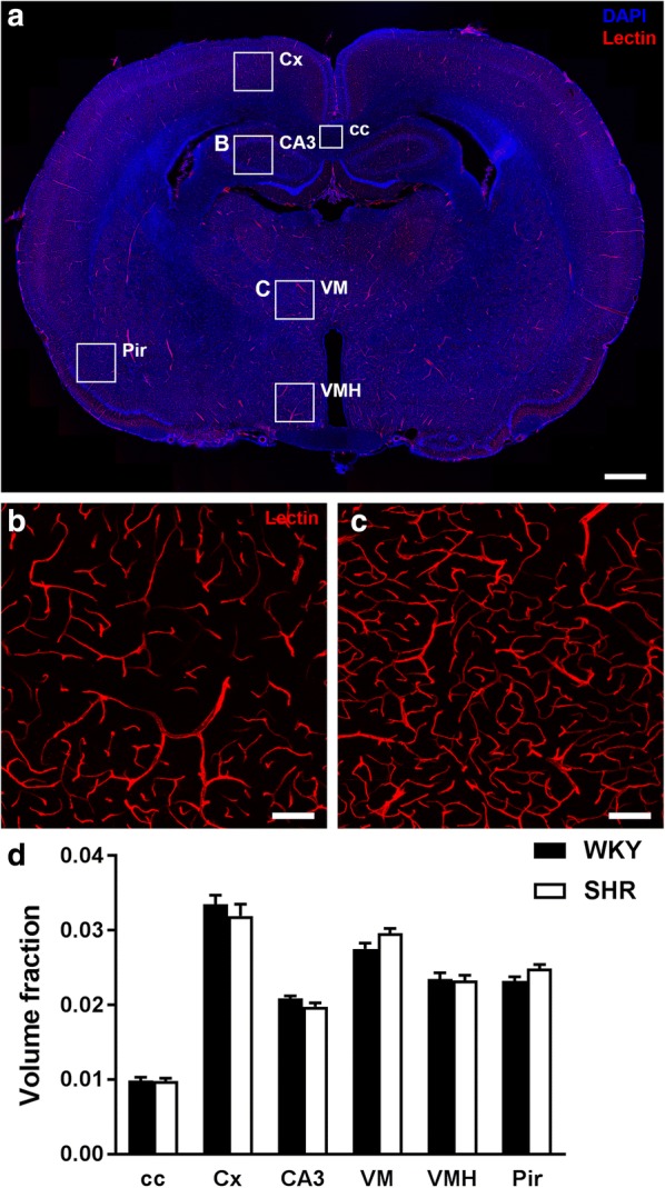
Methods: To study this, we monitored intracranial pressure and estimated the cerebrospinal fluid production rate in spontaneously hypertensive (SHR) and normotensive rats (Wistar Kyoto, WKY) at 10 months of age. Blood-brain barrier and blood-cerebrospinal fluid barrier integrity was determined by measuring the leakage of fluorescein from the circulation into the brain and cerebrospinal fluid compartment. Prior to sacrifice, a fluorescently labelled lectin was injected into the bloodstream to visualise the vasculature and subsequently study a number of specific vascular characteristics in six different brain regions.

Results: Blood and brain fluorescein levels were not different between the two strains. However, cerebrospinal fluid fluorescein levels were significantly lower in SHR. This could not be explained by a difference in cerebrospinal fluid turnover, as cerebrospinal fluid production rates were similar in SHR and WKY, but may relate to a larger ventricular volume in the hypertensive strain. Also, intracranial pressure was not different between SHR and WKY. Morphometric analysis of capillary volume fraction, number of branches, capillary diameter, and total length did not reveal differences between SHR and WKY.

Conclusion: In conclusion, we found no evidence for blood-brain barrier or blood-cerebrospinal fluid barrier leakage to a small solute, fluorescein, in rats with established hypertension.

Keywords: Blood–brain barrier; Blood–cerebrospinal fluid barrier; Cerebrospinal fluid; Hypertension; Interstitial fluid.


Figures

Fig. 1
Fig. 1
Changes in ICP during collection of CSF from the cisterna magna, baseline ICP, and CSF production rate in WKY and SHR. a In this animal, the mean baseline ICP of 4.0 mmHg was measured during the first 5 min of the experiment. Subsequently, a CSF sample was collected over a short period of about 1 min until an ICP of 0.5 mmHg was reached. The animal was then allowed to refill the withdrawn CSF volume for another 30 min, from which the CSF production rate could be calculated. The inset shows a zoom in on the ICP recording, with approximately 1 breath and 4 to 5 heart beats per second. b Mean baseline ICP was not different between WKY (n = 10) and SHR (n = 10). c Also, CSF production rates did not differ between WKY (n = 6) and SHR (n = 10). Values are mean ± SEM (unpaired Student’s t-test)
Fig. 2
Fig. 2
Fluorescein concentrations in plasma, brain and CSF. Fluorescein concentrations were quantified by spectrophotometric analysis in plasma (a), brain (b), and CSF (c) samples of WKY (n = 10 for plasma and brain, n = 7 at CSF 30 min, and n = 5 at CSF 60 min) and SHR (n = 10 for plasma and brain, n = 9 at CSF 30 min, and n = 6 at CSF 60 min). Values are mean ± SEM. *p ≤ 0.05, ***p ≤ 0.001. (unpaired Student’s t-test or Mann–Whitney U test)
Fig. 3
Fig. 3
Visualisation and quantification of the rat brain microvasculature. The vascular endothelium was visualised by a DyLight®594-labelled L. esculentum lectin (red), and cell nuclei by DAPI staining (blue). a Overview of a coronal rat brain section indicating the stereotaxic coordinates and 6 different brain regions used to quantify a number of vascular parameters. b and c Representative images of the lectin staining used for the analysis of vascular parameters in the CA3 and VM respectively. d Volume fraction of capillaries did not differ between WKY (n = 10) and SHR (n = 10) in six different brain regions. Values are mean ± SEM (repeated measures ANOVA, Bonferroni’s post hoc tests). cc, corpus callosum; Cx, cerebral cortex; CA3, field CA3 of the hippocampus; VM, ventromedial thalamic nucleus; VMH, ventromedial hypothalamic nucleus; Pir, piriform cortex. Scale bar in a represents 1 mm, and 100 µm in b and c

Similar articles

Cited by

References

    1. Iadecola C, Yaffe K, Biller J, Bratzke LC, Faraci FM, Gorelick PB, et al. Impact of hypertension on cognitive function: a scientific statement from the American Heart Association. Hypertension. 2016;68(6):e67–e94. doi: 10.1161/HYP.0000000000000053. - DOI - PMC - PubMed
    1. Ishida H, Takemori K, Dote K, Ito H. Expression of glucose transporter-1 and aquaporin-4 in the cerebral cortex of stroke-prone spontaneously hypertensive rats in relation to the blood-brain barrier function. Am J Hypertens. 2006;19(1):33–39. doi: 10.1016/j.amjhyper.2005.06.023. - DOI - PubMed
    1. Dandy WE. Experimental hydrocephalus. Ann Surg. 1919;70(2):129–142. doi: 10.1097/00000658-191908000-00001. - DOI - PMC - PubMed
    1. Hladky SB, Barrand MA. Mechanisms of fluid movement into, through and out of the brain: evaluation of the evidence. Fluids Barriers CNS. 2014;11(1):26. doi: 10.1186/2045-8118-11-26. - DOI - PMC - PubMed
    1. Segal MB, Pollay M. The secretion of cerebrospinal fluid. Exp Eye Res. 1977;25(Suppl):127–148. doi: 10.1016/S0014-4835(77)80012-2. - DOI - PubMed