Review
. 2019 Apr:198:204-216.
doi: 10.1016/j.biomaterials.2018.08.006. Epub 2018 Aug 3.

Multiscale bioprinting of vascularized models

Affiliations
Review

Multiscale bioprinting of vascularized models

Amir K Miri et al. Biomaterials. 2019 Apr.

Abstract

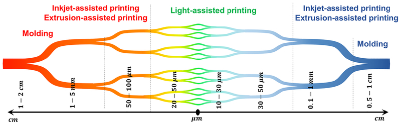
A basic prerequisite for the survival and function of three-dimensional (3D) engineered tissue constructs is the establishment of blood vessels. 3D bioprinting of vascular networks with hierarchical structures that resemble in vivo structures has allowed blood circulation within thick tissue constructs to accelerate vascularization and enhance tissue regeneration. Successful rapid vascularization of tissue constructs requires synergy between fabrication of perfusable channels and functional bioinks that induce angiogenesis and capillary formation within constructs. Combinations of 3D bioprinting techniques and four-dimensional (4D) printing concepts through patterning proangiogenic factors may offer novel solutions for implantation of thick constructs. In this review, we cover current bioprinting techniques for vascularized tissue constructs with vasculatures ranging from capillaries to large blood vessels and discuss how to implement these approaches for patterning proangiogenic factors to maintain long-term, stimuli-controlled formation of new capillaries.

Keywords: Angiogenesis; Core/shell; Sacrificial bioink; Stereolithography; Tissue engineering.


Figures

Fig. 1.
Fig. 1.
The summary of blood vessel hierarchy (from arteries to veins) in the body and the range of diameters at each level, along with current fabrication techniques used to engineer biomimetic tissue construct.
Fig. 2.
Fig. 2.. Sacrificial extrusion-assisted bioprinting:
(A) Step-by-step display of a chip bioprinting: (i) the device is prepared with a bottom lid, (ii) imprinting a matrix material, (iii) bioprinting of the matrix material and (iv) liquefying the infusion bioink. (B) Agarose channels within GelMA hydrogel: (i) printed agarose template fibers, (ii) perfused fluorescent dye in the channel. (C) Pluronic F-127 channels within GelMA hydrogel: (i) bifurcated microchannels in the GelMA hydrogel, (ii) HUVECs CD31 (green) and nuclei (blue) staining. (D) Vascularized gelatin-based tissues, (left) HUVEC-lined vascular channel supporting a fibroblast-laden gelatin within a 3D perfusion chip (right) and long-term perfusion of HUVEC-lined (red) network supporting HNDF-laden (green) gelatin at day 45 (Scale bar: 100 μm). (E) Carbohydrate-glass lattice as the sacrificial element: patterned channels support positive pressure and pulsatile flow of human blood with inter-vessel junctions supporting branched fluid flow (left). Spiral flow patterns (right, 0.4 s; Scale bars: 1 mm for left and 2 mm for right). Reprinted with permission from references Datta et al. [37], Bertassoni et al. [33], Zhang et al. [44], Kolesky et al. [6] and Miller et al. [50].
Fig. 3.
Fig. 3.. Inkjet/Light-assisted strategies for 3D bioprinting:
(A) Inkjet-assisted printed fibrin scaffold, with minor deformations of the printed pattern (as indicated by arrows). (B) Perfusable microfluidic chip: (i) schematics of the setup, and (ii) HUVEC coated (red) long-term culture with human dermal fibroblasts support (red) after 45 second perfusion. (C) Schematic diagram of the DMD system: the UV light illuminates a programmable DMD chip, and is reflected down onto the photosensitive monomer solution. (D) Cell-laden DMD-printed constructs: (i) HUVECs are encapsulated in the channels and (ii) HepG2 are encapsulated in the surrounding area with (iii) overlay image (scale bars: 250 μm); (iv-vi) endothelial network formation after 1-week culture of the prevascularized tissue construct: (iv) showing HUVECs (Green, CD31) and (v) supportive MSCs (Purple, alpha-smooth muscle actin; scale bars: 100 μm). Reprinted with permission from references Cui et al. [57], Lee et al. [45], Huang et al. [58] and Zhu et al. [16].
Fig. 4.
Fig. 4.. Schematic of core/shell bioprinting:
(A) Printed lattices made by coaxial extrusion of alginate and gelatin hydrogels: (i) single layer of hybrid gel with fluorescently labelled cells in the core (Scale bar: 1 mm); (ii) 3D construct made by gelatin strands and cell-laden core/shell strands (Scale bar: 1 cm); (iii) structural integrity after the removal of gelatin; (iv) SEM image of the side profile showing interconnected pores (Scale bar: 1 mm). (B) 3D printing of core/shell cell-laden strands. (C) Three common bioprinting strategies using alginate-based assays. (D) Schematic diagram depicting two independent crosslinking procedures of the bioink, where PEGTA, alginate, and GelMA hydrogel are ionically and covalently crosslinked. (E) Bioprinting of embryonic chick heart using alginate-based bioink: (i) typical embryonic chick heart (ii) stained for nuclei (blue), fibronectin (green), and F-actin (red); (iii) a cross-section of printed chicken heart; (iv) with fluorescent alginate (green); (v) optical microscopy image of the printed trabeculated embryonic chicken heart; (Scale bars: 1 mm). Reprinted with permission from references Mistry et al. [67], Akkineni et al. [75], Jia et al. [73], Axpe et al. [68], and Hilton et al. [40].
Fig. 5.
Fig. 5.. Scaffold-free/freestanding bioprinting of shell constructs:
(A) Schematics showing vascular tissue engineering using scaffold-free bioprinting phase by phase, where agarose rods (pink) and similar multicellular spheroids (orange) were placed layer-by-layer. (B) Scaffold-free fabrication of bifurcated blood vessels using human skin fibroblast (HSF) spheroids that spheroids fuse into tissue after 6 days. (C) Scanning electron micrographs of tubular structure, by two-photon photocrosslinking of α, ω-poly-tetra-hydro-furan-ether-diacrylate resin, with a height of 160 μm (left), an inner diameter of 18 μm and wall thickness of 3 μm (middle), while smaller wall thicknesses around 1 μm collapsed (right). Reprinted with permission from references Datta et al. [37], Tan et al. [78] and Meyer et al. [83].
Fig. 6.
Fig. 6.. Different approaches of 4D bioprinting:
(A) Different flower morphologies created by moisture responsive 4D printing using cellulose nanofiber acrylamide hydrogel (scale bars: 5 mm, inset: 2.5 mm). (B) Water responsive photopatterned PEG-based hydrogel bilayers: (i) the first layer printed, (ii) then added the second layer, and (iii) self-folded into curved hydrogels; (iv) schematic shown for multi-culture of cells in distinct layers of a self-folded hydrogel and (v) fluorescent micrographs indicting fibroblasts (blue: Hoechst) in the inner layer and fibroblasts (green: calcein) in the outer layer. (C) 4D printed self-folding tubes: (i) printing of methacryloyl alginate (AA-MA) or hyaluronic acid (HA-MA) followed by photocrosslinking with green light (530 nm) and instant folding into tubes upon immersion in water or cell culture media; (ii) self-folded methacryloyl alginate (schematics are at the upper panels and representative images are at the lower panels) that folded in water and unfolded by deswelling in calcium chloride, and then refolded in EDTA. (D) Bioprinting of vascular structures: (i) self-assembly of multicellular spheroids into (ii) tubular structures fused after one week showing red- and green-labeled cells; (iii, iv) two double-layered vascular models made of human umbilical vein smooth muscle cells (green) and skin fibroblasts (red) along with histology (center: H&E) and immunocytochemistry (right: smooth muscle α-actin in brown and Caspase-3 in brown) images (scale bars: 500 μm). Reprinted with permission from references Norotte et al. [81], Gladman et al. [93], Kirillova et al. [101], and Jamal et al. [99].
Fig. 7.
Fig. 7.. Bioprinting solutions for drug carriers:
(A) Electrospun shell: i) SEM micrographs of 2.2-mm vascular graft, ii) cross section of the fibrous membrane loaded by vascular endothelial growth factor (VEGF) and platelet-derived growth factor (PDGF), iii) HUVECs stained with anti-CD31 antibody (FITC labeled CD31; Blue, DAPI stained nuclei) double-layered electrospun membranes loading single VEGF (top-right) or PDGF (bottom-left) or without any growth factor (top-left) loading dual VEGF and PDGF (bottom-right). (B) Microfluidic vessel networks: i) schematic cross-sectional view of a section of μVN illustrating (ii) morphology and barrier function of endothelium, iii) endothelial sprouting and perivascular association (Scale bar: 500 μm). (C) Biodegradable electrospun drug-fibers coated stent to inhibit inflammation and scar formation in benign esophageal structures. (D) Skin wound batch designed to release VEGF and other therapeutics into scar region. (E) Potential solutions through different bioprinting techniques. Reprinted with permission from references Zhang et al. [70], Zu et al. [122], Zheng et al. [123] and Mostafalu et al. [117].

References

    1. Laschke MW, Harder Y, Amon M, Martin I, Farhadi J, Ring A, et al. Angiogenesis in tissue engineering: breathing life into constructed tissue substitutes. Tissue engineering. 2006;12:2093–104. - PubMed
    1. Bae H, Puranik AS, Gauvin R, Edalat F, Carrillo-Conde B, Peppas NA, et al. Building vascular networks. Science translational medicine. 2012;4:160ps23–ps23. - PMC - PubMed
    1. Fukuda S, Yoshii S, Kaga S, Matsumoto M, Kugiyama K, Maulik N. Angiogenic strategy for human ischemic heart disease: brief overview. Molecular and cellular biochemistry. 2004;264:143–9. - PubMed
    1. Bennett S, Griffiths G, Schor A, Leese G, Schor S. Growth factors in the treatment of diabetic foot ulcers. British Journal of Surgery. 2003;90:133–46. - PubMed
    1. Kaully T, Kaufman-Francis K, Lesman A, Levenberg S. Vascularization—the conduit to viable engineered tissues. Tissue Engineering Part B: Reviews. 2009;15:159–69. - PubMed

Publication types