. 2018 Oct 18;72(2):263-274.e5.
doi: 10.1016/j.molcel.2018.08.028. Epub 2018 Sep 20.

Mode of Action of Kanglemycin A, an Ansamycin Natural Product that Is Active against Rifampicin-Resistant Mycobacterium tuberculosis

Affiliations

Mode of Action of Kanglemycin A, an Ansamycin Natural Product that Is Active against Rifampicin-Resistant Mycobacterium tuberculosis

Hamed Mosaei et al. Mol Cell. .

Abstract

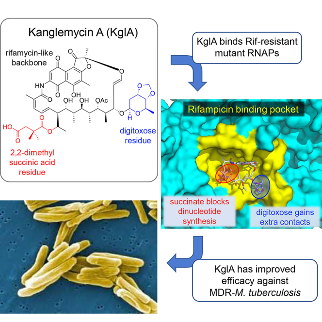
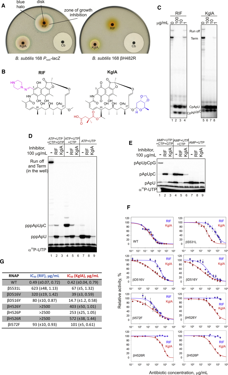
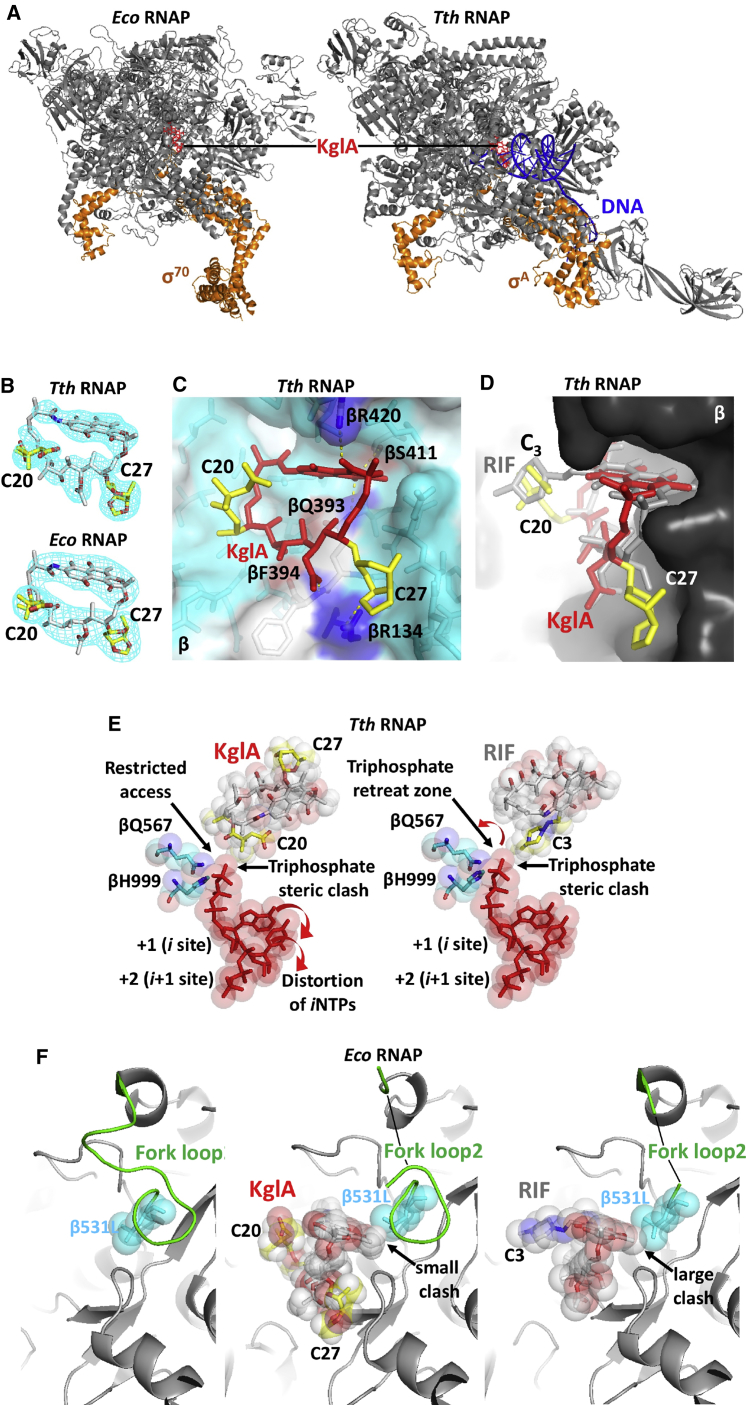
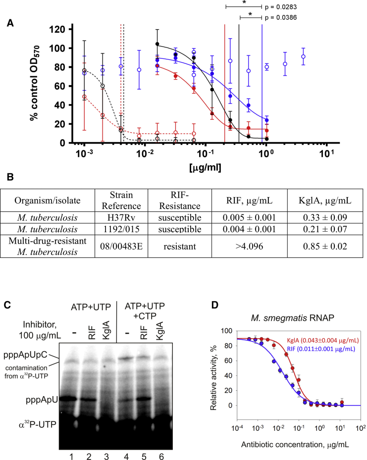
Antibiotic-resistant bacterial pathogens pose an urgent healthcare threat, prompting a demand for new medicines. We report the mode of action of the natural ansamycin antibiotic kanglemycin A (KglA). KglA binds bacterial RNA polymerase at the rifampicin-binding pocket but maintains potency against RNA polymerases containing rifampicin-resistant mutations. KglA has antibiotic activity against rifampicin-resistant Gram-positive bacteria and multidrug-resistant Mycobacterium tuberculosis (MDR-M. tuberculosis). The X-ray crystal structures of KglA with the Escherichia coli RNA polymerase holoenzyme and Thermus thermophilus RNA polymerase-promoter complex reveal an altered-compared with rifampicin-conformation of KglA within the rifampicin-binding pocket. Unique deoxysugar and succinate ansa bridge substituents make additional contacts with a separate, hydrophobic pocket of RNA polymerase and preclude the formation of initial dinucleotides, respectively. Previous ansa-chain modifications in the rifamycin series have proven unsuccessful. Thus, KglA represents a key starting point for the development of a new class of ansa-chain derivatized ansamycins to tackle rifampicin resistance.

Keywords: MDR-TB; RNA polymerase; antibiotic resistance; antibiotics; holo-enzyme crystal structure; kanglemycin A; multidrug-resistant Mycobacterium tuberculosis; promoter complex crystal structure; rifampicin; rifamycin.


Figures

None
Graphical abstract
Figure 1
Figure 1
Identification and Mode of Action of KglA (A) Disk diffusion assay with rifampicin (RIF), carbenicillin (Cb), and kanglemycin A (KglA). Paper disks soaked with antibiotics were placed on the lawns of wild-type B. subtilis carrying the lacZ gene under the PhelD promoter, inducible during partial inhibition of transcription (left), and RIF-resistant B. subtilis (right). Note the blue halo around the growth inhibition zones in the case of transcription inhibitors. (B) Chemical structure of KglA in comparison with RIF. The ansa chain modifications in KglA are highlighted. The semisynthetic group of RIF is shown in purple. See also Figure S1 and Table S1. (C) Transcription in vitro by E. coli RNAP started with CpA on a linear template containing the T7A1 promoter. Runoff, termination, and abortive tri- and tetra-nucleotides are marked. Note that tetra-nucleotides migrate faster than tri-nucleotides under these electrophoretic conditions. (D) Different ratio of the di-nucleotide-long (pppApU) and the tri-nucleotide-long (pppApUpC) abortive products in the presence of RIF and KglA. Transcription was performed in the presence of the nucleotides depicted (in the absence of the CpA primer). Note that, in the 33% gel, runoff and termination products remain in the well. (E) The experiment was performed as in (D) but in the presence of AMP instead of ATP. (F and G) Inhibition of wild-type and RIF-resistant E. coli RNAPs by RIF and KglA. Error bars (F) are ± SD. The brackets (G) contain ± SE and the Hill slope.
Figure 2
Figure 2
KglA Binding to Free and Promoter-Bound RNAP Holoenzyme (A) Overall views of E. coli (left) and T. thermophilus (right) RNAPs with KglA bound in the RIF-binding pocket. RNAPs (gray core and orange σ) and DNA (blue) are shown as ribbon models, and KglA (red) is shown as a stick model. (B) Electron density maps of KglA in complexes with T. thermophilus (top) and E. coli (bottom) RNAPs. Electron densities are shown as a light blue mesh, and KglA molecules are shown as stick models colored by element. The ansa bridge side chains of KglA extending from C20 and C27 are colored in yellow. (C) A close-up view of the RIF-binding pocket of the T. thermophilus RNAP containing KglA (in red, with the side chains in yellow). RNAP is shown as a transparent surface model (light blue), KglA is shown as a stick model with the ansa bridge side chains labeled, and the β subunit residues forming the RIF-binding pocket are shown as stick models. The hydrogen bonds between KglA and β amino acid residues (colored by element) are depicted by yellow dotted lines; the βR134 involved in polar interaction with the C20 sugar of KglA is shown in blue. (D) A side view of the RIF-binding pocket shown in (C). KglA is overlaid on RIF (gray); the side chains of the two molecules are labeled. KglA maintains a larger distance to the wall in the RIF-binding pocket than RIF. (E) Modeling of the initiating nucleotides in the active site of the T. thermophilus RNAP (from PDB: 4Q4Z) in the presence of KglA (left) and RIF (right). The ribonucleotides (red), the βQ567 and βH999 residues coordinating the 5′ end triphosphate (blue), and KglA and RIF (gray) are shown as stick and sphere models. The side chains of the inhibitors and the positions of the initiating NTPs are indicated. Red arrows indicate the translocation directions for the corresponding substrates. (F) Structural rearrangements in the RIF-binding pocket of E. coli βS531L RNAP triggered by KglA and RIF binding. RNAP is shown as a ribbon model in gray, the structured regions of the fork loop2 are shown in green, and the β531L residue (blue), KglA (colored by element with yellow side chains), and RIF (colored by element) are shown as stick and sphere models. The side chains of KglA and RIF are indicated. Arrows mark the clash points between the naphthalene cores of the corresponding compounds with the β531L side chain. See also Figure S2.
Figure 3
Figure 3
KglA Inhibits Mycobacterial RNAP and Retains Antimicrobial Activity against MDR-M. tuberculosis (A) Dose-dependent growth inhibition of the RIF-susceptible M. tuberculosis strains H36Rv (black) and 1192/015 (red) and RIF-resistant M. tuberculosis 08/00483E (blue) by KglA (solid line, solid symbols) and RIF (dashed line, open symbols). Data presented are the mean of four independent experiments ± SD and analyzed using a Student’s t test. (B) MIC values ± SD, as determined using the modified Gompertz function for M. tuberculosis strains H37Rv, 1192/015, and 08/00483E, which were exposed to RIF or KglA. (C) Mode of action of KglA and RIF against M. smegmatis RNAP. The experiment was performed exactly as in Figure 1D. Note that the high radioactive background compared with Figure 1D is due to much lower activity of M. smegmatis RNAP. (D) Inhibition of M. smegmatis RNAP by RIF and KglA. Error bars are ± SD. Shown in brackets are the IC50 values ± SE.

References

    1. Adams P.D., Afonine P.V., Bunkóczi G., Chen V.B., Davis I.W., Echols N., Headd J.J., Hung L.-W., Kapral G.J., Grosse-Kunstleve R.W. PHENIX: a comprehensive Python-based system for macromolecular structure solution. Acta. Cryst. 2010;D66:213–221. - PMC - PubMed
    1. Afonine P.V., Mustyakimov M., Grosse-Kunstleve R.W., Moriarty N.W., Langan P., Adams P.D. Joint X-ray and neutron refinement with phenix.refine. Acta Crystallogr. D Biol. Crystallogr. 2010;66:1153–1163. - PMC - PubMed
    1. Andrews J.M. Determination of minimum inhibitory concentrations. J. Antimicrob. Chemother. 2001;48(Suppl 1):5–16. - PubMed
    1. Aristoff P.A., Garcia G.A., Kirchhoff P.D., Showalter H.D. Rifamycins--obstacles and opportunities. Tuberculosis (Edinb.) 2010;90:94–118. - PubMed
    1. Artsimovitch I., Vassylyeva M.N., Svetlov D., Svetlov V., Perederina A., Igarashi N., Matsugaki N., Wakatsuki S., Tahirov T.H., Vassylyev D.G. Allosteric modulation of the RNA polymerase catalytic reaction is an essential component of transcription control by rifamycins. Cell. 2005;122:351–363. - PubMed

Publication types

MeSH terms