. 2018 Oct 4;175(2):429-441.e16.
doi: 10.1016/j.cell.2018.08.061. Epub 2018 Sep 20.

LC3-Associated Phagocytosis in Myeloid Cells Promotes Tumor Immune Tolerance

Affiliations

LC3-Associated Phagocytosis in Myeloid Cells Promotes Tumor Immune Tolerance

Larissa D Cunha et al. Cell. .

Abstract

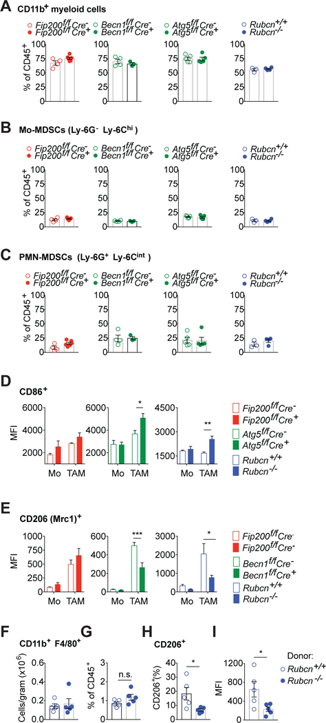
Targeting autophagy in cancer cells and in the tumor microenvironment are current goals of cancer therapy. However, components of canonical autophagy play roles in other biological processes, adding complexity to this goal. One such alternative function of autophagy proteins is LC3-associated phagocytosis (LAP), which functions in phagosome maturation and subsequent signaling events. Here, we show that impairment of LAP in the myeloid compartment, rather than canonical autophagy, induces control of tumor growth by tumor-associated macrophages (TAM) upon phagocytosis of dying tumor cells. Single-cell RNA sequencing (RNA-seq) analysis revealed that defects in LAP induce pro-inflammatory gene expression and trigger STING-mediated type I interferon responses in TAM. We found that the anti-tumor effects of LAP impairment require tumor-infiltrating T cells, dependent upon STING and the type I interferon response. Therefore, autophagy proteins in the myeloid cells of the tumor microenvironment contribute to immune suppression of T lymphocytes by effecting LAP.

Keywords: LC3-associated phagocytosis; STING; anti-cancer immunity; autophagy; efferocytosis; immune tolerance; macrophage polarization; tumor microenvironment; tumor-associated macrophages; type I interferon.


Conflict of interest statement

COMPETING INTERESTS

The authors declare no competing financial interests.

Figures

Figure 1.
Figure 1.. LAP in myeloid cells promotes tumor growth.
A. Tumor growth in wild-type and deficient littermates subcutaneously injected with B16F10 cells (Atg14f/f Cre n=3, Cre+ n=4 mice/ Fip200f/f Cre n=4, Cre+ n=6 mice/ Becn1f/f Cre n=4, Cre+ n=5 mice/ Atg5f/f Cre n=4, Cre+ n=5 mice/ Rubcn+/+ n=7, Rubcn− /− n=4 mice). Color scheme represents autophagy-deficient, LAP-sufficient (red), autophagy-deficient, LAP-deficient (green) and autophagy-sufficient, LAP-deficient (blue) mice. B. Tumor growth in wild-type and deficient littermates subcutaneously injected with LLC cells (Fip200f/f Cre n=7, Cre+ n=10 mice/ Becn1f/f Cre n=4, Cre+ n=4 mice/ Atg7f/f Cre n=4, Cre+ n=5 mice/ Rubcn+/+ n=3, Rubcn−/− n=4 mice). Colors as in (A). A-B. Data are expressed as mean ±SEM. Significance was calculated with ANOVA. Data are representative of two independent experiments per genotype. C. Micrometastatic lesions on the lungs of wild-type and deficient littermate mice intravenously injected with B16F10 cells. Data are pooled from two separate experiments. Dots represent the total number of lesions per mouse, line indicates mean ±SEM. Significance was calculated with Student’s t-test. Colors as in (A). D. Schematic of the K-rasLSL-G12D, p53f/f bone-marrow chimeric mice transplanted with bone-marrow cells from either Rubcn+/+ or Rubcn−/− donor mice. E. Quantification of tumor burden by MRI (expressed as % of total lobe volume) in K-rasLSL-G12D, p53f/f chimeras inoculated with Adv-cre. Data are pooled from two separate experiments. Dots represent data for each individual mouse (n= 10 mice per genotype), line indicates mean ±SEM. Significance was calculated using Student’s t-test. F. Representative MRI images from the left lung lobe of the indicated K-rasLSL-G12D, p53f/f bone-marrow chimera. G. Representative micrographs of H&E staining of the right lung cranial lobe for the indicated K-rasLSL-G12D, p53f/f bone-marrow chimera. Scale bar = 1 mm. *p<0.05, **p<0.01, ***p<0.001, ****p<0.0001. See also Figure S1.
Figure 2.
Figure 2.. LAP regulates TAM function.
A-C. Percentage of total (A), monocytic (B) and granulocytic (C) myeloid cells within the CD45+ population of tumor infiltrating cells isolated from LLC tumors engrafted in Rubcn−/−, Fip200f/f Cre+, Becn1f/f Cre+, Atg7f/f Cre+ and respective littermate controls, estimated by flow cytometry. Data are expressed as individual data points (n = 3–5 mice per genotype as shown) and mean bar ±SEM. D-E. MFI of CD86 (D) and CD206 (E) expression on the surface of monocytes (Mo) and mature TAM (TAM) isolated from LLC tumor engrafted into the indicated wild-type and deficient littermates (n=4 per group), measured by flow cytometry. Data are expressed as mean ±SEM. Significance was calculated with ANOVA. F-G. Absolute number (F) and percentage (G) of macrophages (CD45+ Ly-6G CD11b+ F4/80+) in the lungs of K-rasLSL-G12D, p53f/f bone-marrow chimeras, estimated by flow cytometry. H-I. Percentage (H) and MFI (I) of CD206 expression on the surface of macrophages in the lungs of K-rasLSL-G12D, p53f/f bone marrow chimeras (n= 5 mice per group), measured by flow cytometry. F-I. Data are expressed as individual data points and mean bar ±SEM. Significance was calculated with Student’s t-test. A-I. Data are representative of two independent experiments per genotype. *p<0.05, **p<0.01, ***p<0.001 See also Figure S2.
Figure 3.
Figure 3.. LAP-deficiency induces differential gene expression in TAM
A. tSNE plots of the expression profiles for monocytes and mature TAM from Rubcn+/− (red) or Rubcn−/− (blue) tumor-infiltrating myeloid cells isolated from LLC subcutaneous grafts. Each circle represents an individual cell. B. Same as (A) but colors represent cells clustered together based on similarity of global gene expression. C. tSNE plots showing the normalized absolute UMI count (log2) of categorizing markers (Mafb, Ly6c2, H2Aa) of monocytes and mature TAM for the clusters in (B). D. Quantification of proportion of Rubcn+/− (red) and Rubcn−/− (blue) cells composing clusters in (B). E. Expression of Mrc1 and Arginase1 in single myeloid cells for the clusters in (B). F. Heat map showing the proportion of cells expressing type I IFN pathway target genes (GO term type I interferon pathway, access number GO:0060337, was used for categorization) for the clusters in (B). Clusters are shown in columns and genes in rows. G. Gene ontology (GO) and Jensen assignments of top 100 uniquely expressed genes in cluster 5 of (B). Dash lines indicate p<0.001. H. qPCR of type I IFN target genes in isolated Rubcn−/− (n=4 mice) tumor-infiltrating myeloid cells relative to Rubcn+/+ (n=5 mice) counterparts. Data are expressed as individual data points and mean bar ±SEM. Significance was calculated with Student’s t-test, *p<0.05. Data are representative of two independent experiments. See also Figure S3.
Figure 4.
Figure 4.. Impaired LAP of engulfed dying tumor cells triggers type I IFN signaling in TAM
A. Confocal image of harvested subcutaneous mCherry-spectrin+ (red) LLC tumor engrafted in GFP-LC3Tg wild-type mouse. Tumor section was stained with anti-CD11b (blue). Arrows point to GFP-LC3+ phagosomes containing LLC debris. Scale bar = 10 μm. B. qPCR of the expression of type I IFN target genes and Ifn β in tumor-associated myeloid cells that engulfed LLC tumor cells (mCherry-spectrin+) isolated from Rubcn+/− and Rubcn−/− mice (n=3 mice per genotype). Data are expressed as mean ±SEM. C. Volcano plot for log2 fold change (LFC) in the expression of genes in Rubcn−/− versus Rubcn+/+ bone marrow-derived macrophages (BMDM) cultured with apoptotic thymocytes for 16 hours. Representative genes associated with M1 TAM phenotype are in blue, type I IFN targets are in green. Horizontal dashed line corresponds to an adjusted p value of 0.01 and vertical dashed line to logFC of 0.33. D. MFI of the expression of CD206 (CD11b+ CD206+) on the surface of untreated control BMDM (cntrl) or BMDM stimulated with apoptotic thymocytes for 24 hours, as measured by flow cytometry. Data are expressed as mean ±SEM of triplicate BMDM samples. E. Quantification of the engulfment of apoptotic mCherry-spectrin+ LLC cells by Tim4+/+ and Tim4−/− BMDM, measured by flow cytometry. The percentage of engulfment (CD11b+ mCherry+ double-positive cells) was normalized to Tim4+/+ BMDM. Data are expressed as mean ±SEM of triplicate BMDM samples. F. Quantification of LAP in Tim4+/+ and Tim4−/− GFP-LC3Tg BMDM cultured with apoptotic mCherry-spectrin+ LLC cells by flow cytometry. The MFI of GFP-LC3 in CD11b+ mCherry+ double-positive cells was normalized to untreated control BMDM. Data are expressed as mean ±SEM of triplicate BMDM samples. G. Micrometastatic lesions on the lungs of Tim4−/− and littermate Tim4+/+ mice intravenously injected with B16F10 cells. Dots represent the total number of lesions per mouse, line indicates mean ±SEM H. Tumor growth in Tim4+/− (n=6 mice) and Tim4−/− deficient (n=8 mice) littermates injected with LLC cells subcutaneously. Data are expressed as mean ±SEM. I. Tumor growth in Tim4+/− (n=5), Tim4−/− (n=5) and Tim4−/− Rubcn−/− (n=3) mice injected with LLC cells subcutaneously. Data are expressed as mean ±SEM. B-I. Significance was calculated with Student’s t-test (B, D-G) or ANOVA (H, I). *p<0.05, **p<0.01, ***p<0.001, ****p<0.0001. Data are from one (C, G) or are representative of at least two independent experiments (B, D-F, H-I). See also Figure S4.
Figure 5.
Figure 5.. STING and type I IFN promote anti-tumor responses in the absence of LAP.
A. Tumor growth in Atg7f/f Cre (n=7), Atg7f/f Cre+ (n=4), Atg7f/f Cre Ifnar−/− (n=10), Atg7f/f Cre+ Ifnar−/− (n=10) mice injected with LLC cells subcutaneously. Data are expressed as mean ±SEM. B. Tumor growth in Atg5f/f Cre (n=6), Atg5f/f Cre+ (n=5), Atg5f/f Cre Ifnar−/− (n=6), Atg5f/f Cre+ Ifnar−/− (n=9) mice injected with LLC cells subcutaneously. Data are expressed as mean ±SEM. C-E. qPCR of the expression of Ifnβ in Rubcn+/+, Rubcn−/− and Rubcn−/− Tmem173−/− BMDM untreated (Cntrl), transfected with 1μg/ml LLC tumor cell DNA for 8h (C) or co-cultured with apoptotic LLC cells (BMDM/LLC 1:2 ratio) (D) or apoptotic thymocytes (BMDM/thymocyte 1:10 ratio) (E) for 20 h. Data are expressed as mean ±SEM of triplicate BMDM samples. F. Tumor growth in Rubcn+/+ (n=4), Rubcn−/− (n=4) and Rubcn−/− Tmem173−/− (n=7) mice injected with LLC cells subcutaneously. Data are expressed as mean ±SEM. G. Tumor growth in Tmem173+/+ (n=8), Tmem173−/− (n=4) and Rubcn−/− Tmem173−/− (n=6) mice injected with LLC cells subcutaneously. Data are expressed as mean ±SEM. H. Tumor growth in Atg5f/f Cre (n=5), Atg5f/f Cre+ (n=5), Atg5f/f Cre+Tmem173−/− (n=8), mice injected with LLC cells subcutaneously. Data are expressed as mean ±SEM. I-J. MFI of CD206 expression on the surface of monocytes (Mo) and mature TAM (TAM), as assessed by flow cytometry. Cells were isolated from subcutaneous LLC tumor engrafted into Rubcn+/+, Rubcn−/− and Rubcn−/− Tmem173−/− (I) or Atg5f/f Cre, Atg5f/f Cre+ and Atg5f/f Cre+Tmem173−/− (n=4–5 mice per group as shown). Data are expressed as mean ±SEM. A-J. Data are representative of two independent experiments. Significance was calculated with ANOVA. *p<0.05, **p<0.01, ***p<0.001, ****p<0.0001.
Figure 6.
Figure 6.. LAP in TAM suppresses T cell function in the tumor microenvironment.
A. Representative flow cytometry plots of CD8+ IFNγ+ T cells (gated on CD45+ NK1.1 B220 CD8+), isolated from subcutaneous LLC tumors engrafted in wild-type and deficient mice as indicated. B. Percentage (upper panels) and MFI (lower panels) of IFNγ+ in CD8+ TIL (n=4–5 mice per genotype as shown). Data are expressed as individual data points and mean bar ±SEM. C. Representative flow cytometry plots of IFNγ+ CD4+ T cells (gated on CD45+ NK1.1 B220 CD4+), isolated from subcutaneous LLC tumors engrafted in wild-type and deficient mice as indicated. D. Percentage (upper panels) and MFI (lower panels) of IFNγ+ in CD4+ TIL (n=4–5 mice per genotype as shown). Data are expressed as individual data points and mean bar ±SEM. E-F. Absolute number (E) and percentage (F) of CD8+ T cells (CD45+ TCRβ+ CD8+) in the lungs of K-rasLSL-G12D, p53f/f bone-marrow chimeras, estimated by flow cytometry (n =5 mice per group). Data are expressed as individual data points and mean bar ±SEM. G. Percentage of IFNγ+ CD8+ T cells in the lungs of K-rasLSL-G12D, p53f/f bone-marrow chimeras, as assessed by flow cytometry (n=5 mice per genotype). Data are expressed as individual data points and mean bar ±SEM. H. Tumor growth in Atg5f/f Cre- and Atg5f/f Cre+ mice upon depletion of T cells. Mice were injected with LLC cells subcutaneously and T cells were depleted with anti-CD4, anti-CD8 or treated with isotype control antibodies (Cre-, n=4, Cre+ n=5, mice per treatment). Data are expressed as mean ±SEM. I. Tumor growth in Rubcn+/+ and Rubcn−/− mice upon depletion of CD8+ T cells. Mice were injected with LLC cells subcutaneously and T cells were depleted with anti-CD8 or treated with isotype control antibodies (n=5 mice per treatment). Data are expressed as mean ±SEM. J. Percentage of IFNγ+ production in CD8+ T cells isolated from subcutaneous LLC tumors engrafted in Atg7f/f Cre-/Cre+ and Atg7f/f Cre-/Cre+ Ifnar−/− mice, by flow cytometry (n=3–5 mice per genotype as shown). Data are expressed as individual data points and mean bar ±SEM. K, L. Flow cytometry analysis of IFNγ+ (K) and Granzyme B (L) production in CD8+ T cells isolated from subcutaneous LLC tumors engrafted in Rubcn+/+, Rubcn−/− and Rubcn−/− Tmem173−/− mice (n=3–4 mice per genotype as shown). Data are expressed as mean bar and individual data points; error bars represent s.e.m. M. Percentage of IFNγ+ production in CD8+ T cells isolated from subcutaneous LLC tumors engrafted in Atg5f/f Cre, Atg5f/f Cre+ and Atg5f/f Cre+ Tmem173−/− mice, by flow cytometry (n=4 mice per genotype). Data are expressed as individual data points and mean bar ±SEM. A-G, J-M. Data are representative of two independent experiments. Significance was calculated using Student’s t-test (B, D-G) or ANOVA (J-M); *p<0.05, **p<0.01, ***p<0.001. See also Figures S5 and S6.

References

    1. Abram CL, Roberge GL, Hu Y, and Lowell CA (2014). Comparative analysis of the efficiency and specificity of myeloid-Cre deleting strains using ROSA-EYFP reporter mice. J Immunol Methods 408, 89–100. - PMC - PubMed
    1. Ahn J, Gutman D, Saijo S, and Barber GN (2012). STING manifests self DNA-dependent inflammatory disease. Proc Natl Acad Sci U S A 109, 19386–19391. - PMC - PubMed
    1. Ahn J, Xia T, Rabasa Capote A, Betancourt D, and Barber GN (2018). Extrinsic Phagocyte-Dependent STING Signaling Dictates the Immunogenicity of Dying Cells. Cancer Cell 33, 862–873 e865. - PMC - PubMed
    1. Amaravadi R, Kimmelman AC, and White E (2016). Recent insights into the function of autophagy in cancer. Genes Dev 30, 1913–1930. - PMC - PubMed
    1. Benjamini Y, and Hochberg Y (1995). Controlling the False Discovery Rate – a Practical and Powerful Approach to Multiple Testing. J Roy Stat Soc B Met 57, 289–300.

Publication types

Substances

LinkOut - more resources