. 2018 Oct 4;103(4):484-497.
doi: 10.1016/j.ajhg.2018.08.006. Epub 2018 Sep 20.

Genomic Landscape and Mutational Signatures of Deafness-Associated Genes

Affiliations

Genomic Landscape and Mutational Signatures of Deafness-Associated Genes

Hela Azaiez et al. Am J Hum Genet. .

Abstract

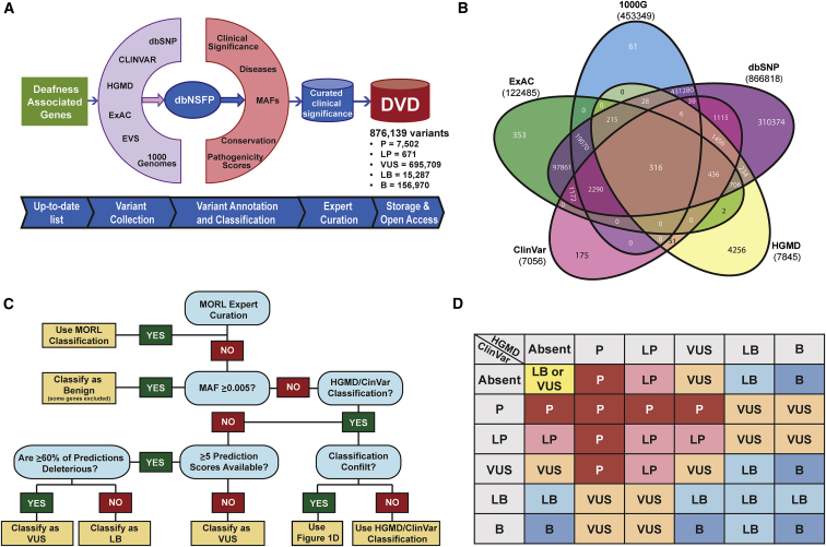
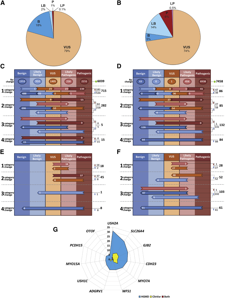
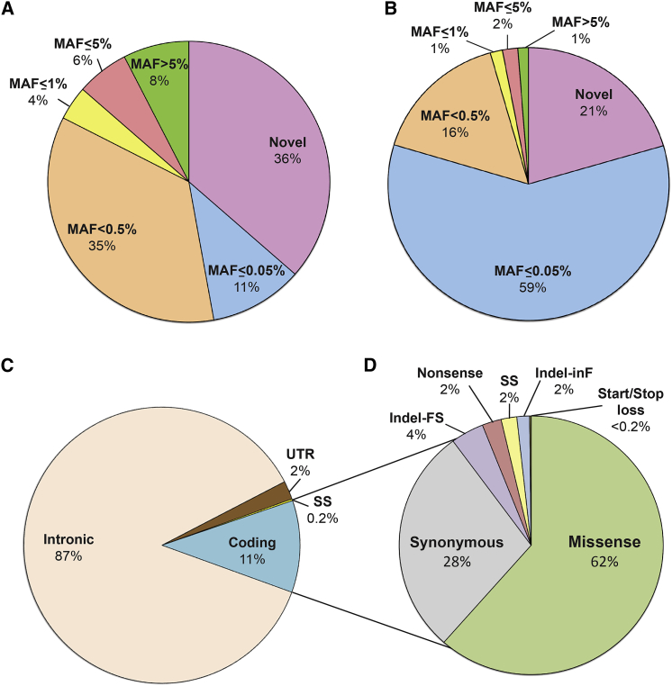
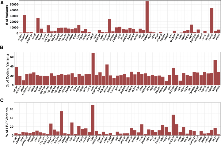
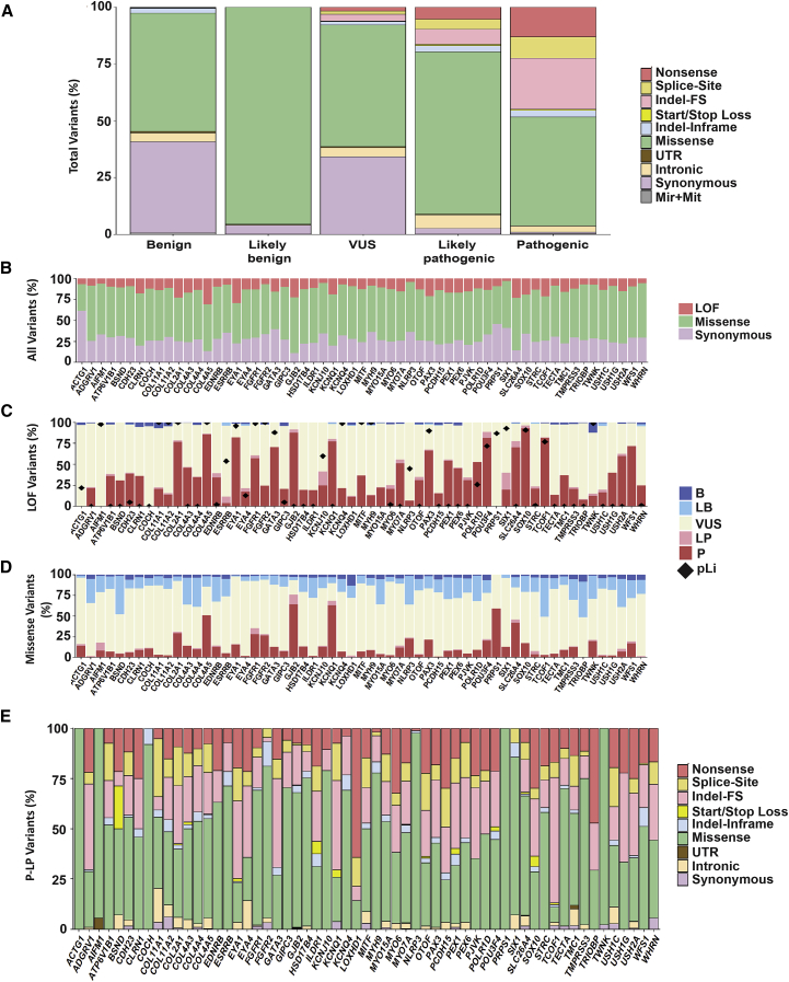
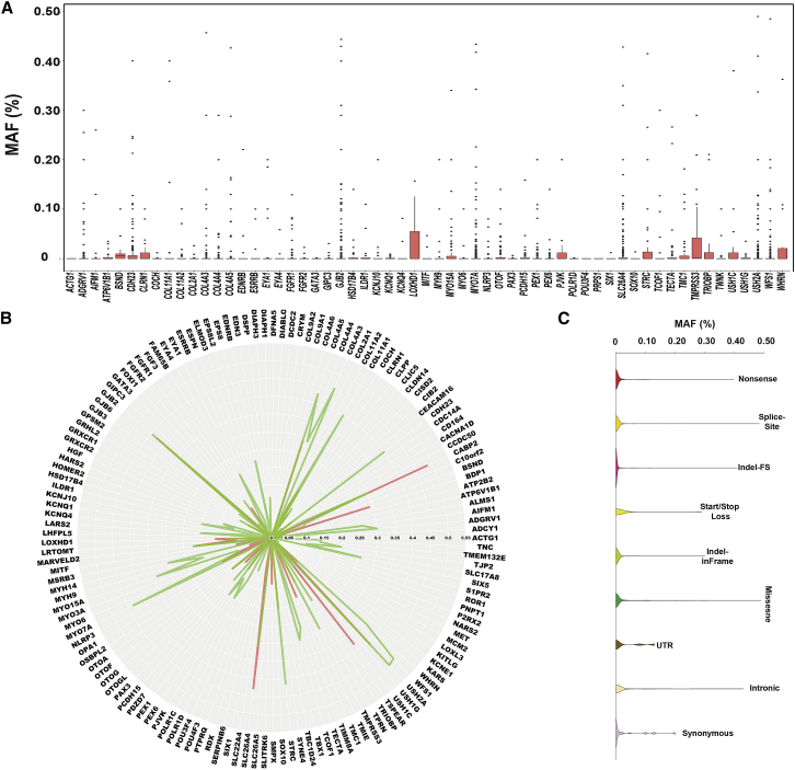
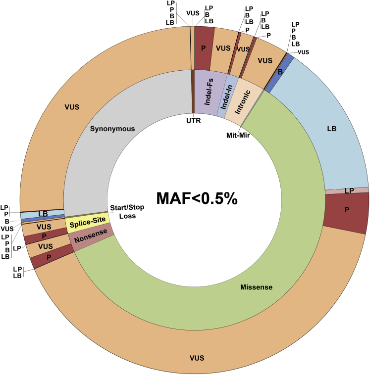
The classification of genetic variants represents a major challenge in the post-genome era by virtue of their extraordinary number and the complexities associated with ascribing a clinical impact, especially for disorders exhibiting exceptional phenotypic, genetic, and allelic heterogeneity. To address this challenge for hearing loss, we have developed the Deafness Variation Database (DVD), a comprehensive, open-access resource that integrates all available genetic, genomic, and clinical data together with expert curation to generate a single classification for each variant in 152 genes implicated in syndromic and non-syndromic deafness. We evaluate 876,139 variants and classify them as pathogenic or likely pathogenic (more than 8,100 variants), benign or likely benign (more than 172,000 variants), or of uncertain significance (more than 695,000 variants); 1,270 variants are re-categorized based on expert curation and in 300 instances, the change is of medical significance and impacts clinical care. We show that more than 96% of coding variants are rare and novel and that pathogenicity is driven by minor allele frequency thresholds, variant effect, and protein domain. The mutational landscape we define shows complex gene-specific variability, making an understanding of these nuances foundational for improved accuracy in variant interpretation in order to enhance clinical decision making and improve our understanding of deafness biology.

Keywords: database; deafness; genetic variant; genomic landscape; mutational signature; precision medicine; variant classification.


Figures

Figure 1
Figure 1
The Deafness Variation Database (A) Kafeen, a custom internal pipeline, gathers data for the DVD by collecting variants and annotations from multiple data sources. Deleteriousness predictions collected from dbNSFP and MAF data are extracted from our local database, EVS, 1000 Genomes, and ExAC to inform the DVD classification. A comparison between DVD versus ClinVar and HGMD classifications captures all changes that result in medically significant differences (defined as up-grading a variant to P/LP or down-grading a variant from P/LP), each of which is manually curated to ensure that the DVD reclassification is appropriate. (B) Venn diagram showing number of variants collated from major population-scale MAF databases and the count of variants that are shared among them and those that are database specific. (C) Decision tree for Kafeen classification. (D) Decision matrix detailing Kafeen logic regarding variants classified in ClinVar and HGMD.
Figure 2
Figure 2
Variant Classification by the DVD (A) Fractions of different classification categories for variants in the whole DVD. (B) A slightly different picture emerges when only clinically relevant regions and deafness-associated variants (variants that were associated with other non-related deafness phenotypes are excluded) are considered. (C) Comparative overview of DVD versus ClinVar. 7,056 classifications from ClinVar were identified within our specified gene regions (each variant in ClinVar with multiple submissions for pathogenicity has been represented by its most pathogenic submission). Of this number, 6,039 ClinVar classifications agreed with the corresponding DVD classification whereas there was disagreement for 1,017 variants. (D) Comparative overview of DVD versus HGMD. 7,845 classifications from HGMD were identified within our specified gene regions. Of this number, 7,458 classifications agreed with the corresponding DVD classification and discrepancies were found for 387 variants. (E) There were 72 major categorical changes between ClinVar and DVD that resulted in medically significant differences (53 up-classifications and 19 down-classifications). (F) 244 medically significant reclassifications were found when DVD was compared to HGMD (2 up-classifications and 242 down-classifications). (G) Of the 20% of genes carrying the greatest numbers of medically significant changes, 6 are implicated in Usher syndrome. For (C) through (F), the horizontal arrows show discordant calls, with the number of discordant classifications shown within each arrow; totals are listed to the right of the colored columns.
Figure 3
Figure 3
Distribution of Variants by Location, MAF, and Type (A and B) MAF (all variants in DVD including intronic) (A) and only variants in gene coding regions (B). Most coding variants (96%) in deafness-associated genes are novel or rare (MAF < 0.5%). (C) Distribution of variant by their gene location. (D) Coding variant breakdown by type showing that missense variants constitute the major set of all coding variants. Abbreviations: FS, frameshift; SS, splice-site; inF, in-frame.
Figure 4
Figure 4
Variation Rate for Deafness-Associated Genes (A) Total number of variants per gene. (B) Normalized number of coding variants based on the size of the coding and splice regions. (C) Normalized number of deafness-associated variants (P+LP) based on the total number of coding variants. Only genes with ≥14 reported deafness-associated variants are included in this figure; the remaining genes are shown in Figures S2 and S3.
Figure 5
Figure 5
Genomic Landscape of Deafness-Associated Genes (A) Variant architecture by each classification category shows a strikingly distinct distribution of variant types across the five classifications. (B) Distribution of LoF, missense, and synonymous variants is different across genes. (C) Most LoF variants are P/LP and some genes are highly enriched in this type of variant. (D) The contribution of missense variants to the mutational pool of hearing loss is variable across genes. However, in most genes, the majority of missense variants are VUSs. (E) The mutational spectrum is gene specific. Splice-site indicates variants in canonical splice sites. Only genes with ≥14 reported deafness-associated variants are included in this figure; the remaining genes are shown in Figures S4 and S5.
Figure 6
Figure 6
MAFs Thresholds for Deafness-Associated Variants Are Gene and Type Specific (A) Plot of MAFs of all P/LP variants in each deafness-associated gene. (B) Maximum MAF is gene specific and there is a clear distinction between LoF versus missense variants. (C) Overall, missense variants exhibit the highest MAFs when compared to all other variants. Only genes with ≥14 reported deafness-associated variants are included in this figure; the remaining genes are shown in Figure S5.
Figure 7
Figure 7
The Challenge of VUSs Variant architecture correlating variant type (inner ring) and clinical significance (outer ring) for variants with MAF less than 0.5% and located within the clinically relevant regions. Of all coding variants with MAF < 0.5%, missense variants represent the majority at 61.5%; of these missense variants, 70% are classified as VUSs. Abbreviations: Indel-In, in-frame indel; Indel-Fs, frameshift indel; Mit-Mir, mitochondrial and microRNA.

References

    1. Collins F.S., Varmus H. A new initiative on precision medicine. N. Engl. J. Med. 2015;372:793–795. - PMC - PubMed
    1. Patel R.Y., Shah N., Jackson A.R., Ghosh R., Pawliczek P., Paithankar S., Baker A., Riehle K., Chen H., Milosavljevic S., ClinGen Resource ClinGen Pathogenicity Calculator: a configurable system for assessing pathogenicity of genetic variants. Genome Med. 2017;9:3. - PMC - PubMed
    1. Richards S., Aziz N., Bale S., Bick D., Das S., Gastier-Foster J., Grody W.W., Hegde M., Lyon E., Spector E., ACMG Laboratory Quality Assurance Committee Standards and guidelines for the interpretation of sequence variants: a joint consensus recommendation of the American College of Medical Genetics and Genomics and the Association for Molecular Pathology. Genet. Med. 2015;17:405–424. - PMC - PubMed
    1. Landrum M.J., Lee J.M., Benson M., Brown G., Chao C., Chitipiralla S., Gu B., Hart J., Hoffman D., Hoover J. ClinVar: public archive of interpretations of clinically relevant variants. Nucleic Acids Res. 2016;44(D1):D862–D868. - PMC - PubMed
    1. Stenson P.D., Mort M., Ball E.V., Shaw K., Phillips A., Cooper D.N. The Human Gene Mutation Database: building a comprehensive mutation repository for clinical and molecular genetics, diagnostic testing and personalized genomic medicine. Hum. Genet. 2014;133:1–9. - PMC - PubMed

Publication types