. 2018 Oct 4;103(4):592-601.
doi: 10.1016/j.ajhg.2018.08.013. Epub 2018 Sep 20.

Bi-allelic Mutations in NDUFA6 Establish Its Role in Early-Onset Isolated Mitochondrial Complex I Deficiency

Affiliations

Bi-allelic Mutations in NDUFA6 Establish Its Role in Early-Onset Isolated Mitochondrial Complex I Deficiency

Charlotte L Alston et al. Am J Hum Genet. .

Abstract

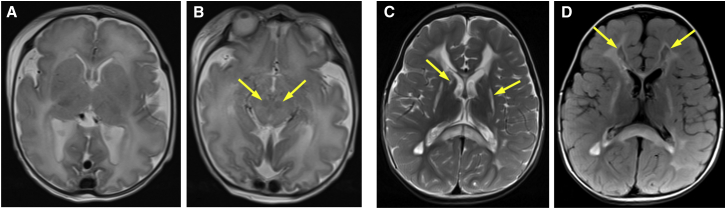
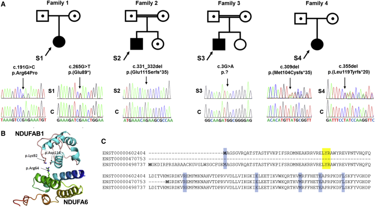
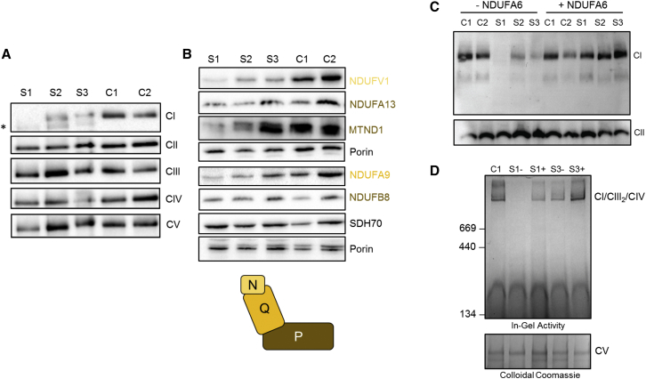
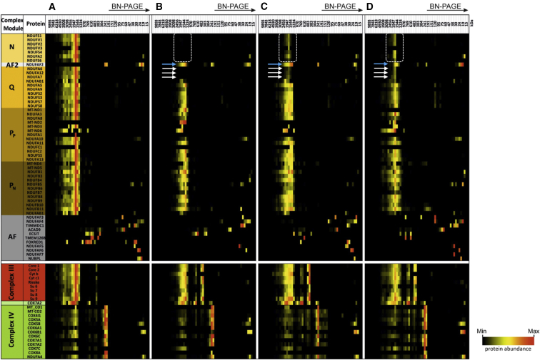
Isolated complex I deficiency is a common biochemical phenotype observed in pediatric mitochondrial disease and often arises as a consequence of pathogenic variants affecting one of the ∼65 genes encoding the complex I structural subunits or assembly factors. Such genetic heterogeneity means that application of next-generation sequencing technologies to undiagnosed cohorts has been a catalyst for genetic diagnosis and gene-disease associations. We describe the clinical and molecular genetic investigations of four unrelated children who presented with neuroradiological findings and/or elevated lactate levels, highly suggestive of an underlying mitochondrial diagnosis. Next-generation sequencing identified bi-allelic variants in NDUFA6, encoding a 15 kDa LYR-motif-containing complex I subunit that forms part of the Q-module. Functional investigations using subjects' fibroblast cell lines demonstrated complex I assembly defects, which were characterized in detail by mass-spectrometry-based complexome profiling. This confirmed a marked reduction in incorporated NDUFA6 and a concomitant reduction in other Q-module subunits, including NDUFAB1, NDUFA7, and NDUFA12. Lentiviral transduction of subjects' fibroblasts showed normalization of complex I. These data also support supercomplex formation, whereby the ∼830 kDa complex I intermediate (consisting of the P- and Q-modules) is in complex with assembled complex III and IV holoenzymes despite lacking the N-module. Interestingly, RNA-sequencing data provided evidence that the consensus RefSeq accession number does not correspond to the predominant transcript in clinically relevant tissues, prompting revision of the NDUFA6 RefSeq transcript and highlighting not only the importance of thorough variant interpretation but also the assessment of appropriate transcripts for analysis.

Keywords: NDUFA6; complex I; complexome profiling; mitochondrial disease.


Figures

Figure 1
Figure 1
Neuroimaging of Subjects 1 and 2 (A and B) Axial T2 imaging of subject 1 shows diffusely abnormal hyperintensity of the entire white matter and decreased cortical folding for her age (A). Signal abnormalities within the brain stem and strikingly low signal affecting red nuclei (arrowheads) are also apparent (B). (C) Axial T2 imaging of subject 2 shows extensive signal abnormalities of the cerebral white matter, including the corpus callosum. In addition, basal ganglia are affected, especially the putamen and caudate nucleus (arrowheads). (D) Axial FLAIR imaging of subject 2 shows partial cystic degeneration of the affected cerebral white matter (arrowheads).
Figure 2
Figure 2
Bi-allelic NDUFA6 Variants Are Identified in Four Unrelated Subjects (A) Family pedigrees of subjects 1–4 and corresponding sequencing chromatograms presenting compound-heterozygous c.191G>C (p.Arg64Pro) and c.265G>T (p.Glu89) NDUFA6 variants in subject 1, the homozygous NDUFA6 deletion c.331_332del (p.Glu111Serfs35) in subject 2, the homozygous c.3G>A (p.?) NDUFA6 variant in subject 3, and compound-heterozygous c.309del (p.Met104Cysfs35) and c.355del (p.Leu119Tyrfs20) NDUFA6 frameshift mutations in subject 4. Abbreviations are as follows: S, subject; C, wild-type control. (B) Analysis of the resolved crystal structure of complex I revealed spatial proximity of the Arg64 NDUFA6 residue with two negatively charged and highly conserved NDUFAB1 residues, Lys92 and Asp114. These most likely represent critical binding interactions between the two proteins during late complex I assembly. (C) Alignment of the three in-frame NDUFA6 transcripts demonstrates that ENST00000602404 (GenBank: NM_002490.5) uses an initiator methionine downstream of that used by ENST00000498737 (GenBank: NM_002490.4). The shortest transcript, ENST00000470753, initiates downstream of the highly conserved LYR motif (shaded yellow) that is reported to be critical for binding the mitochondrial acyl carrier (encoded by NDUFAB1); the function of this isoform therefore remains unknown. Subject variants are shaded blue.
Figure 3
Figure 3
BN-PAGE, SDS-PAGE, and Complementation Studies (A) Mitochondria isolated from cultured skin fibroblasts from subjects 1–3 and age-matched control subjects were solubilized in n-dodecyl β-d-maltoside (DDM) and subjected to BN-PAGE and immunoblotting analysis using antibodies directed to various OXPHOS complexes as indicated. The blot probed with an antibody raised against NDUFB8 revealed the presence of additional, partially assembled complex I intermediates in the samples from subjects 1–3 (indicated by an asterisk) but not in control samples. (B) Whole-fibroblast cell lysates from subjects 1–3 and age-matched control subjects were analyzed by SDS-PAGE. Immunoblotting was performed with antibodies against complex I subunits or control proteins (SDHA and porin) as indicated. Complex I structural subunits are color coded according to their corresponding complex I modules, as illustrated in the complex I pictogram. (C) Wild-type NDUFA6 cDNA was generated and introduced into control and subject cell lines via retroviral expression. Whole-cell lysates were solubilized in 1% Triton X-100 (immunoblotting) before BN-PAGE analysis. Immunoblotting using antibodies against the complex I subunit NDUFA9 (top) and the complex II subunit SDHA (bottom) as a loading control revealed less complex I in subject cell lines than in control cell lines. After transfection with NDUFA6, complex I levels were restored. (D) In-gel activity analysis was performed according to Zerbetto et al. with mitochondria isolated from the fibroblasts of subjects 1 (S1) and 3 (S3) with (+) and without (−) lentiviral transduction with NDUFA6 cDNA and an aged-matched control subject; enriched mitochondria were solubilized in 1% digitonin for BN-PAGE analysis, which demonstrated restoration of complex I activity in subjects 1 and 3 after the introduction of NDUFA6 (top). The gel was then stained with colloidal Coomassie according to Neuhoff et al. as a loading control (bottom). All antibodies used are documented in Table S1.
Figure 4
Figure 4
Complexome Profiling of Fibroblasts from Subjects 1–3 Identifies Stalled Complex I Assembly Intermediates Control (A), subject 1 (B), subject 2 (C), and subject 3 (D). The left lane indicates assembly factors (gray) and structural subunits (orange-brown) of complex I modules, complex III (red), and complex IV (green). Dashed boxes indicate the loss or reduction of N-module subunits in subject fibroblast lines. White arrows highlight the loss or reduction of NDUFA6, NDUFA7 and NDUFA12 from complex I in subjects 1–3, and blue arrows indicate the location of NDUFAF2, still bound to the stalled complex I intermediate in subject complexomes but not in the control. Assignment of complex I subunits to modules was according to Wirth et al. Intensity-based absolute quantification (iBAQ) values were normalized to the sum of all values of the control. Native mass calibration was performed according to Fuhrmann et al.

References

    1. Formosa L.E., Dibley M.G., Stroud D.A., Ryan M.T. Building a complex complex: Assembly of mitochondrial respiratory chain complex I. Semin. Cell Dev. Biol. 2018;76:154–162. - PubMed
    1. Calvo S.E., Clauser K.R., Mootha V.K. MitoCarta2.0: An updated inventory of mammalian mitochondrial proteins. Nucleic Acids Res. 2016;44(D1):D1251–D1257. - PMC - PubMed
    1. Frazier A.E., Thorburn D.R., Compton A.G. Mitochondrial energy generation disorders: Genes, mechanisms and clues to pathology. J. Biol. Chem. 2017 Published online December 12, 2017. - PMC - PubMed
    1. Piekutowska-Abramczuk D., Assouline Z., Mataković L., Feichtinger R.G., Koňařiková E., Jurkiewicz E., Stawiński P., Gusic M., Koller A., Pollak A. NDUFB8 mutations cause mitochondrial complex I deficiency in individuals with Leigh-like encephalomyopathy. Am. J. Hum. Genet. 2018;102:460–467. - PMC - PubMed
    1. Haack T.B., Kopajtich R., Freisinger P., Wieland T., Rorbach J., Nicholls T.J., Baruffini E., Walther A., Danhauser K., Zimmermann F.A. ELAC2 mutations cause a mitochondrial RNA processing defect associated with hypertrophic cardiomyopathy. Am. J. Hum. Genet. 2013;93:211–223. - PMC - PubMed

Publication types

Substances

Supplementary concepts