. 2018 Sep 19:7:64.
doi: 10.1038/s41377-018-0065-2. eCollection 2018.

Metasurface-based multi-harmonic free-electron light source

Affiliations

Metasurface-based multi-harmonic free-electron light source

Gilles Rosolen et al. Light Sci Appl. .

Abstract

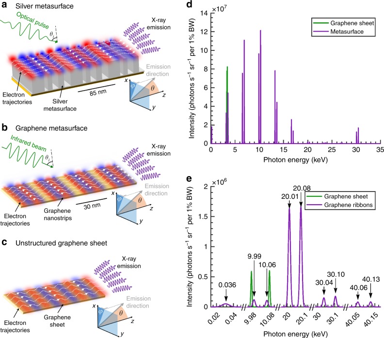
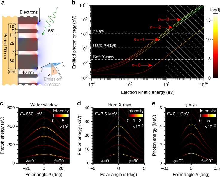
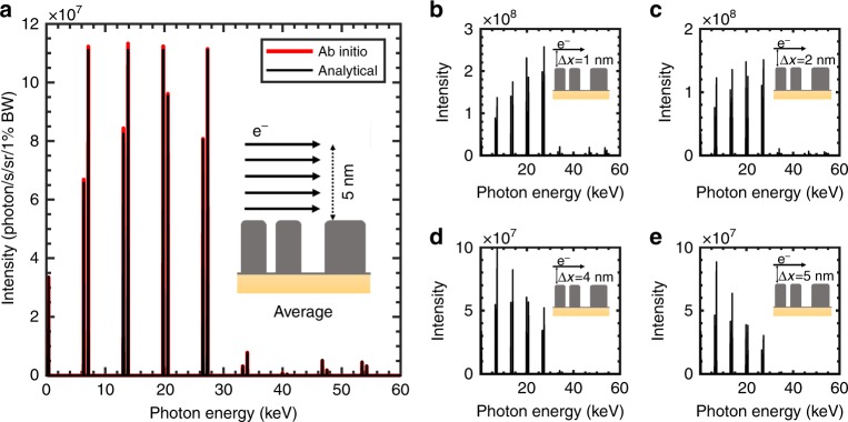
Metasurfaces are subwavelength spatial variations in geometry and material where the structures are of negligible thickness compared to the wavelength of light and are optimized for far-field applications, such as controlling the wavefronts of electromagnetic waves. Here, we investigate the potential of the metasurface near-field profile, generated by an incident few-cycle pulse laser, to facilitate the generation of high-frequency light from free electrons. In particular, the metasurface near-field contains higher-order spatial harmonics that can be leveraged to generate multiple higher-harmonic X-ray frequency peaks. We show that the X-ray spectral profile can be arbitrarily shaped by controlling the metasurface geometry, the electron energy, and the incidence angle of the laser input. Using ab initio simulations, we predict bright and monoenergetic X-rays, achieving energies of 30 keV (with harmonics spaced by 3 keV) from 5-MeV electrons using 3.4-eV plasmon polaritons on a metasurface with a period of 85 nm. As an example, we present the design of a four-color X-ray source, a potential candidate for tabletop multicolor hard X-ray spectroscopy. Our developments could help pave the way for compact multi-harmonic sources of high-energy photons, which have potential applications in industry, medicine, and the fundamental sciences.


Conflict of interest statement

The authors declare that they have no conflict of interest.

Figures

Fig. 1
Fig. 1. Higher-harmonic X-rays from metasurface-enhanced plasmon-based free-electron light source:
The presence of higher spatial harmonics in the near-field profiles leads to output of higher-order X-ray harmonics, allowing the spatiotepmoral characteristics to be tailored via the metasurface geometry. a Plasmon polaritons (red and blue) of a silver metasurface with 85-nm periodicity are excited by an optical pulse with a wavelength of 370 nm (3.4 eV) and a peak field amplitude of E0 = 1 GV/m. An electron beam with a kinetic energy of 5 MeV and an average current of 100 µA is sent parallel to the metasurface at a distance of 1 nm. These electrons are modulated by the plasmonic field, resulting in undulating electron trajectories (dashed white lines) that generate highly directional photons. b Plasmon polaritons of a 20-nm wide graphene nanoribbon array (with periods of 28.5 nm) excited by an infrared Gaussian beam with a wavelength of 2 µm and a field amplitude of E0 = 0.1 GV/m. c The process in (a) and (b) is compared to the unstructured graphene sheet in ref. using a plasmon wavelength of 85 nm. d The sinusoidal trajectory of the electrons modulated by the graphene plasmons (situation (c)) results in a single harmonic (green lines), whereas the nonsinusoidal trajectory of the electrons in the metasurface near-field (situation (a)) results in multiple monoenergetic harmonics in addition to the free-space Compton harmonic at 0.3 keV and the fundamental harmonics at 3.1 keV and 3.7 keV (purple line). e The 5-MeV electron beam with an average current of 100 µA generates multiple harmonics dominated by the second harmonic at a photon energy of 20 keV (situation (b)). In both cases, the angle of incidence for the laser is θi = 50°
Fig. 2
Fig. 2. Tunability of the emitted photon energy via the electron energy.
a Metasurface optimized to enhance the first five orders. The plasmon polaritons are excited by a laser pulse with an energy of 3.4 eV (370 nm), and electrons are shot parallel to the metasurface at a distance of 1 nm. The emission direction is described by φ and θ in the bottom right inset. b At a fixed electron energy, the on-axis output photon energy is higher for higher orders (for visibility, we only present orders n = 0, −1, −2, −3) and the source is brighter (intensity in units of number of photons/s/sr/1%BW). At high energies (denoted by the hatched region), our analytical formulation begins to differ from the results predicted by energy–momentum conservation (denoted by green lines), since the analytical formulation does not take quantum recoil into account. ce Highly monochromatic harmonic peaks (fractional bandwidths (c) < 0.4% (d) < 0.2% (e) < 0.19%) for c soft X-rays from 550 keV electrons (already accessible with a TEM), d hard X-rays from 7.5-MeV electrons (can be obtained using a tabletop setup), and e γ-rays from 0.1-GeV electrons. Note that the line thickness has been artificially increased for clarity
Fig. 3
Fig. 3. Enhancing X-ray intensity by velocity matching the laser intensity peak and electrons.
The maximum intensity for positive and negative emission orders are presented in (a) and (b), respectively. The inset shows the output spectrum for θi  =0, and the arrow colors are used to distinguish the different emission orders: order 0 (green), order 1 (blue), order 2 (red), and order 3 (black). Solid (dashed) lines represent the 7.5-MeV (100 keV) electron energy and the dots are approximated in Equations 3 and 4. The logarithmic spectrum in (c) shows the peak intensities and energies for the ± 1 emission orders at different angles of incidence of the laser pulse. The metasurface considered has a period of 30 nm consisting of silver blocks 15 nm in width and 40 nm in height laying on a substrate with a refractive index of 1.5, and the spectrum is measured in the direction of the electrons
Fig. 4
Fig. 4. Control of multi-harmonic output radiation via metasurface geometry and input wavelength.
In the plots of output intensity as a function of input laser wavelength for various metasurface designs, we see that the silver tips (a) and triangular grating structure (b) enhance the negative orders (denoted by dashed lines) compared with the positive orders (denoted by solid lines), as opposed to the rectangular grating structure (c), where positive and negative orders are much more comparable in intensity. d Using the first (second) plasmon mode of the graphene nanoribbon leads to an enhanced first (second) order emission. The silver metasurface with periods of 30 nm and containing 40-nm high structures ((a) to (c)) and 30- nm periods of 20- nm-wide graphene nanoribbons (d). The incident Gaussian beam has an angle of θi = 65°, a spot size of 12 µm FWHM on the metasurface, and an energy of 7.5 MeV. The tips in (a) are 6 nm wide and the blocks in (b) are 15 nm wide. The incident fields in(a)–(c) are E0  = 1 GV/m, and the field in (d) is E0  = 0.1 GV/m
Fig. 5
Fig. 5. Optimized multicolor hard X-ray source with analytical formulations, showing excellent agreement with ab initio simulations.
a The average intensity spectrum for a uniformly distributed electron beam, optimizing the metasurface profile with a procedure in which only five-test particle trajectories have to be considered. These five test particles are spaced between 1 and 5 nm away from the metasurface. The optimization results for the five-test particle model show excellent agreement with ab initio simulations of the actual multiparticle beam. The inset shows a period of the metasurface with the electron beam. be The intensity spectra of electrons launched 1 nm (b), 2 nm (c), 4 nm (d), and 5 nm (e) from the top of the metasurface. The electron beam energy is 7.5 MeV with an assumed current of 100 µA; the 10-fs laser pulse impinges the metasurface at θi = 50°

Similar articles

Cited by

References

    1. Zhou ZP, Yin B, Michel J. Corrigendum: on-chip light sources for silicon photonics. Light Sci. Appl. 2016;4:e16098. doi: 10.1038/lsa.2016.98. - DOI - PMC - PubMed
    1. Lukianova-Hleb EY, et al. On-demand intracellular amplification of chemoradiation with cancer-specific plasmonic nanobubbles. Nat. Med. 2014;20:778–784. doi: 10.1038/nm.3484. - DOI - PMC - PubMed
    1. Hara T, et al. Two-colour hard x-ray free-electron laser with wide tunability. Nat. Commun. 2013;4:2919. doi: 10.1038/ncomms3919. - DOI - PubMed
    1. Bleuet P, et al. Probing the structure of heterogeneous diluted materials by diffraction tomography. Nat. Mater. 2008;7:468–472. doi: 10.1038/nmat2168. - DOI - PubMed
    1. Bargheer M, Zhavoronkov N, Woerner M, Elsaesser T. Recent progress in ultrafast X-ray diffraction. Chemphyschem. 2006;7:783–792. doi: 10.1002/cphc.200500591. - DOI - PubMed