. 2018 Sep 11:4:50.
doi: 10.1038/s41421-018-0053-y. eCollection 2018.

Human embryonic stem cell-derived retinal pigment epithelium transplants as a potential treatment for wet age-related macular degeneration

Affiliations

Human embryonic stem cell-derived retinal pigment epithelium transplants as a potential treatment for wet age-related macular degeneration

Yong Liu et al. Cell Discov. .

Abstract

Stem cell therapy may provide a safe and promising treatment for retinal diseases. Wet age-related macular degeneration (wet-AMD) is a leading cause of blindness in China. We developed a clinical-grade human embryonic stem cell (hESC) line, Q-CTS-hESC-2, under xeno-free conditions that differentiated into retinal pigment epithelial cells (Q-CTS-hESC-2-RPE). A clinical trial with three wet-AMD patients was initiated in order to study the safety and tolerance to Q-CTS-hESC-2-RPE cell transplants. The choroidal neovascularization membrane was removed and then a suspension of 1 × 10⁶ Q-CTS-hESC-2-RPE cells were injected into a subfoveal pocket. The patients were followed for 12 months during which no adverse effects resulting from the transplant were observed. Anatomical evidence suggested the existence of new RPE-like cell layer in the previously damaged area. Visual and physiological testing indicated limited functional improvement, albeit to different degrees between patients. This study provides some promising early results concerning the use of transplanted hESC-RPE cells to alleviate wet-AMD.


Conflict of interest statement

The authors declare that they have no conflict of interest.

Figures
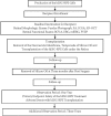
Fig. 1
Fig. 1
Flow chart showing the experimental protocol
Fig. 2
Fig. 2. Morphological changes in patient #1 following CNV removal and hESC-RPE cell transplantation.
ad show pre- and postoperative color fundus photographs (column 1); en-face structural OCT (column 2) in which the green arrows correspond to the highly-sampled OCT b-scans vertical sectional views in column 3; column 4 shows 3D reconstruction images of the same retinal region. a A large fibrotic CNV and hemorrhage were observed in the subfoveal space preoperatively. b 1 month after surgery the fundus images show a complete removal of the CNV, and an almost intact Bruch’s membrane. Note the cell clusters (indicated by white arrows) evident as a dense medium reflective mass in the region of baseline RPE atrophy. c At 6 months, macular edema was not observed in the region of the fovea. A highly reflective RPE-like cell layer was observed in the transplant region (white arrows). This layer was similar to the healthy RPE outside the region of the transplant (white asterisks). d At 12 months the highly reflective RPE-like cell layer was still present in the transplant area (white arrows). The photoreceptor layer above this region shows better anatomical preservation compared to adjacent regions. Panel e shows the mapping strategy using OCT scanning to determine the distribution of grafted cells; 61 horizontal lines were evenly scanned while focusing on the transplantation area. An RPE-like cell layer extended from the 20th to the 38th scan (red dashed lines in the OCT images). SD-OCT spectral domain ocular coherence tomography, CNV choroidal neovascularization, RPE retinal pigment epithelial
Fig. 3
Fig. 3. Angiographic characteristics and physiological changes in Patient #1.
a Retinal and choroidal vascular changes as seen with fundus fluorescein FA (left side of pair) or ICGA (right side of pair). The preoperative test showed the subfoveal choroidal neovessels beneath the CNV membrane (early phase) and leakage with hyperfluorescence that obscured the boundaries of the lesion (late phase). One year later, new vessel growth was not observed in the ICGA, nor any additional leakage observed in the FA. b The mfERG waves before (baseline) and after (4 and 12 months follow-up) transplantation. c Analysis of amplitude changes for the central hexagon (ring 1) that mainly represents foveal visual function. The standard measurement for mfERG amplitude is the trough-to-peak amplitude (nv) measured between N1 and P and given as the amplitude density nv over its hexagon in degrees (nv/deg2). d The waveform of FVEP at different time points after transplantation. e Graph shows P2 amplitude changes induced by FVEP. FA fluorescein angiography, ICGA indocyanine green fluorescein angiography, mfERG multifocal electroretinography, FVEP flash visual evoked potentials
Fig. 4
Fig. 4. Morphological changes after CNV removal and hESC-RPE transplantation in Patients #2 and #3.
ad Patient #2: pre- and postoperative color fundus photographs (left panel), en-face structural SD-OCT images (middle panel) in which the green arrow corresponds to the highly sampled SD-OCT b-scan in the right-most panel. a The subretinal neovascular membrane could be clearly seen prior to the transplant (white arrow). b Four months after surgery, the subretinal neovascular membrane was gone and there is no indication of macular edema. Cell clusters (white arrows) could be observed within and adjacent to macular area. c Six months post-transplant, cell clusters (white arrows) were seen in the graft region. d By the 12 month follow-up, the transplanted cell clusters appeared to have merged (white arrows). eh Patient #3: (e) A subretinal neovascular membrane was seen before surgery (white arrow). f 4 months after surgery; no hemorrhage or edema was observed in the fovea. An RPE-like cell layer had formed under the macular zone (white arrows) that was similar to the host RPE layer (white asterisks). g 6 months later the highly reflective RPE-like cell layer was still present in the graft area. h 12 months later there still appeared to be evidence for an RPE-like cell layer in the graft area (white arrows). Scale bar = 1 mm. SD-OCT spectral domain ocular coherence tomography, RPE retinal pigment epithelial

Similar articles

Cited by

References

    1. Fine SL, B J, Maguire MG, Ho AC. Age-related macular degeneration. N. Engl. J. Med. 2000;342:483–492. doi: 10.1056/NEJM200002173420707. - DOI - PubMed
    1. Ding X, P M, Chan CC. Molecular pathology of age-related macular degeneration. Prog. Retin Eye Res. 2009;28:1–18. doi: 10.1016/j.preteyeres.2008.10.001. - DOI - PMC - PubMed
    1. Strauss O. The retinal pigment epithelium in visual function. Physiol. Rev. 2005;85:845–881. doi: 10.1152/physrev.00021.2004. - DOI - PubMed
    1. Ye H, et al. Prevalence of age-related macular degeneration in an elderly urban chinese population in China: the Jiangning Eye Study. Invest. Ophthalmol. & Vis. Sci. 2014;55:6374–6380. doi: 10.1167/iovs.14-14899. - DOI - PubMed
    1. Wong CW, et al. Age-related macular degeneration and polypoidal choroidal vasculopathy in Asians. Prog. Retin Eye Res. 2016;53:107–139. doi: 10.1016/j.preteyeres.2016.04.002. - DOI - PubMed