Rehabilitative exercise and spatially patterned nanofibrillar scaffolds enhance vascularization and innervation following volumetric muscle loss
- PMID: 30245849
- PMCID: PMC6141593
- DOI: 10.1038/s41536-018-0054-3
Rehabilitative exercise and spatially patterned nanofibrillar scaffolds enhance vascularization and innervation following volumetric muscle loss
Abstract
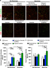
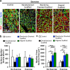
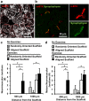
Muscle regeneration can be permanently impaired by traumatic injuries, despite the high regenerative capacity of skeletal muscle. Implantation of engineered biomimetic scaffolds to the site of muscle ablation may serve as an attractive off-the-shelf therapeutic approach. The objective of the study was to histologically assess the therapeutic benefit of a three-dimensional spatially patterned collagen scaffold, in conjunction with rehabilitative exercise, for treatment of volumetric muscle loss. To mimic the physiologic organization of skeletal muscle, which is generally composed of myofibers aligned in parallel, three-dimensional parallel-aligned nanofibrillar collagen scaffolds were fabricated. When implanted into the ablated murine tibialis anterior muscle, the aligned nanofibrillar scaffolds, in conjunction with voluntary caged wheel exercise, significantly improved the density of perfused microvessels, in comparison to treatments of the randomly oriented nanofibrillar scaffold, decellularized scaffold, or in the untreated control group. The abundance of neuromuscular junctions was 19-fold higher when treated with aligned nanofibrillar scaffolds in conjunction with exercise, in comparison to treatment of aligned scaffold without exercise. Although, the density of de novo myofibers was not significantly improved by aligned scaffolds, regardless of exercise activity, the cross-sectional area of regenerating myofibers was increased by > 60% when treated with either aligned and randomly oriented scaffolds, in comparison to treatment of decellularized scaffold or untreated controls. These findings demonstrate that voluntary exercise improved the regenerative effect of aligned scaffolds by augmenting neurovascularization, and have important implications in the design of engineered biomimetic scaffolds for treatment of traumatic muscle injury.
Conflict of interest statement
The authors declare no competing interests.
Figures


Similar articles
-
Transplantation of insulin-like growth factor-1 laden scaffolds combined with exercise promotes neuroregeneration and angiogenesis in a preclinical muscle injury model.Biomater Sci. 2020 Oct 7;8(19):5376-5389. doi: 10.1039/d0bm00990c. Epub 2020 Sep 2. Biomater Sci. 2020. PMID: 32996916 Free PMC article.
-
Comparative Effects of Basic Fibroblast Growth Factor Delivery or Voluntary Exercise on Muscle Regeneration after Volumetric Muscle Loss.Bioengineering (Basel). 2022 Jan 14;9(1):37. doi: 10.3390/bioengineering9010037. Bioengineering (Basel). 2022. PMID: 35049746 Free PMC article.
-
Treatment of volumetric muscle loss in mice using nanofibrillar scaffolds enhances vascular organization and integration.Commun Biol. 2019 May 7;2:170. doi: 10.1038/s42003-019-0416-4. eCollection 2019. Commun Biol. 2019. PMID: 31098403 Free PMC article.
-
Electrospun three-dimensional aligned nanofibrous scaffolds for tissue engineering.Mater Sci Eng C Mater Biol Appl. 2018 Nov 1;92:995-1005. doi: 10.1016/j.msec.2018.06.065. Epub 2018 Jun 30. Mater Sci Eng C Mater Biol Appl. 2018. PMID: 30184829 Review.
-
Challenges to acellular biological scaffold mediated skeletal muscle tissue regeneration.Biomaterials. 2016 Oct;104:238-46. doi: 10.1016/j.biomaterials.2016.07.020. Epub 2016 Jul 20. Biomaterials. 2016. PMID: 27472161 Review.
Cited by
-
Elastin-like protein hydrogels with controllable stress relaxation rate and stiffness modulate endothelial cell function.J Biomed Mater Res A. 2023 Jul;111(7):896-909. doi: 10.1002/jbm.a.37520. Epub 2023 Mar 2. J Biomed Mater Res A. 2023. PMID: 36861665 Free PMC article.
-
Biomaterial-Based Regenerative Strategies for Volumetric Muscle Loss: Challenges and Solutions.Adv Wound Care (New Rochelle). 2025 Mar;14(3):159-175. doi: 10.1089/wound.2024.0079. Epub 2024 Jul 10. Adv Wound Care (New Rochelle). 2025. PMID: 38775429 Review.
-
3D printing a biocompatible elastomer for modeling muscle regeneration after volumetric muscle loss.Biomater Adv. 2022 Nov;142:213171. doi: 10.1016/j.bioadv.2022.213171. Epub 2022 Oct 24. Biomater Adv. 2022. PMID: 36341746 Free PMC article.
-
Influence of Physical Exercise on the Rehabilitation of Volumetric Muscle Loss Injury Reconstructed with Autologous Adipose Tissue.J Funct Morphol Kinesiol. 2024 Oct 8;9(4):188. doi: 10.3390/jfmk9040188. J Funct Morphol Kinesiol. 2024. PMID: 39449482 Free PMC article.
-
Engineering skeletal muscle: Building complexity to achieve functionality.Semin Cell Dev Biol. 2021 Nov;119:61-69. doi: 10.1016/j.semcdb.2021.04.016. Epub 2021 May 11. Semin Cell Dev Biol. 2021. PMID: 33994095 Free PMC article. Review.
References
Grants and funding
LinkOut - more resources
Full Text Sources
Other Literature Sources

