. 2018 Sep 11:4:31.
doi: 10.1038/s41420-018-0094-x. eCollection 2018.

KLLN-mediated DNA damage-induced apoptosis is associated with regulation of p53 phosphorylation and acetylation in breast cancer cells

Affiliations

KLLN-mediated DNA damage-induced apoptosis is associated with regulation of p53 phosphorylation and acetylation in breast cancer cells

Madhav Sankunny et al. Cell Death Discov. .

Erratum in

Abstract

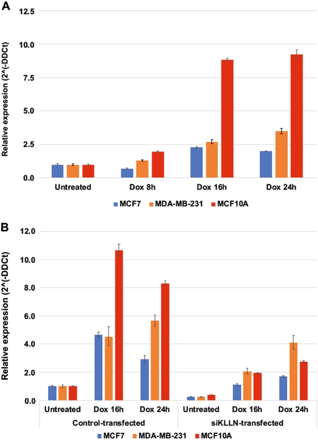
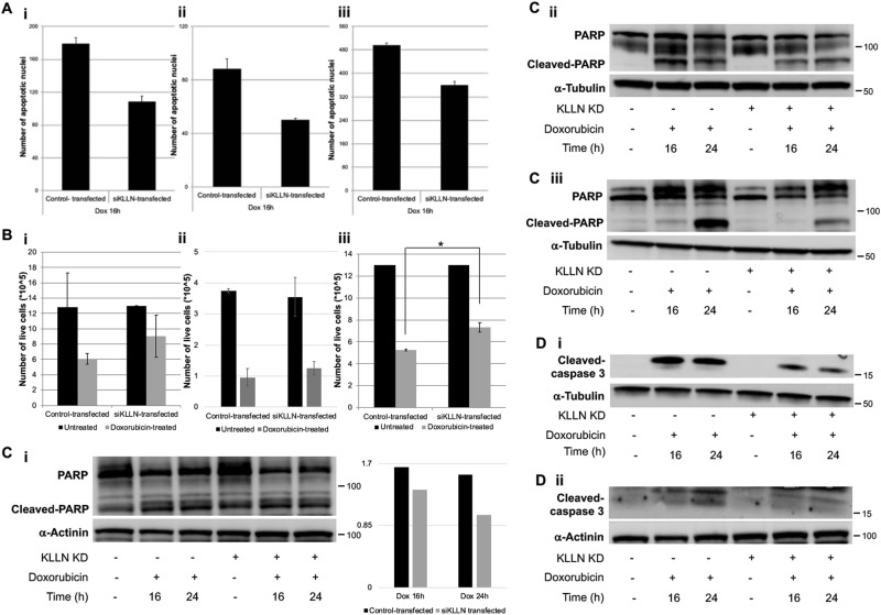
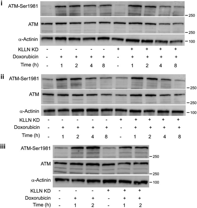
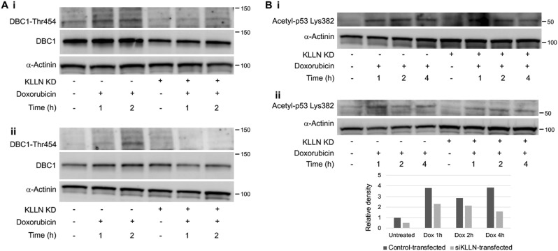
KLLN is a target of p53 involved in S-phase cell cycle regulation deemed necessary and sufficient for p53-mediated apoptosis. Germline promoter hypermethylation of KLLN is associated with a cancer-predisposition syndrome, Cowden syndrome. KLLN's DNA-binding ability is associated with transcription regulation and maintenance of genomic stability. Here, we report on KLLN's role in DNA damage response (DDR) mediated through apoptosis in breast cells with and without a cancer phenotype. KLLN expression was upregulated after doxorubicin-induced DNA damage and this upregulation can be abrogated using RNAi-mediated gene silencing. Silencing KLLN after doxorubicin treatment effected DDR shown by decreased γ-H2AX foci and expression, and apoptosis assessed by decreased frequency of apoptotic nuclei and decreased expression of definitive markers of apoptosis. Contrary to expectations, there was no change in cell cycle regulation after KLLN silencing. These results were observed in breast cells with wildtype and mutant p53. At early timepoints after doxorubicin treatment, knocking down KLLN resulted in decreased Ser15-phosphorylation of p53 but not Thr68-phosphorylation of CHK2 or the phosphorylation of upstream regulators such as ATM and ATR. Interestingly, a second pathway for p53 activation was also affected by knockdown of KLLN. After doxorubicin treatment, Thr454-phosphorylation of DBC1, required to inhibit deacetylation of p53 by SIRT1, was decreased and therefore acetylation of p53 was also decreased with KLLN knockdown. Therefore, our observations suggest that KLLN's role in DNA damage-induced apoptosis is likely independent of p53 and is associated with a two-pronged regulation of p53 activation.


Conflict of interest statement

The authors declare that they have no conflict of interest.

Figures

Fig. 1
Fig. 1. Expression of KLLN in response to DNA damage-induced by doxorubicin with and without RNAi-mediated gene silencing.
a Graph of relative KLLN expression at different timepoints after doxorubicin treatment shows marked increase in KLLN expression at 16 h and 24 h after treatment. Values represent mean ± SD. b Graph of relative KLLN expression after KLLN knockdown and doxorubicin treatment. The increase observed with doxorubicin treatment alone was decreased after RNAi-mediated KLLN knockdown. Values represent mean ± SD
Fig. 2
Fig. 2. Knockdown of KLLN expression results in diminished response to DNA damage in breast cells.
a RNAi-based knockdown of KLLN results in decreased frequency of γ-H2AX foci after doxorubicin-induced DNA damage in MCF7 (i), MCF10A (ii), and MDA-MB-231 (iii) cells. Values represent mean ± SD. **p value < 0.005, *p value < 0.05. b γ-H2AX expression in response to DNA damage-induced by doxorubicin was also decreased after knockdown of KLLN expression in MCF7 (i), MCF10A (ii), and MDA-MB-231 (iii) cells
Fig. 3
Fig. 3. KLLN has a role in apoptosis regulation but not cell cycle regulation after DNA damage.
a TUNEL assay measuring frequency of apoptotic nuclei showed that after doxorubicin-induced DNA damage, there are fewer apoptotic nuclei in breast cells [(i) MCF7, (ii) MCF10A and (iii) MDA-MB-231] with knockdown of KLLN expression. Values represent mean ± SD. b Cell viability assays showed increased proliferation of breast cells [(i) MCF7, (ii) MCF10A and (iii) MDA-MB-231] with knockdown of KLLN expression after DNA damage. Values represent mean ± SD. *p value < 0.05. c Immunoblotting for cleaved PARP, a marker of apoptosis showed decrease in cleavage after doxorubicin-induced DNA damage in breast cells [(i) MCF7, (ii) MCF10A and (iii) MDA-MB-231] with knockdown of KLLN expression. d Immunoblotting for cleaved caspase-3 after doxorubicin-induced DNA damage in breast cells MCF10A (i) and MDA-MB-231 (ii) showed decrease in cleavage with knockdown of KLLN expression
Fig. 4
Fig. 4. KLLN effects phosphorylation of p53 but not phosphorylation of CHK2 after DNA damage in breast cells.
a RNAi-mediated silencing of KLLN was able to maintain decreased KLLN expression through early timepoints (1, 2, 4, and 8 h) after doxorubicin-induced DNA damage in all three breast cell lines. Values represent mean ± SD. b Immunoblotting for Ser15-phosphorylation of p53 showed that knockdown of KLLN expression resulted in decreased p53 phosphorylation in MCF7 (i) and MCF10A (ii) cells. Total p53 expression was assessed and found to be unaffected due to knockdown of KLLN expression. c Immunoblotting for Ser15-phosphorylation of p53 in MDA-MB-231 cells after doxorubicin-induced DNA damage showed no effect on this phosphorylation with knockdown of KLLN expression
Fig. 5
Fig. 5. KLLN regulation of p53 phosphorylation is not through regulation of ATM phosphorylation.
Immunoblotting for Ser1981-phosphorylation of ATM after doxorubicin-induced DNA damage showed that ATM phosphorylation was unaffected in MCF7 (i) and MDA-MB-231 (ii) cells after knockdown of KLLN expression. In MCF10A (iii) cells, knocking down KLLN expression resulted in a 30% decrease in phosphorylation of ATM after DNA damage
Fig. 6
Fig. 6. KLLN’s role in p53 acetylation after DNA damage is through the regulation of DBC1 phosphorylation.
a Immunoblotting for Thr454-phosphorylation after doxorubicin-induced DNA damage showed that knocking down KLLN expression resulted in decreased DBC1 phosphorylation in MCF10A (i) and MCF7 (ii) cells. b Immunoblotting for Lys382-acetylation of p53 after doxorubicin-induced DNA damage showed decreased p53 acetylation after knockdown of KLLN expression in MCF7 (i) and MCF10A (ii) cells
Fig. 7
Fig. 7. Schematic for KLLN regulation of p53 activation after DNA damage (modified from Zannini et al.,).
Knocking down KLLN results in the abrogation of p53 activation after DNA damage by decreasing both the phosphorylation and acetylation of p53

References

    1. Eng C. Will the real Cowden syndrome please stand up: revised diagnostic criteria. J. Med. Genet. 2000;37:828–830. doi: 10.1136/jmg.37.11.828. - DOI - PMC - PubMed
    1. Tan MH, et al. A clinical scoring system for selection of patients for PTEN mutation testing is proposed on the basis of a prospective study of 3042 probands. Am. J. Hum. Genet. 2011;88:42–56. doi: 10.1016/j.ajhg.2010.11.013. - DOI - PMC - PubMed
    1. Bennett KL, et al. Germline and somatic DNA methylation and epigenetic regulation of KILLIN in renal cell carcinoma. Genes Chromosomes Cancer. 2011;50:654–661. doi: 10.1002/gcc.20887. - DOI - PMC - PubMed
    1. Bennett KL, Mester J, Eng C. Germline epigenetic regulation of KILLIN in Cowden and Cowden-like syndrome. JAMA. 2010;304:2724–2731. doi: 10.1001/jama.2010.1877. - DOI - PMC - PubMed
    1. Nizialek EA, Peterson C, Mester JL, Downes-Kelly E, Eng C. Germline and somatic KLLN alterations in breast cancer dysregulate G2 arrest. Hum. Mol. Genet. 2013;22:2451–2461. doi: 10.1093/hmg/ddt097. - DOI - PMC - PubMed