

NDRG2 suppression as a molecular hallmark of photoreceptor-specific cell death in the mouse retina
- PMID: 30245855
- PMCID: PMC6135825
- DOI: 10.1038/s41420-018-0101-2
NDRG2 suppression as a molecular hallmark of photoreceptor-specific cell death in the mouse retina
Erratum in
-
Erratum: Publisher Correction: articles initially published in wrong volume.Cell Death Discov. 2019 Jul 10;5:116. doi: 10.1038/s41420-019-0186-2. eCollection 2019. Cell Death Discov. 2019. PMID: 31312525 Free PMC article.
Abstract
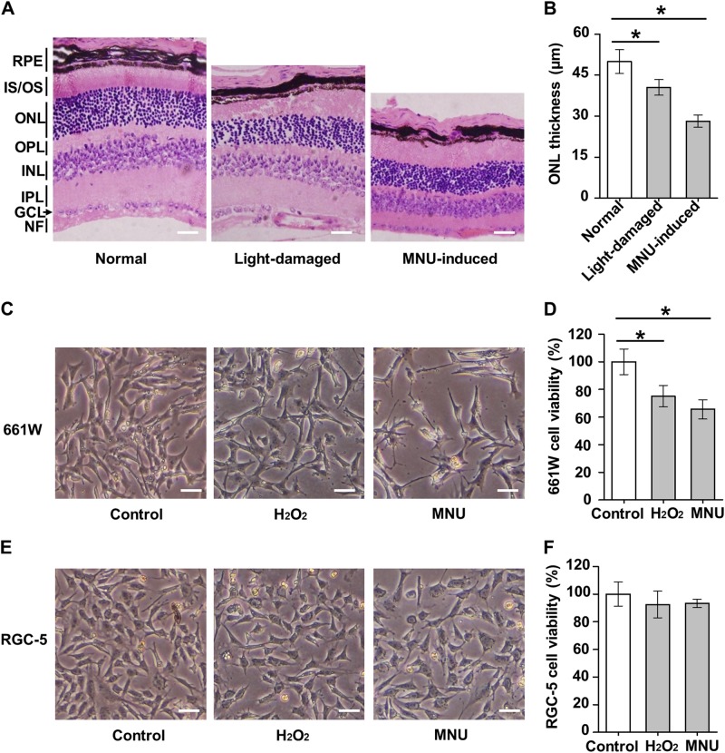
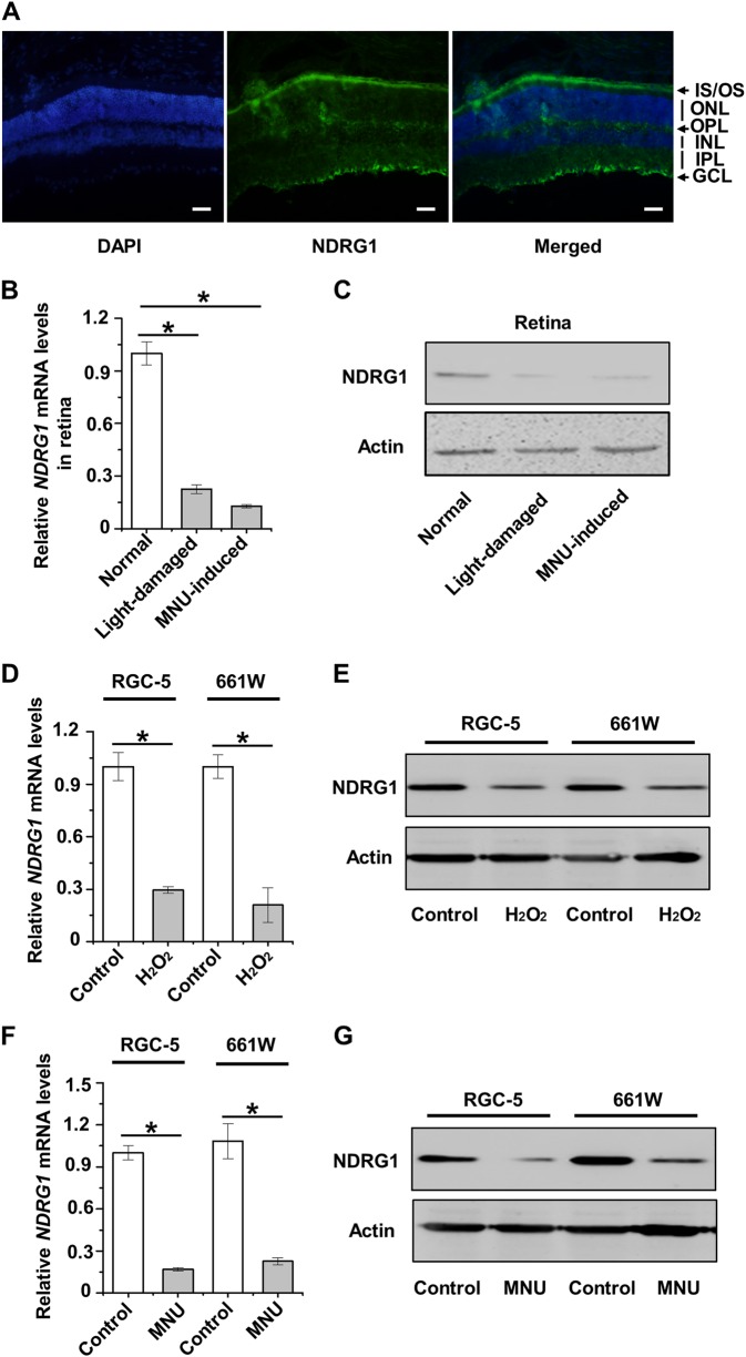
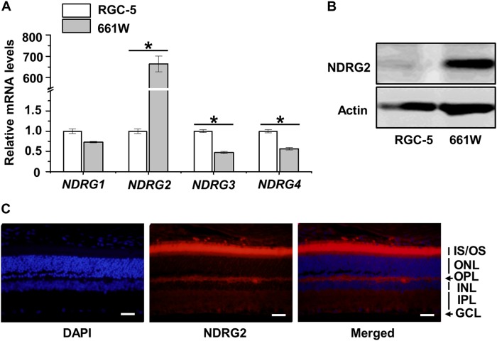
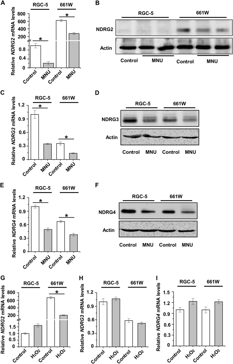
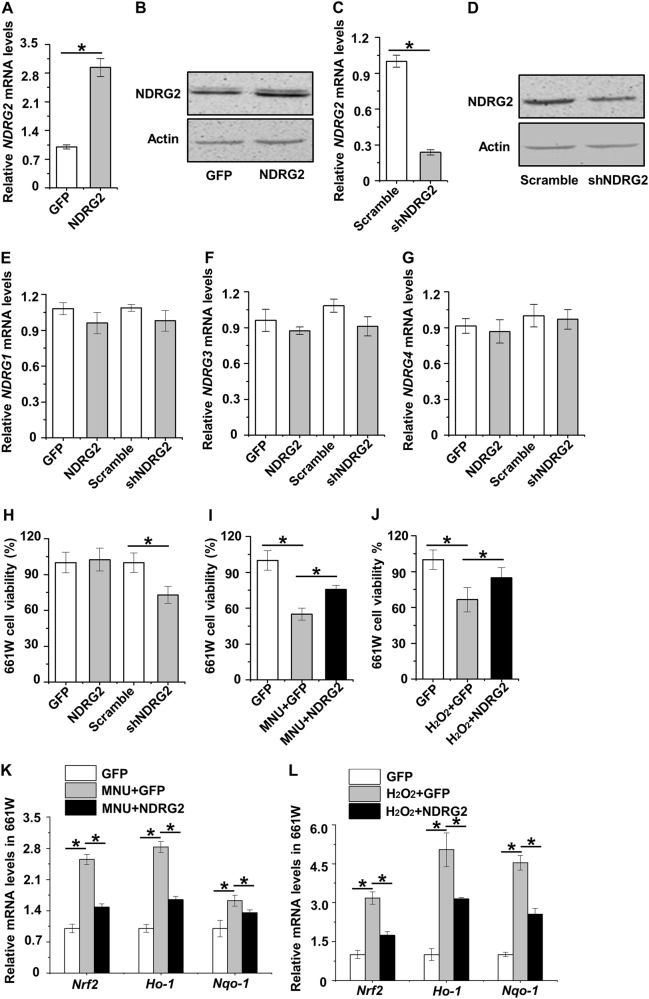
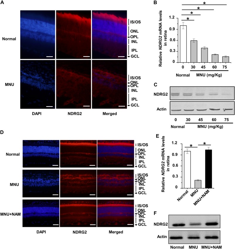
Photoreceptor cell death is recognized as the key pathogenesis of retinal degeneration, but the molecular basis underlying photoreceptor-specific cell loss in retinal damaging conditions is virtually unknown. The N-myc downstream regulated gene (NDRG) family has recently been reported to regulate cell viability, in particular NDRG1 has been uncovered expression in photoreceptor cells. Accordingly, we herein examined the potential roles of NDRGs in mediating photoreceptor-specific cell loss in retinal damages. By using mouse models of retinal degeneration and the 661 W photoreceptor cell line, we showed that photoreceptor cells are indeed highly sensitive to light exposure and the related oxidative stress, and that photoreceptor cells are even selectively diminished by phototoxins of the alkylating agent N-Methyl-N-nitrosourea (MNU). Unexpectedly, we discovered that of all the NDRG family members, NDRG2, but not the originally hypothesized NDRG1 or other NDRG subtypes, was selectively expressed and specifically responded to retinal damaging conditions in photoreceptor cells. Furthermore, functional experiments proved that NDRG2 was essential for photoreceptor cell viability, which could be attributed to NDRG2 control of the photo-oxidative stress, and that it was the suppression of NDRG2 which led to photoreceptor cell loss in damaging conditions. More importantly, NDRG2 preservation contributed to photoreceptor-specific cell maintenance and retinal protection both in vitro and in vivo. Our findings revealed a previously unrecognized role of NDRG2 in mediating photoreceptor cell homeostasis and established for the first time the molecular hallmark of photoreceptor-specific cell death as NDRG2 suppression, shedding light on improved understanding and therapy of retinal degeneration.
Conflict of interest statement
The authors declare that they have no conflict of interest.
Figures
Similar articles
-
NDRG2 alleviates photoreceptor apoptosis by regulating the STAT3/TIMP3/MMP pathway in mice with retinal degenerative disease.FEBS J. 2024 Mar;291(5):986-1007. doi: 10.1111/febs.17021. Epub 2023 Dec 10. FEBS J. 2024. PMID: 38037211
-
17β-Oestradiol Attenuates the Photoreceptor Apoptosis in Mice with Retinitis Pigmentosa by Regulating N-myc Downstream Regulated Gene 2 Expression.Neuroscience. 2021 Jan 1;452:280-294. doi: 10.1016/j.neuroscience.2020.11.010. Epub 2020 Nov 24. Neuroscience. 2021. PMID: 33246060
-
Role of oxidative stress in retinal photoreceptor cell death in N-methyl-N-nitrosourea-treated mice.J Pharmacol Sci. 2012;118(3):351-62. doi: 10.1254/jphs.11110fp. Epub 2012 Feb 23. J Pharmacol Sci. 2012. PMID: 22362184
-
Nervous NDRGs: the N-myc downstream-regulated gene family in the central and peripheral nervous system.Neurogenetics. 2019 Oct;20(4):173-186. doi: 10.1007/s10048-019-00587-0. Epub 2019 Sep 4. Neurogenetics. 2019. PMID: 31485792 Free PMC article. Review.
-
NDRGs in Breast Cancer: A Review and In Silico Analysis.Cancers (Basel). 2024 Mar 29;16(7):1342. doi: 10.3390/cancers16071342. Cancers (Basel). 2024. PMID: 38611020 Free PMC article. Review.
Cited by
-
NDRG2 Deficiency Exacerbates UVB-Induced Skin Inflammation and Oxidative Stress Damage.Inflammation. 2025 Jun;48(3):1313-1325. doi: 10.1007/s10753-024-02121-3. Epub 2024 Aug 15. Inflammation. 2025. PMID: 39145786
-
The ndrg2 Gene Regulates Hair Cell Morphogenesis and Auditory Function during Zebrafish Development.Int J Mol Sci. 2023 Jun 11;24(12):10002. doi: 10.3390/ijms241210002. Int J Mol Sci. 2023. PMID: 37373150 Free PMC article.
-
Photoreceptor protection by mesenchymal stem cell transplantation identifies exosomal MiR-21 as a therapeutic for retinal degeneration.Cell Death Differ. 2021 Mar;28(3):1041-1061. doi: 10.1038/s41418-020-00636-4. Epub 2020 Oct 20. Cell Death Differ. 2021. PMID: 33082517 Free PMC article.
-
Analysis of shared ceRNA networks and related-hub genes in rats with primary and secondary photoreceptor degeneration.Front Neurosci. 2023 Sep 21;17:1259622. doi: 10.3389/fnins.2023.1259622. eCollection 2023. Front Neurosci. 2023. PMID: 37811327 Free PMC article.
-
Salvianolic acid B inhibits the growth and metastasis of A549 lung cancer cells through the NDRG2/PTEN pathway by inducing oxidative stress.Med Oncol. 2024 Jun 7;41(7):170. doi: 10.1007/s12032-024-02413-6. Med Oncol. 2024. PMID: 38847902
References
-
- Fletcher EL. Mechanisms of photoreceptor death during retinal degeneration. Optom. Vis. Sci. 2010;87:269–275. - PubMed
LinkOut - more resources
Full Text Sources
Other Literature Sources
