. 2018 Sep 12:4:35.
doi: 10.1038/s41420-018-0100-3. eCollection 2018.

PIDD-dependent activation of caspase-2-mediated mitochondrial injury in E1A-induced cellular sensitivity to macrophage nitric oxide-induced apoptosis

Affiliations

PIDD-dependent activation of caspase-2-mediated mitochondrial injury in E1A-induced cellular sensitivity to macrophage nitric oxide-induced apoptosis

Jay R Radke et al. Cell Death Discov. .

Erratum in

Abstract

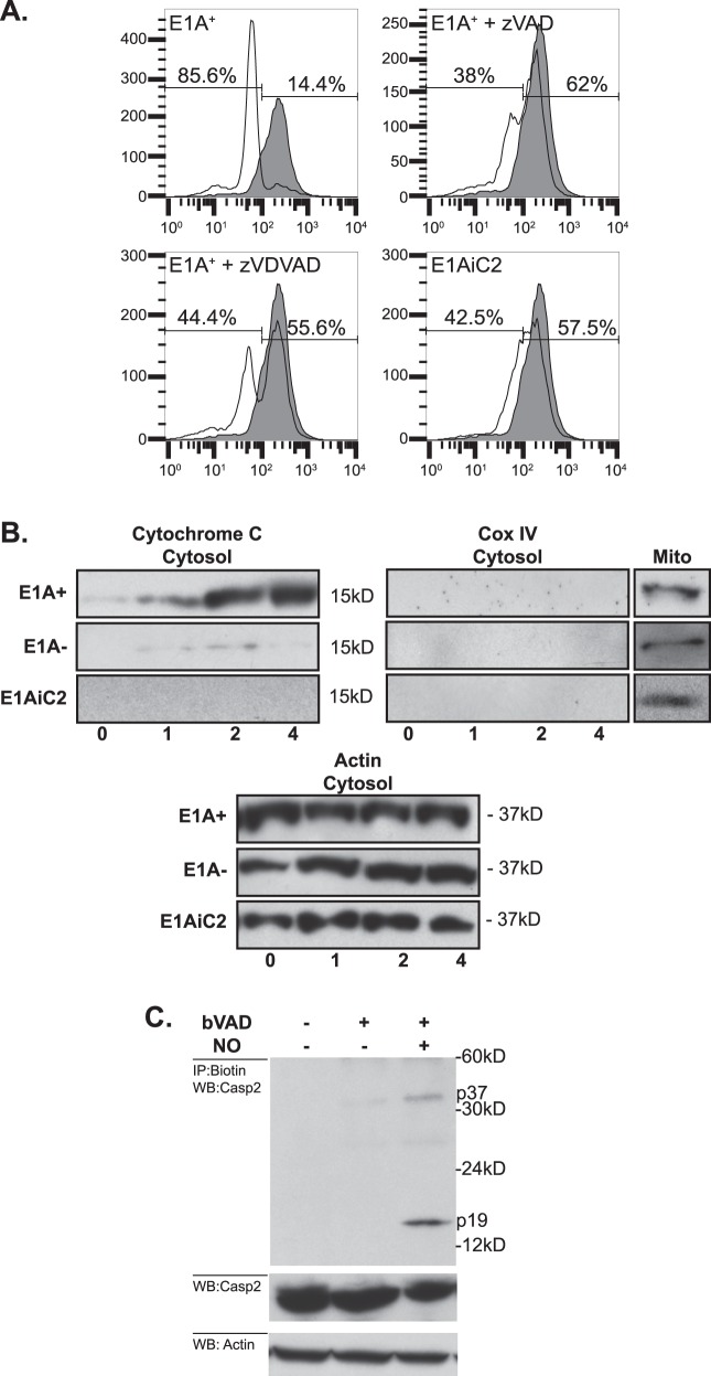
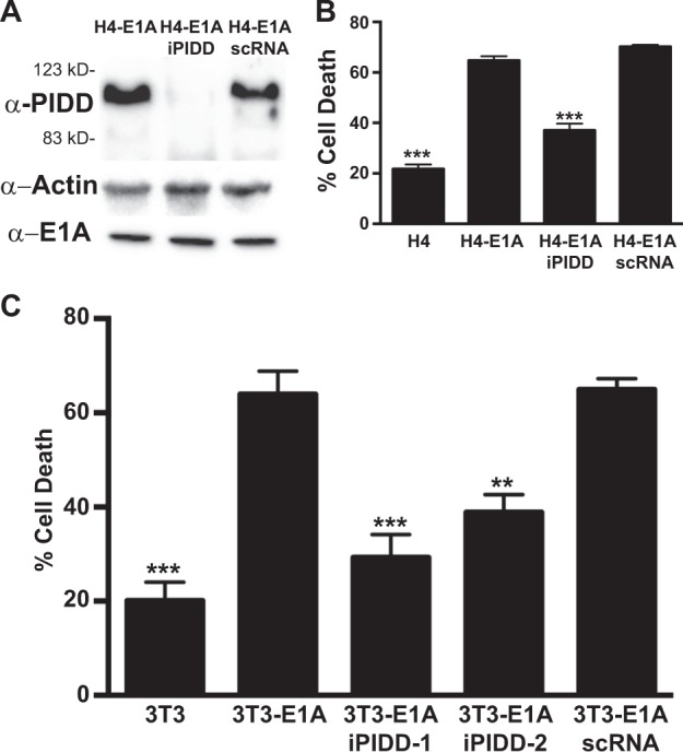
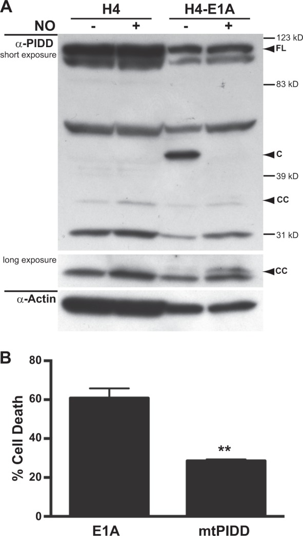
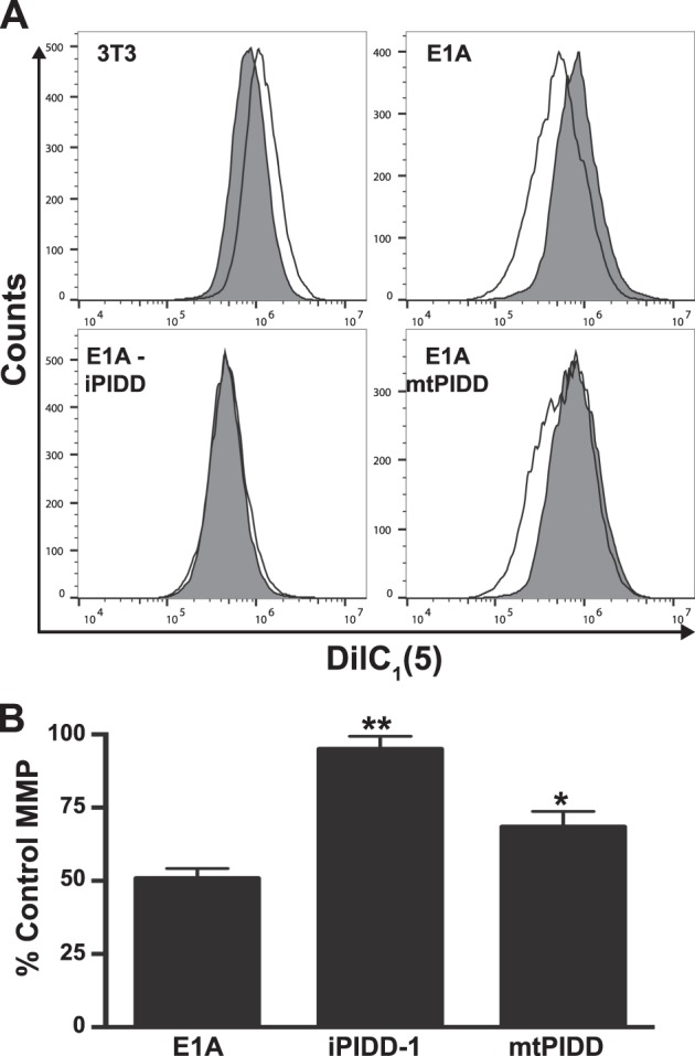
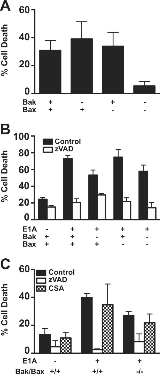
Expression of the adenovirus E1A oncogene sensitizes tumor cells to innate immune rejection by apoptosis induced by macrophage-produced tumor necrosis factor (TNF)-α and nitric oxide (NO). E1A sensitizes cells to TNF-α and NO through two distinct mechanisms, by repressing NF-κB-dependent antiapoptotic responses and enhancing caspase-2 activation and mitochondrial injury, respectively. The mechanisms through which E1A enhances caspase-2 activation in response to NO were unknown. Here, we report that E1A-induced sensitization to NO-induced apoptosis is dependent on expression of PIDD (p53-inducible protein with a death domain) and enhancement of primary immunodeficiency diseases (PIDD) processing for formation of the PIDDosome, the core component of the caspase-2 activation complex. NO-induced apoptosis in E1A-expressing cells did not require expression Bak or Bax, indicating that NO-induced caspase-2-mediated mitochondrial injury does not proceed through the activities of typical, proapoptotic Bcl-2 family members that induce mitochondrial cytochrome C release. These results define a PIDD-dependent pathway, through which E1A enhances casapse-2-mediated mitochondrial injury, resulting in increased sensitivity of mammalian cells to macrophage-induced, NO-mediated apoptosis.


Conflict of interest statement

The authors declare that they have no conflict of interest.

Figures

Fig. 1
Fig. 1. Caspase-2 mediated mitochondrial injury in response to NO.
a Role of caspase-2 in NO-induced loss of MMP. E1A+ cells were treated with 250 μM DETA-NONOate for 6 h then stained with TMRE to estimate MMP. Loss of MMP by NO-injured (open histogram) compared to uninjured (shaded histogram) E1A-positive cells was dependent on caspase activity (E1A+ zVAD), caspase-2 activity (E1A+ zVDVAD) and expression of caspase-2 (E1AiC2). b Mitochondrial release of cytochrome c into the cytosol in E1A-positive (E1A+), E1A-negative, (E1A−) or caspase-2 siRNA-expressing E1A-positive (E1AiC2) cells following treatment with 250 μM DETA-NONOate for the times (h) indicated below the figures. c Role of caspase-2 as the initiator caspase in response to NO injury. E1A-positive cells pretreated with or without biotin-VAD (bVAD) and treated with or without 250 μM DETA-NONOate for 4 h
Fig. 2
Fig. 2. Requirement for PIDD in NO-induced apoptosis.
a Western blot for the expression of human PIDD, actin and E1A in E1A-positive (H4-E1A), PIDD shRNA E1A+ cells (H4-E1AiPIDD) and E1A+ cells expressing scrambled control shRNA (H4-E1A scRNA). b Human fibrosarcoma cells H4, H4-E1A, H4-E1A iPDD, and H4-E1A scRNA treated with 750 μM DETA NONOate for 18 h. Cell viability was determined by MTS staining and expressed as % cell death (mean ± SEM; n = 4, ***P < 0.001, one-way ANOVA). c Mouse NIH-3T3 cells 3T3, 3T3-E1A, 3T3-E1AiPIDD-1 and −2, and 3T3-E1A scRNA treated with 250 μM DETA-NONOate for 18 h. Cell viability was determined by MTS staining and expressed as % cell death (mean ± SEM; n = 3, ***P < 0.001, **P = 0.0028, one-way ANOVA)
Fig. 3
Fig. 3. Role of NO-induced PIDD cleavage.
a Western blot analysis of PIDD and actin in E1A-negative (H4) and E1A-positive (H4-E1A) cells in the absence or presence of DETA-NONOate. b Mouse NIN-3T3-E1A+ or E1A+ mtPIDD (S588A) cells were treated with DETA-NONOate for 18 h. Cell viability was determined by MTS staining and expressed as % cell death (mean ± SEM; n = 3, **P = 0.003, Student’s two-tailed t test)
Fig. 4
Fig. 4. PIDD-mediated mitochondrial injury in response to NO.
a DilC1 (5) staining of NIH-3T3, E1A-positive (E1A), E1A siRNA PIDD (E1A-iPIDD) and E1A PIDD-S588A (E1A-mtPIDD) cells, following treatment with DETA-NONOate for 18 h. Histograms for DETA-NONOate treated cells (unshaded histograms) are overlayed on those for untreated cells (shaded histograms). b PIDD-related changes in MMP in response to NO, expressed as a percentage of untreated control cells (mean ± SEM; n = 3, ***P < 0.001, *P < 0.03, one-way ANOVA)
Fig. 5
Fig. 5. Role of Bak/Bax in macrophage and NO-induced apoptosis.
a [3H]-thymidine-labeled BMK-E1A, BMK-E1A-Bak−/−, BMK-E1A-Bax−/−, or BMK-E1A-Bak/Bax−/−/−/− cells were incubated with ceramide (100 µM) overnight. Supernatants were collected and % specific thymidine release was assessed (mean ± SEM; n = 5, ***P = 0.001, one-way ANOVA). b [3H]-thymidine-labeled BMK, BMK-E1A, BMK-E1A-Bak−/−, BMK-E1A-Bax−/−, or BMK-E1A-Bak/Bax−/−/−/− cells were incubated with activated bone marrow-derived macrophages at E:T of 30:1 in the absence or presence of 100 μM zVAD-fmk. After 48 h supernatants were collected and % specific thymidine release was assessed (mean ± SEM; n = 3, ***P ≤ 0.0001, **P = 0.029, one-way ANOVA). c [3H]-thymidine-labeled BMK, BMK-E1A, or BMK-E1A-Bak/Bax−/−/−/− cells were incubated with DETA-NONOate at 650 µM alone (black bars), with 100 μM zVAD-fmk (white bars) or with cyclosporine A (100 µM) (CSA, hatched bars). After 18 h supernatants were collected and % specific thymidine release was assessed (mean ± SEM; n = 3, ***P ≤ 0.0005, **P = 0.01, one-way ANOVA)

References

    1. Ben-Israel H, Kleinberger T. Adenovirus and cell cycle control. Front. Biosci. 2002;7:d1369–d1395. doi: 10.2741/ben. - DOI - PubMed
    1. Felsani A, Mileo AM, Paggi MG. Retinoblastoma family proteins as key targets of the small DNA virus oncoproteins. Oncogene. 2006;25:5277–5285. doi: 10.1038/sj.onc.1209621. - DOI - PubMed
    1. Ralston R, Bishop M. The protein products of the myc and myb oncogenes and adenovirus E1a are structurally related. Nature. 1983;306:803–806. doi: 10.1038/306803a0. - DOI - PubMed
    1. Cook, J. L. & Routes, J. M. Role of the innate immune response in determining the tumorigenicity of neoplastic cells. Dev Biol 99–107 (2001) [discussion 107-08-143-60]. - PubMed
    1. Cook JL, Routes JM. Adenovirus E1A gene-induced tumor cell rejection through cellular sensitization to immune and nonimmune apoptotic injuries. Front. Biosci. 2005;10:1396–1414. doi: 10.2741/1628. - DOI - PubMed