Propagation and Molecular Characterization of Bioreactor Adapted Very Virulent Infectious Bursal Disease Virus Isolates of Malaysia
- PMID: 30245887
- PMCID: PMC6139196
- DOI: 10.1155/2018/1068758
Propagation and Molecular Characterization of Bioreactor Adapted Very Virulent Infectious Bursal Disease Virus Isolates of Malaysia
Abstract
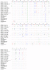
Two Malaysian very virulent infectious bursal disease virus (vvIBDV) strains UPM0081 (also known as B00/81) and UPM190 (also known as UPM04/190) isolated from local IBD outbreaks in 2000 and 2004, respectively, were separately passaged for 12 consecutive times in 11-day-old specific pathogen free (SPF) chicken embryonated eggs (CEE) via the chorioallantoic membrane (CAM) route. The CEE passage 8 (EP8) isolates were passaged once in BGM-70 cell line yielding UPM0081EP8BGMP1 and UPM190EP8BGMP1, while the EP12 isolates were passaged 15 times in BGM-70 cell line yielding UPM0081EP12BGMP15 and UPM190EP12BGMP15 using T25 tissue culture flask. These isolates were all propagated once in bioreactor using cytodex 1 as microcarrier at 3 g per liter (3 g/L) yielding UPM0081EP8BGMP1BP1, UPM190EP8BGMP1BP1, UPM0081EP12BGMP15BP1, and UPM190EP12BGMP15BP1 isolates. The viruses were harvested at 3 days after inoculation, following the appearance of cytopathic effects (CPE) characterized by detachment from the microcarrier using standard protocol and filtered using 0.2 μm syringe filter. The filtrates were positive for IBDV by RT-PCR and immunofluorescence. Sequence and phylogenetic tree analysis indicated that the isolates were of the vvIBDV strains and were not different from the flask propagated parental viruses.
Figures


Similar articles
-
Adaptation and Molecular Characterization of Two Malaysian Very Virulent Infectious Bursal Disease Virus Isolates Adapted in BGM-70 Cell Line.Adv Virol. 2017;2017:8359047. doi: 10.1155/2017/8359047. Epub 2017 Nov 5. Adv Virol. 2017. PMID: 29230245 Free PMC article.
-
Characterization of Chicken Splenic-Derived Dendritic Cells Following Vaccine and Very Virulent Strains of Infectious Bursal Disease Virus Infection.Avian Dis. 2016 Dec;60(4):739-751. doi: 10.1637/11275-091315-Reg.1. Avian Dis. 2016. PMID: 27902915
-
Characteristics of a very virulent infectious bursal disease virus from California.Avian Dis. 2009 Dec;53(4):592-600. doi: 10.1637/8957-061109-Reg.1. Avian Dis. 2009. PMID: 20095162
-
Pathogenicity of cell culture-derived and bursa-derived infectious bursal disease viruses in specific-pathogen-free chickens.Avian Dis. 2001 Oct-Dec;45(4):844-52. Avian Dis. 2001. PMID: 11785889
-
Isolation of Infectious Bursal Disease Virus Using Indigenous Chicken Embryos in Kenya.Int Sch Res Notices. 2015 Nov 23;2015:464376. doi: 10.1155/2015/464376. eCollection 2015. Int Sch Res Notices. 2015. PMID: 27347520 Free PMC article.
Cited by
-
Efficacy, humoral, and cell-mediated immune response of inactivated fowl adenovirus 8b propagated in chicken embryo liver cells using bioreactor in broiler chickens.Vet World. 2022 Nov;15(11):2681-2692. doi: 10.14202/vetworld.2022.2681-2692. Epub 2022 Nov 26. Vet World. 2022. PMID: 36590109 Free PMC article.
-
Isolation and genomic characteristics of the novel variant infectious bursal disease virus in China.Front Vet Sci. 2023 Dec 11;10:1314903. doi: 10.3389/fvets.2023.1314903. eCollection 2023. Front Vet Sci. 2023. PMID: 38146498 Free PMC article.
-
Molecular characterization and phylogenetic analysis of orf virus isolated from goats in Sokoto metropolis, Nigeria.Future Sci OA. 2021 Apr 20;7(6):FSO700. doi: 10.2144/fsoa-2020-0162. Future Sci OA. 2021. PMID: 34046202 Free PMC article.
References
-
- Rodriguez-Chavez I. R., Rosenberger J. K., Cloud S. S., Pope C. R. Characterization of the antigenic, immunogenic, and pathogenic variation of infectious bursal disease virus due to propagation in different host systems (bursa, embryo, and cell culture). III. Pathogenicity. Avian Pathology. 2002;31(5):485–492. doi: 10.1080/0307945021000005851. - DOI - PubMed
-
- Lukert P. D., Leonard J., Davis R. B. Infectious Bursal Disease Virus: Antigen Production and Immunity. American Journal of Veterinary Research. 1975;36:539–540. - PubMed
LinkOut - more resources
Full Text Sources
Other Literature Sources
Research Materials
Miscellaneous

