Review
. 2018 Aug 20;8(4):247-267.
eCollection 2018.

Targeted α-therapy of prostate cancer using radiolabeled PSMA inhibitors: a game changer in nuclear medicine

Affiliations
Review

Targeted α-therapy of prostate cancer using radiolabeled PSMA inhibitors: a game changer in nuclear medicine

Rubel Chakravarty et al. Am J Nucl Med Mol Imaging. .

Abstract

Prostate cancer (PCa) is one of the most common malignancies in men and is a major contributor to cancer related deaths worldwide. Metastatic spread and disease progression under androgen deprivation therapy signify the onset of metastatic castration resistant prostate cancer (mCRPCa)-the lethal form of the disease, which severely deteriorates the quality of life of patients. Over the last decade, tremendous progress has been made toward identifying appropriate molecular targets that could enable efficient in vivo targeting for non-invasive imaging and therapy of mCPRCa. In this context, a promising enzymatic target is prostate specific membrane antigen (PSMA), which is overexpressed on PCa cells, in proportion to the stage and grade of the tumor progression. This is especially relevant for mCRPCa, which has significant overexpression of PSMA. For therapy of mCRPCa, several nuclear medicine clinics all over the world have confirmed that 177Lu-labeled-PSMA enzyme inhibitors (177Lu-PSMA-617 and 177Lu-PSMA I&T) have a favorable dosimetry and convincing therapeutic response. However, ~30% of patients were found to be short or non-responders and dose escalation was severely limited by chronic hematological toxicity. Such limitations could be better overcome by targeted alpha therapy (TAT) which has the potential to bring a paradigm shift in treatment of mCRPCa patients. This concise review presents an overview of the successes and challenges currently faced in TAT of mCRPCa using radiolabeled PSMA inhibitors. The preclinical and clinical data reported to date are quite promising, and it is expected that this therapeutic modality will play a pivotal role in advanced stage PCa management in the foreseeable future.

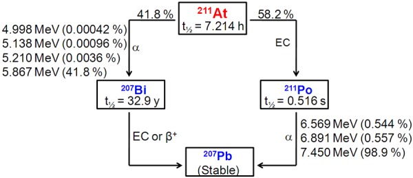
Keywords: 211At; 213Bi; 225Ac; PSMA; metastasis; prostate cancer; targeted alpha therapy.


Figures

Figure 1
Figure 1
Structure of PSMA. A. Ribbon diagrams of side and top view of PSMA. B. A surface rendering in which the apical domain is light green, the helical domain is light red, the protease domain is light blue, and zinc ions are orange. The residues facing the substrate-binding cavity are indicated in a darker version of the color matching to the domain from which the residue derives. PSMA active site is encircled. C. Stereoview of the PSMA active site. Zinc ions are orange spheres, and a water molecule is shown as a red sphere. Zinc binding residues are yellow sticks, water- or substrate-binding ligands are purple sticks, and residues with structural roles are light blue sticks. Adapted from Ref. [39] with permission. Copyright 2005 National Academy of Sciences.
Figure 2
Figure 2
The enzymatic action of PSMA. A. N-Acetyl-L-aspartyl-L-glutamate (NAAG) is hydrolyzed to aspartate and glutamate. B. Glutamic acid is released from folate polyglutamate resulting in the release of folic acid. After successive release of glutamate, folate is released. Adapted from Ref. [10] with permission. Copyright 2016 Elsevier.
Figure 3
Figure 3
Structures of representative urea-based PSMA ligands used in clinical context. Adapted from Ref. [50] with permission. Copyright 2017 Elsevier.
Figure 4
Figure 4
Simplified decay scheme of 211At.
Figure 5
Figure 5
(A) Excitation function for the 209Bi (a, 2n) 211At reaction, (B) Excitation function for the 209Bi (a, 3n) 211At reaction. Adapted from Ref. [104] with permission. Copyright 2009 International Atomic Energy Agency.
Figure 6
Figure 6
Decay chain of 233U indicating production and decay of 225Ac and 213Bi. Adapted from Ref. [111] with permission. Copyright 2005 American Chemical Society.
Figure 7
Figure 7
Excitation function for the 226Ra (p, 2n) 225Ac reaction. Adapted from Ref. [104] with permission. Copyright 2009 International Atomic Energy Agency.
Figure 8
Figure 8
Synthesis of 211At-labeled PSMA inhibitor. R = 5 para-methoxybenzyl. Adapted from Ref. [127] with permission. Copyright 2016 Society of Nuclear Medicine and Molecular Imaging.
Figure 9
Figure 9
(A) α-camera images at 1 h showing relative 211At-labeled PSMA inhibitor activity concentrations for PSMA+ and PSMA- tumors and kidneys. Scale shows activity concentration relative to whole tumor/kidney average concentration. (B) Renal histopathology from non-treated mouse (i and ii) and mouse treated with 1.5 MBq of 211At-labeled PSMA inhibitor (iii and iv). Treated kidney showed subcortical atrophy and degenerative loss of proximal tubules (arrows) consistent with late nephropathy due to α-particle irradiation. Adapted from Ref. [127] with permission. Copyright 2016 Society of Nuclear Medicine and Molecular Imaging.
Figure 10
Figure 10
(A) 68Ga-PSMA-11 PET/CT scans of patient A. Pretherapeutic tumor spread (i), restaging 2 months after third cycle of 225Ac-PSMA-617 (ii), and restaging 2 months after one additional consolidation therapy (iii). (B) 68Ga-PSMA-11 PET/CT scans of patient B. In comparison to initial tumor spread (i), restaging after 2 cycles of β-emitting 177Lu-PSMA-617 presented progression (ii). In contrast, restaging after second (iii) and third (iv) cycles of 225Ac-PSMA-617 therapy presented impressive response. Adapted from Ref. [96] with permission. Copyright 2016 Society of Nuclear Medicine and Molecular Imaging.
Figure 11
Figure 11
68Ga-PSMA-11 PET/CT scans of patient. (A) Pretherapeutic tumor spread, (B) restaging 11 months after therapy with 213Bi-PSMA-617. Adapted from Ref. [133] with permission. Copyright 2017 Springer.

References

    1. Domachevsky L, Goldberg N, Bernstine H, Nidam M, Groshar D. Quantitative characterisation of clinically significant intra-prostatic cancer by prostate-specific membrane antigen (PSMA) expression and cell density on PSMA-11. Eur Radiol. 2018 [Epub ahead of print] - PubMed
    1. Goolam AS, la Rosa AH, Manoharan M. Surgical management of organ-confined prostate cancer with review of literature and evolving evidence. Indian J Surg Oncol. 2018;9:225–231. - PMC - PubMed
    1. Rahbar K, Afshar-Oromieh A, Jadvar H, Ahmadzadehfar H. PSMA theranostics: current status and future directions. Mol Imaging. 2018;17:1536012118776068. - PMC - PubMed
    1. Ruggero K, Farran-Matas S, Martinez-Tebar A, Aytes A. Epigenetic regulation in prostate cancer progression. Curr Mol Biol Rep. 2018;4:101–115. - PMC - PubMed
    1. Smith RA, Andrews KS, Brooks D, Fedewa SA, Manassaram-Baptiste D, Saslow D, Brawley OW, Wender RC. Cancer screening in the United States, 2018: a review of current American cancer society guidelines and current issues in cancer screening. CA Cancer J Clin. 2018;68:297–316. - PubMed

LinkOut - more resources