

doi: 10.1212/NXI.0000000000000503.
eCollection 2018 Nov.
MOG antibodies in combined central and peripheral demyelination syndromes
Affiliations
- PMID: 30246057
- PMCID: PMC6147156
- DOI: 10.1212/NXI.0000000000000503
Item in Clipboard
MOG antibodies in combined central and peripheral demyelination syndromes
Neurol Neuroimmunol Neuroinflamm.
.
No abstract available
Figures
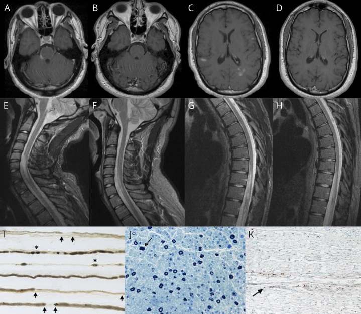
(A) Gadolinium-enhanced axial T1-weighted MRI of the brain, showing several enhancing lesions in the right pons/middle cerebellar peduncle (trigeminal nerve entry zone), (C) the left periatral region and subcortical white matter of both cerebral hemispheres; and (B and D) at 3-month follow-up, showing resolution of the previously observed contrast-enhancing supratentorial and infratentorial lesions. (E and G) Sagittal T2 short tau inversion recovery sequence MRI of the cervical and thoracic spine, showing a longitudinally extensive lesion from C5 through T1, lesions involving T10-T11 segments, and conus medullaris; and (F and H) at 3-month follow-up, showing a marked decrease in the size of previously observed cervical, thoracic, and conus medullaris lesions. (I) Teased nerve fiber preparation (×16) of the left sural nerve, revealing increased demyelination and remyelination (arrows) and axonal degeneration (asterisks). (J) Semithin epoxy-embedded section (×40), showing moderately to severely reduced myelinated fiber density and rare degenerating profiles (arrow). (K) CD45 (leukocyte common antigen) preparation (×16), showing reactive individual cells within the endoneurium (arrow) and a single small perivascular epineurial collection (not shown).
References
-
- Ogata H, Matsuse D, Yamasaki R, et al. . A nationwide survey of combined central and peripheral demyelination in Japan. J Neurol Neurosurg Psychiatry 2016;87:29–36. - PubMed
-
- Cortese A, Franciotta D, Alfonsi E, et al. . Combined central and peripheral demyelination: clinical features, diagnostic findings, and treatment. J Neurol Sci 2016;15:182–187. - PubMed
-
- Wang Y, Chen H, Zhuang W, Li H. The clinical features of combined central and peripheral demyelination in Chinese patients. J Neuroimmunol 2018;15:32–36. - PubMed
LinkOut - more resources
Full Text Sources
Other Literature Sources