. 2019 Mar;29(2):205-216.
doi: 10.1111/bpa.12659. Epub 2018 Nov 11.

Childhood supratentorial ependymomas with YAP1-MAMLD1 fusion: an entity with characteristic clinical, radiological, cytogenetic and histopathological features

Affiliations

Childhood supratentorial ependymomas with YAP1-MAMLD1 fusion: an entity with characteristic clinical, radiological, cytogenetic and histopathological features

Felipe Andreiuolo et al. Brain Pathol. 2019 Mar.

Abstract

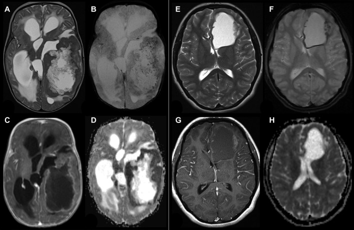
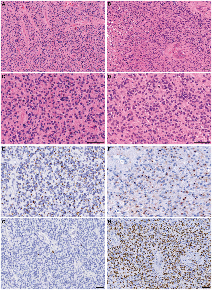
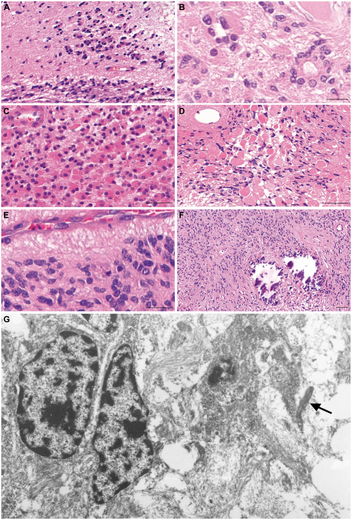
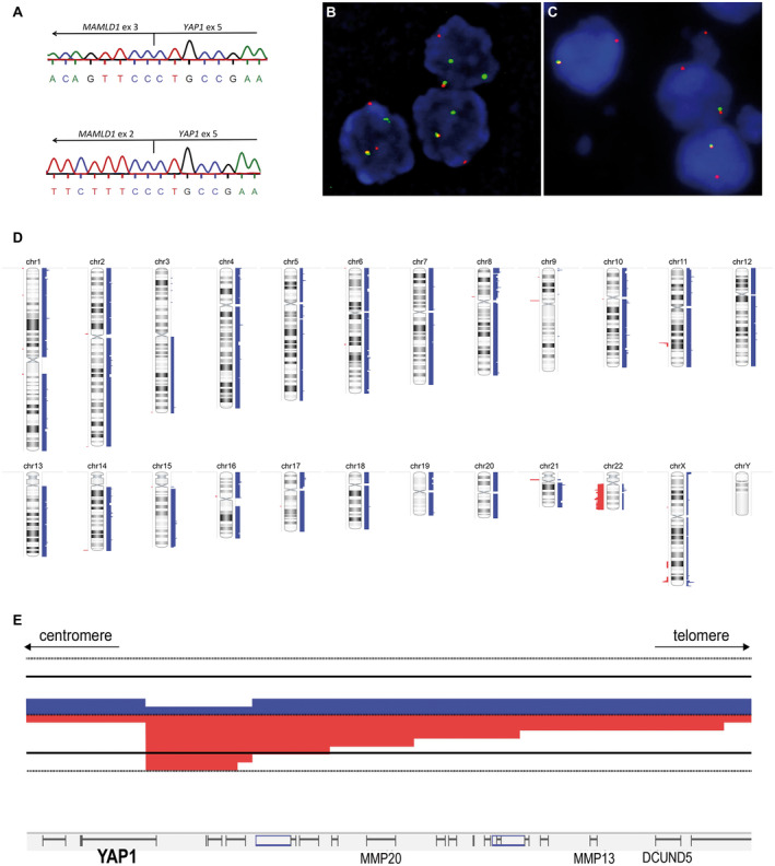
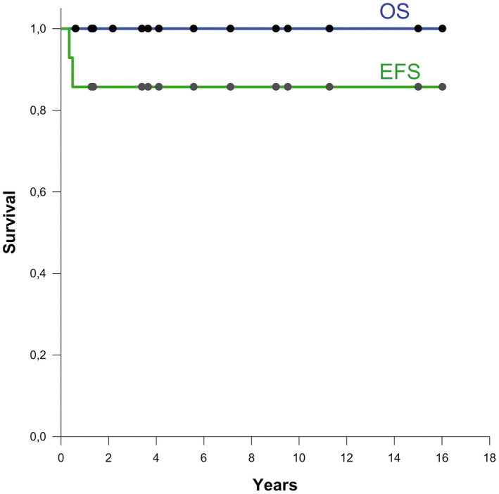
Ependymoma with YAP1-MAMLD1 fusion is a rare, recently described supratentorial neoplasm of childhood, with few cases published so far. We report on 15 pediatric patients with ependymomas carrying YAP1-MAMLD1 fusions, with their characteristic histopathology, immunophenotype and molecular/cytogenetic, radiological and clinical features. The YAP1-MAMLD1 fusion was documented by RT-PCR/Sanger sequencing, and tumor genomes were studied by molecular inversion probe (MIP) analysis. Significant copy number alterations were identified by GISTIC (Genomic Identification of Significant Targets in Cancer) analysis. All cases showed similar histopathological features including areas of high cellularity, presence of perivascular pseudo-rosettes, small to medium-sized nuclei with characteristic granular chromatin and strikingly abundant cells with dot-like cytoplasmic expression of epithelial membrane antigen. Eleven cases presented features of anaplasia, corresponding to WHO grade III. MRI showed large supratentorial multinodular tumors with cystic components, heterogeneous contrast enhancement, located in the ventricular or periventricular region. One of two variants of YAP1-MAMLD1 fusions was detected in all cases. The MIP genome profiles showed balanced profiles, with focal alterations of the YAP1 locus at 11q22.1-11q21.2 (7/14), MAMLD1 locus (Xp28) (10/14) and losses of chromosome arm 22q (5/14). Most patients were female (13/15) and younger than 3 years at diagnosis (12/15; median age, 8.2 months). Apart from one patient who died during surgery, all patients are alive without evidence of disease progression after receiving different treatment protocols, three without postoperative further treatment (median follow-up, 4.84 years). In this to date, largest series of ependymomas with YAP1-MAMLD1 fusions we show that they harbor characteristic histopathological, cytogenetic and imaging features, occur mostly in young girls under 3 years and are associated with good outcome. Therefore, this genetically defined neoplasm should be considered a distinct disease entity. The diagnosis should be confirmed by demonstration of the specific fusion. Further studies on large collaborative series are warranted to confirm our findings.

Keywords: YAP1-MAMLD1 fusion; childhood; ependymoma; supratentorial.


Figures

Figure 1
Figure 1
Radiological findings in two patients with YAP1‐MAMLD1‐fused ependymoma. (A–D; Patient #9) 4 months old infant with a huge left temporal tumor, a large cystic part surrounded by solid tumor, which is isointense to gray matter on T2‐weigthed image (A) and punctuated hemorrhages in the solid part a small rim along the cyst wall (T2*, B). The solid part of the tumor shows a garland‐shaped enhancement (C) and ADC values are decreased compared to gray matter. T2 and ADC changes are compatible with a higher cell density of the tumor. (E–H, patient # 6) 15‐year‐old girl with a left frontal tumor without edema consisting of a large cyst and a small solid part at the lateral border. The solid part is nearly isointense to gray matter in T2‐weighted image (E). T2* (F) reveals subtle punctuated hemorrhages in the solid part a small rim along the cyst wall. The solid part of the tumor shows a faint enhancement on T1‐weighted image (G) and the apparent diffusion coefficient (ADC) is comparable to the gray matter.
Figure 2
Figure 2
Representative morphology and immunophenotype of ependymomas with YAP1‐MAMLD1 fusion. Panels on the left (A, C, E, G) are from patient #1, and panels on the right (B, D, F, H) from patient #10. All tumors in this series showed densely populated areas and perivascular pseudorosettes, as can be observed in H&E stained slides from the two tumors (A) and (B). In (B) vascular proliferation and necrosis can be observed. At higher magnification (C–D) the characteristic tumor cells with small nuclei, and eosinophilic cytoplasms are seen, as well as a brisk mitotic activity in (D). Panels (E) and (F) show the strong and diffuse dot‐like epithelial membrane antigen (EMA) expression. Panels (G) and (H) highlight the strikingly variable proliferation index (Ki67 expression), very low in the areas shown from tumor on the left (G) and high in the tumor on the right (H). bar, 50 µm.
Figure 3
Figure 3
Morphological and ultrastructural features of YAP1‐MAMLD1‐fused ependymomas. Although generally well demarcated from surrounding brain parenchyma, 7/15 tumors showed focally invasive tumor cells or tumor cell clusters as depicted in panel (A). True ependymal rosettes were also often present (B). Another common feature were tumor cells with eosinophilic, granular cytoplasms, sometimes with well‐defined boundaries, peripherally placed nuclei and tendency to form small groupings. (C). Eosinophilic granular bodies were also found, in the case shown in (D) these structures were very abundant. In (E), one observes the small nuclei with homogenous chromatin, yet pleomorphic often angulated contours. Calcifications were also frequent, as shown in (F). Similar nuclear features to those depicted in (E) are observed by electron microscopy performed in a sample from patient #4 (G). In addition, abortive cilia (arrow) can be observed as an indicator of ependymal differentiation. Bars in (A), (C), (D), (F) 50 µm, in (B), (E), 20 µm.
Figure 4
Figure 4
Molecular and cytogenetic features of ependymomas with YAP1‐MAMLD1 fusions. (A) shows cDNA sequencing of two distinct fusions. On the top the most common type is shown, between YAP1‐exon 5 and MAMLD1‐exon3 seen in 14 cases and on the bottom the fusion YAP1‐exon 5 and MAMLD1‐exon 2 detected in a single case (#6). In (B) and (C), two different patterns observed by YAP1 FISH, confirming the YAP1‐rearrangement. In (B) a classic rearrangement with one fused (normal) and one break‐apart signal in each nucleus (3′YAP1: green signal; 5YAP1: red signal) is shown (patient #4). In (C) images of a rearrangement with one fused signal and a single 5YAP1 (unbalanced rearrangement with loss of the 3′locus during the translocation; 3′YAP1: green signal; 5YAP1: red signal) (patient #14). In (D) a virtual karyotype (summary plot) of 14 ependymoma with YAP1‐MAMLD1 fusion analyzed by MIP assay is shown. Gains are indicated by blue bars on the right side of each chromosome, losses are shown in red on the left side. Thickness of the bars indicates the frequency of alterations. chr, chromosome. In (E), a detailed view on the YAP1 locus is shown at higher magnification revealing focal copy number alterations. Six cases had a breakpoint within the YAP1 gene with copy number loss of 3′ regions of YAP1.
Figure 5
Figure 5
Overall survival (OS) and event‐free survival (EFS) (n = 14).

References

    1. Antonescu CR, Le Loarer F, Mosquera JM, Sboner A, Zhang L, Chen CL et al (2013) Novel YAP1‐TFE3 fusion defines a distinct subset of epithelioid hemangioendothelioma. Genes Chrom Cancer 52:775–784. - PMC - PubMed
    1. Bao Y, Hata Y, Ikeda M, Withanage K (2011) Mammalian Hippo pathway: from development to cancer and beyond. J Biochem (Tokyo) 149:361–379. - PubMed
    1. Beroukhim R, Getz G, Nghiemphu L, Barretina J, Hsueh T, Linhart D et al (2007) Assessing the significance of chromosomal aberrations in cancer: methodology and application to glioma. Proc Natl Acad Sci U S A 104:20007–20012. - PMC - PubMed
    1. Figarella‐Branger D, Lechapt‐Zalcman E, Tabouret E, Jünger S, de Paula AM, Bouvier C et al (2016) Supratentorial clear cell ependymomas with branching capillaries demonstrate characteristic clinicopathological features and pathological activation of nuclear factor‐kappaB signaling. Neuro‐Oncol 18:919–927. - PMC - PubMed
    1. Fukami M, Wada Y, Okada M, Kato F, Katsumata N, Baba T et al (2008) Mastermind‐like domain‐containing 1 (MAMLD1 or CXorf6) transactivates the Hes3 promoter, augments testosterone production, and contains the SF1 target sequence. J Biol Chem 283:5525–5532. - PubMed

Publication types

MeSH terms

LinkOut - more resources