. 2019 Jan;5(1):25-39.
doi: 10.1002/cjp2.118. Epub 2018 Oct 22.

Breast cancer metastasis to gynaecological organs: a clinico-pathological and molecular profiling study

Affiliations

Breast cancer metastasis to gynaecological organs: a clinico-pathological and molecular profiling study

Jamie R Kutasovic et al. J Pathol Clin Res. 2019 Jan.

Abstract

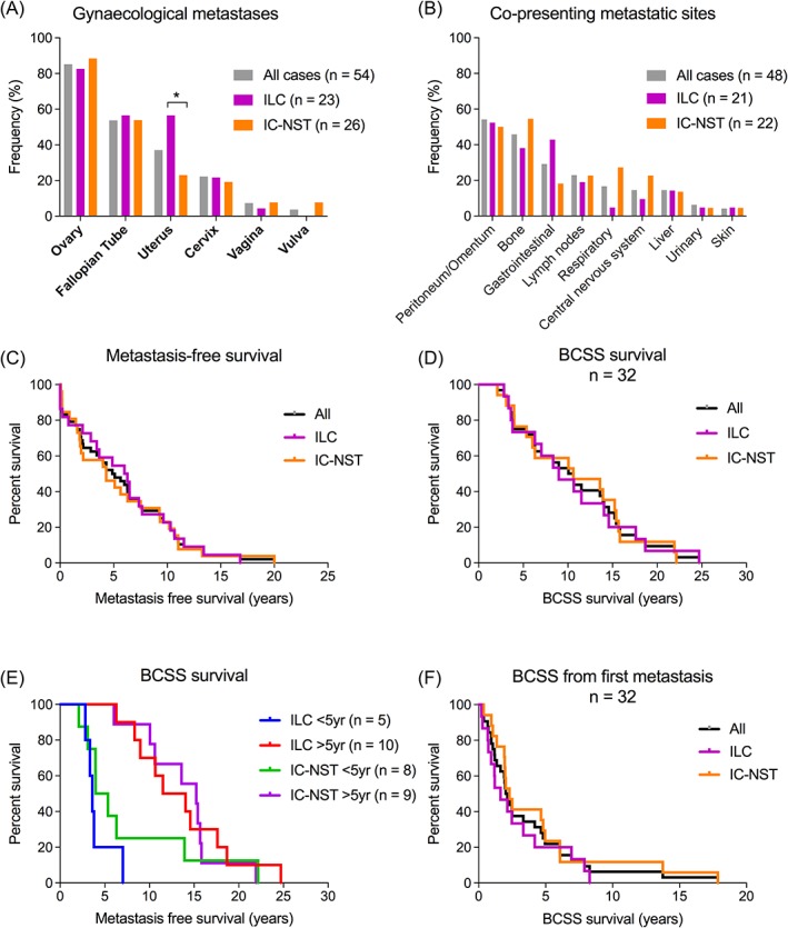
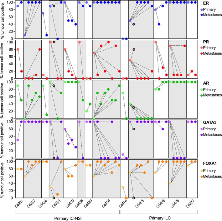
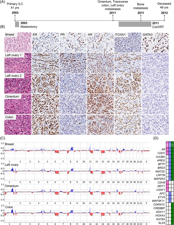
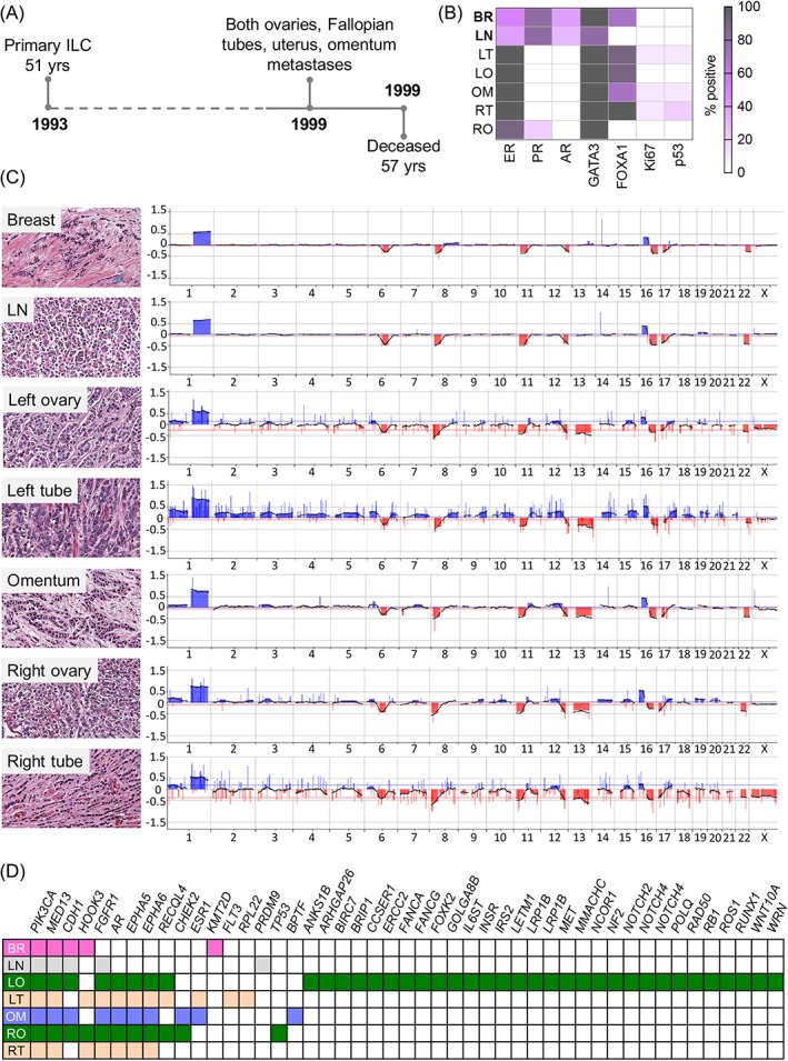
Breast cancer metastasis to gynaecological organs is an understudied pattern of tumour spread. We explored clinico-pathological and molecular features of these metastases to better understand whether this pattern of dissemination is organotropic or a consequence of wider metastatic dissemination. Primary and metastatic tumours from 54 breast cancer patients with gynaecological metastases were analysed using immunohistochemistry, DNA copy-number profiling, and targeted sequencing of 386 cancer-related genes. The median age of primary tumour diagnosis amongst patients with gynaecological metastases was significantly younger compared to a general breast cancer population (46.5 versus 60 years; p < 0.0001). Median age at metastatic diagnosis was 54.4, time to progression was 4.8 years (range 0-20 years), and survival following a diagnosis of metastasis was 1.95 years (range 0-18 years). Patients had an average of five involved sites (most frequently ovary, fallopian tube, omentum/peritoneum), with fewer instances of spread to the lungs, liver, or brain. Invasive lobular histology and luminal A-like phenotype were over-represented in this group (42.8 and 87.5%, respectively) and most patients had involved axillary lymph nodes (p < 0.001). Primary tumours frequently co-expressed oestrogen receptor cofactors (GATA3, FOXA1) and harboured amplifications at 8p12, 8q24, and 11q13. In terms of phenotype conversion, oestrogen receptor status was generally maintained in metastases, FOXA1 increased, and expression of progesterone receptor, androgen receptor, and GATA3 decreased. ESR1 and novel AR mutations were identified. Metastasis to gynaecological organs is a complication frequently affecting young women with invasive lobular carcinoma and luminal A-like breast cancer, and hence may be driven by sustained hormonal signalling. Molecular analyses reveal a spectrum of factors that could contribute to de novo or acquired resistance to therapy and disease progression.

Keywords: breast cancer; genomics; immunophenotyping; luminal subtype; metastasis; ovary.


Figures

Figure 1
Figure 1
(A) Distribution of gynaecological organs involved by metastatic disease, stratified by primary tumour type. The ovary was the most common site involved (85.1%, 46/54 patients). Metastasis to the uterus was more frequent in ILC than IC‐NST (p = 0.0214). Forty‐eight cases (88%) presented with multiple metastases. (B) Frequencies of co‐presenting metastatic sites. The most common sites involved were peritoneum/omentum (54.2%, 27/48 patients), followed by bone (45.8%, 22/48), and GI tract (29.2%, 12/48). Common sites of breast cancer metastasis (lungs, liver, brain) rarely coincided with GMs. Bone, lung, and brain metastases were more frequent in IC‐NST and GI metastases were more frequent in ILC, but the differences were not statistically significant. (C) Metastasis‐free survival in the GM cohort. The median time to metastasis was 5 years. Approximately 25% of patients developed metastases after 10 years from primary tumour diagnosis. (D, E) BCSS in the GM cohort (n = 36): 50% of patients were alive at 10 years post‐primary diagnosis, and BCSS was not affected by primary tumour type or time to metastasis (<5 years versus >5 years). (F) Median survival after first metastasis diagnosis was 1.9 years (ILC: 1.6 years, IC‐NST: 2.3 years). 75% of patients died within 5 years of metastatic diagnosis.
Figure 2
Figure 2
Immunophenotyping of endocrine‐related biomarkers between the primary tumour and matched metastases in 13 cases. Each case displayed a variety of changes in expression of these biomarkers within metastatic deposits in a case; the changes in expression also varied between cases. Data points with a black border indicate lymph node metastases that were removed at primary tumour diagnosis in cases GM59 and GM63.
Figure 3
Figure 3
Case GM78. (A) The patient presented with a lymph node positive, grade 2 ILC at age 41; metastases to the left ovary, transverse colon, and omentum were diagnosed 8 years later. (B) Morphology and immunophenotype of tumours: the ovarian metastasis displayed two distinct morphological patterns; PR and GATA3 were lost during metastasis, while ER, AR, and FOXA1 remained positive. (C) Copy‐number profiling highlights the clonal nature of all tumours, together with evidence of intra‐tumour heterogeneity (e.g. 1p−, 1q−, and 20q+ in the metastases). (D) Targeted gene sequencing demonstrated shared mutations included CDH1 and AR (white boxes indicate that no mutation was detected). Of note, AR protein was still expressed at 3+ intensity in 100% of the tumour cells. The colon sample failed sequencing. BR, breast; Cxt, chemotherapy; LO, left ovary; OM, Omentum; XRT, radiation therapy.
Figure 4
Figure 4
Case GM63. (A) The patient presented with a lymph node positive, grade 2 ILC at age 51; metastases to the left and right ovaries and Fallopian tubes, uterus, and omentum were detected 6 years later. (B) Immunophenotype of the tumours: white = negative, grey = 100% positive. (C) Copy‐number analysis highlights clonal relatedness of all tumours (shared alterations include 1+, 6q−, 8p−, 16p+, 16q−, 17q−). The axillary lymph node metastasis (detected at the time of primary tumour diagnosis) is more similar to the primary tumour than to the distant metastases; note 13q loss in distant metastases but not the primary tumour or lymph node. (D) Targeted gene sequencing reveals shared mutations between tumours, including PIK3CA, MED13, and CDH1 (not detected in the Fallopian tube metastases; may be due to technical limitations of the DNA in this tumour). Note: (1) an AR mutation in the metastases but not the primary tumour mirrors loss of protein expression during progression; (2) an ESR1 mutation (S463P) detected in the left Fallopian tube and omental tumours. The metastasis in the uterus was too small for analysis. BR, breast; BSO, bilateral salpingo‐oophorectomy; LN, lymph node; LO, left ovary; LT, left Fallopian tube; OM, omentum; RO, right ovary; RT, right Fallopian tube; TAH, total abdominal hysterectomy.

References

    1. Chiang AC, Massague J. Molecular basis of metastasis. N Engl J Med 2008; 359: 2814–2823. - PMC - PubMed
    1. Lee YT. Breast carcinoma: pattern of metastasis at autopsy. J Surg Oncol 1983; 23: 175–180. - PubMed
    1. Hess KR, Varadhachary GR, Taylor SH, et al Metastatic patterns in adenocarcinoma. Cancer 2006; 106: 1624–1633. - PubMed
    1. Borst MJ, Ingold JA. Metastatic patterns of invasive lobular versus invasive ductal carcinoma of the breast. Surgery 1993; 114: 637–641 discussion 41‐2. - PubMed
    1. Lamovec J, Bracko M. Metastatic pattern of infiltrating lobular carcinoma of the breast: an autopsy study. J Surg Oncol 1991; 48: 28–33. - PubMed

Publication types

MeSH terms