. 2018 Aug 21:9:1160.
doi: 10.3389/fphys.2018.01160. eCollection 2018.

The Cephalopod Large Brain Enigma: Are Conserved Mechanisms of Stem Cell Expansion the Key?

Affiliations

The Cephalopod Large Brain Enigma: Are Conserved Mechanisms of Stem Cell Expansion the Key?

Astrid Deryckere et al. Front Physiol. .

Abstract

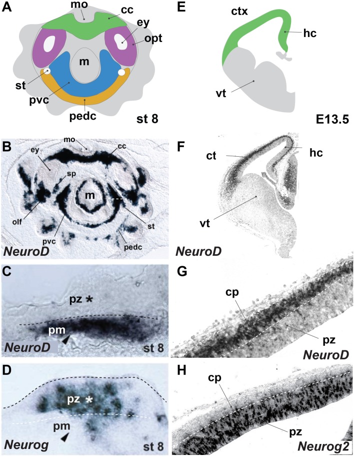
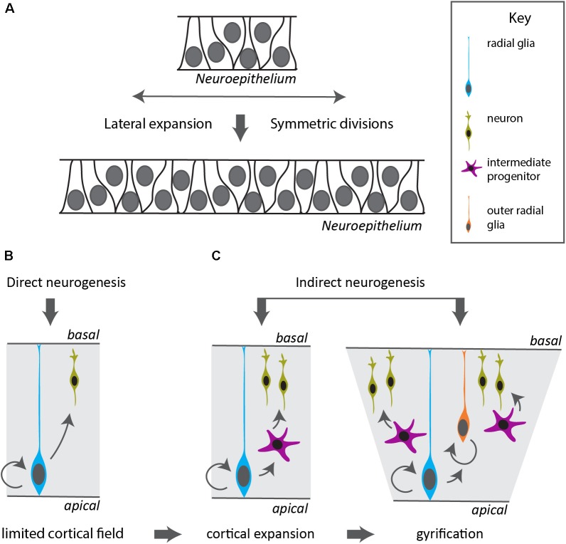
Within the clade of mollusks, cephalopods have developed an unusually large and complex nervous system. The increased complexity of the cephalopod centralized "brain" parallels an amazing amount of complex behaviors that culminate in one order, the octopods. The mechanisms that enable evolution of expanded brains in invertebrates remain enigmatic. While expression mapping of known molecular pathways demonstrated the conservation of major neurogenesis pathways and revealed neurogenic territories, it did not explain why cephalopods could massively increase their brain size compared to other mollusks. Such an increase is reminiscent of the expansion of the cerebral cortex in mammalians, which have enlarged their number and variety of neurogenic stem cells. We hypothesize that similar mechanisms might be at play in cephalopods and that focusing on the stem cell biology of cephalopod neurogenesis and genetic innovations might be smarter strategies to uncover the mechanism that has driven cephalopod brain expansion.

Keywords: brain development; cephalopod; invertebrate neuron; neurogenesis; stem cell.


Figures

FIGURE 1
FIGURE 1
Octopus bimaculoides and mouse neurogenesis occurs in similarly laminated neuroectoderm. (A) Schematic top–down overview of the neurogenic territories in the stage 8 Octopus bimaculoides embryo. All color-marked areas are neurogenic, cord-like regions. (B) Whole-mount in situ hybridization for NEUROD, a marker of young post-mitotic neurons. (C,D) Higher magnification of the neurogenic area (white dashed line in B) demarcating a laminated structure with post-mitotic neurons (pm, arrowhead, marked by NEUROD, C) separated from progenitors (pz, star, marked by NEUROG, D) (dashed line). (E) Schematic view of a coronal section through the mouse telencephalon at E13.5, demarcating the ventral telencephalon (vt, gray) and dorsally placed cortex (ctx) and hippocampal (hc) areas (green). (F) In situ hybridization of Neurod, a post-mitotic neuron proneural transcription factor. (G,H) Higher magnification of the cortical laminated structure (dashed lines), with a progenitor zone (pz, marked by Neurog2, H) lining the ventricle and a post-mitotic cortical plate (cp, marked by NeuroD, G). (B–D) Adapted from Shigeno et al. (2015). cc, cerebral cord; cp, cortical plate; ctx, cerebral cortex; ey, eye; hc, hippocampus; m, mantle; mo, mouth; olf, olfactory organ; opt, optic lobe; pedc, pedal cord; pvc, palliovisceral cord; pz, progenitor zone; sp, subpedunculate tissue; st, statocyst; vt, ventral telencephalon; ve, ventricle.
FIGURE 2
FIGURE 2
Modes of neurogenesis in the vertebrate cerebral cortex. (A) Before the onset and during early stages of neurogenesis, the neuroepithelium divides symmetrically to expand in a lateral fashion, increasing the neurogenic domain. (B) In vertebrates with a small cortical field, the radial glia divide asymmetrically to generate neurons in a direct manner. (C) Indirect neurogenesis generates intermediate progenitors that divide symmetrically resulting in increased neuronal output and expansion of the cerebral cortex. The appearance of a duplication of the radial glia layer in outer radial glia allows further radial and lateral expansion and gyrification of the cortex.

References

    1. Abdel-Mannan O., Cheung A. F. P., Molnár Z. (2008). Evolution of cortical neurogenesis. Brain Res. Bull. 75 398–404. 10.1016/j.brainresbull.2007.10.047 - DOI - PubMed
    1. Albertin C. B., Simakov O., Mitros T., Wang Z. Y., Pungor J. R., Edsinger-Gonzales E., et al. (2015). The octopus genome and the evolution of cephalopod neural and morphological novelties. Nature 524 220–224. 10.1038/nature14668 - DOI - PMC - PubMed
    1. Altman J. (2011). “The discovery of adult mammalian neurogenesis,” in Neurogenesis in the Adult Brain I, eds Seki T., Sawamoto K., Parent J. M., Alvarez-Buylla A. (Dordrecht: Springer; ), 10.1007/978-4-431-53933-9_1 - DOI
    1. Arendt D., Tosches M. A., Marlow H. (2016). From nerve net to nerve ring, nerve cord and brain-evolution of the nervous system. Nat. Rev. Neurosci. 17 61–72. 10.1038/nrn.2015.15 - DOI - PubMed
    1. Baratte S., Bonnaud L. (2009). Evidence of early nervous differentiation and early catecholaminergic sensory system during Sepia officinalis embryogenesis. J. Compar. Neurol. 517 539–549. 10.1002/cne.22174 - DOI - PubMed

LinkOut - more resources