. 2018 Oct 15;526(15):2462-2481.
doi: 10.1002/cne.24505. Epub 2018 Sep 24.

Differential response of pineal microglia to surgical versus pharmacological stimuli

Affiliations

Differential response of pineal microglia to surgical versus pharmacological stimuli

María P Ibañez Rodriguez et al. J Comp Neurol. .

Abstract

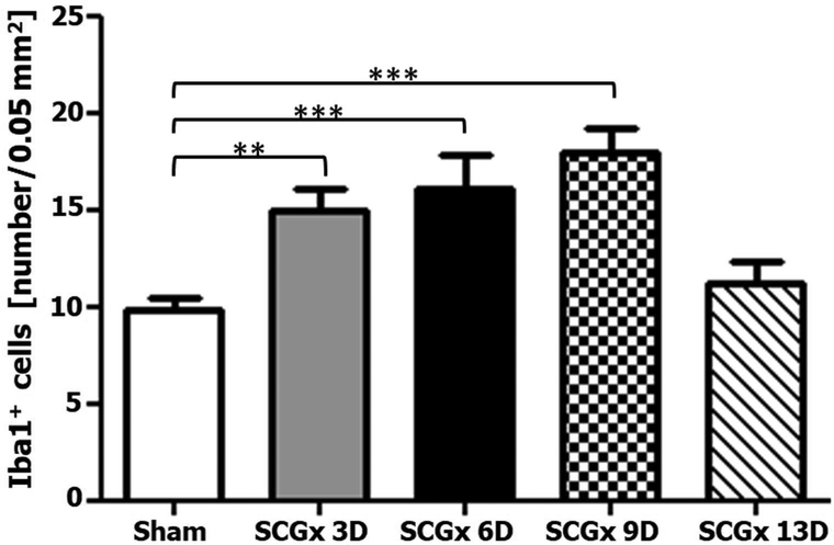
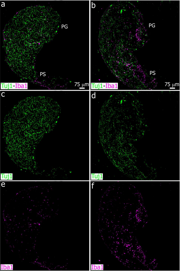
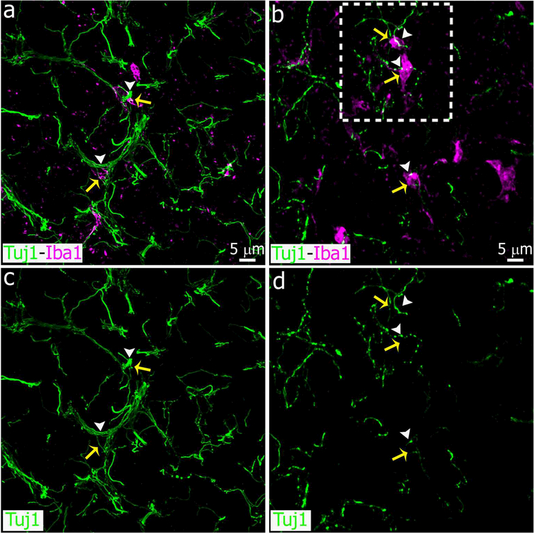
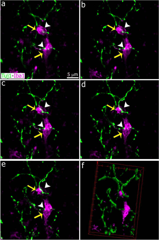
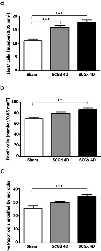
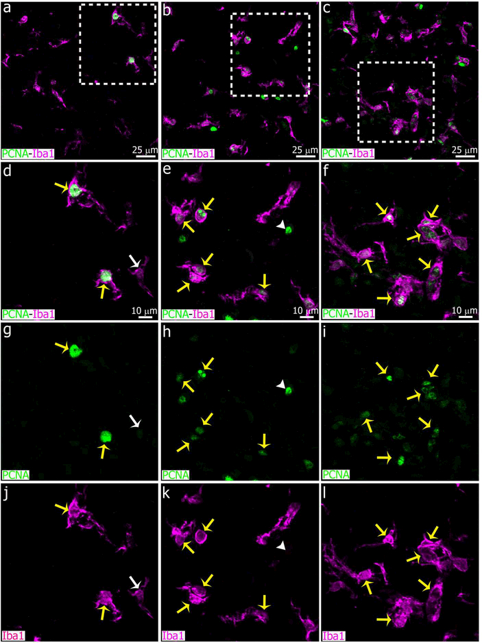
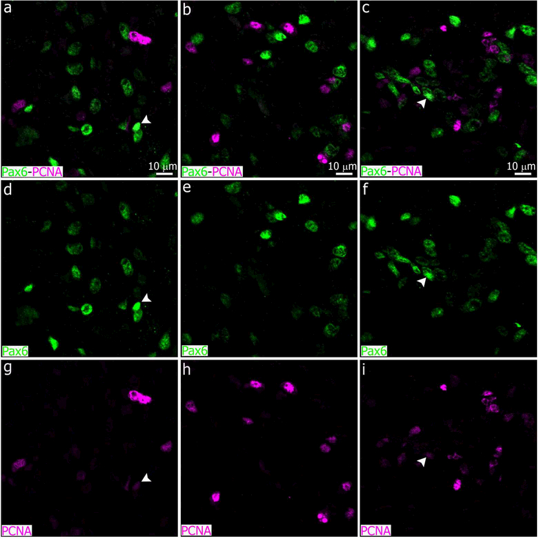
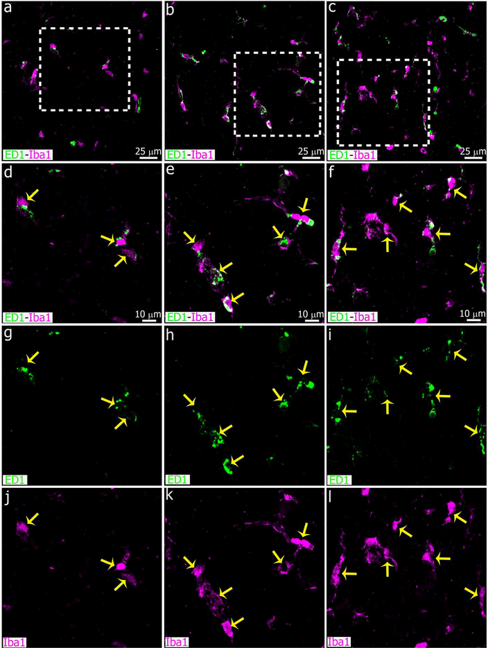
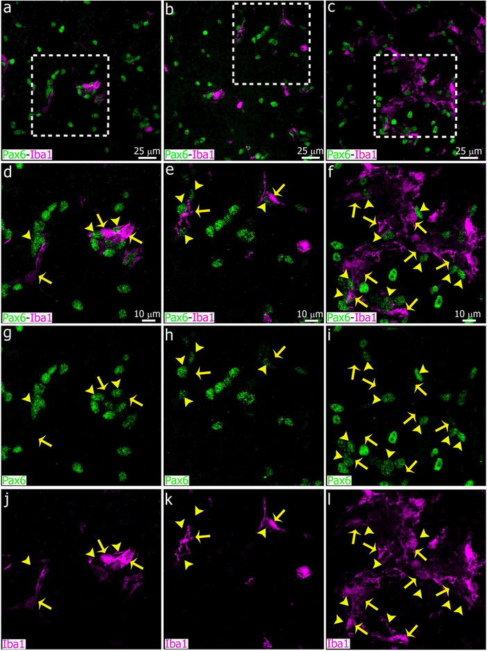
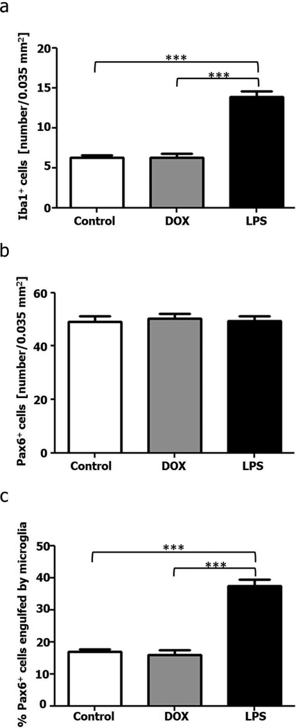
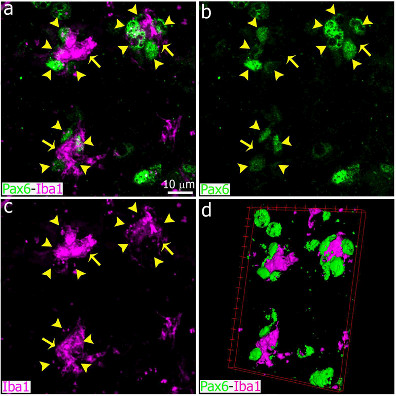
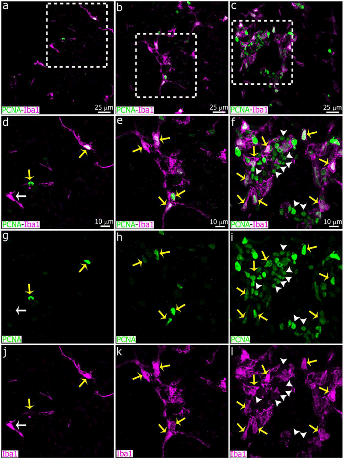
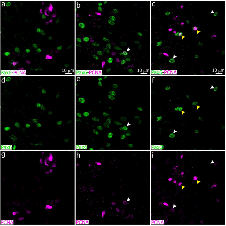
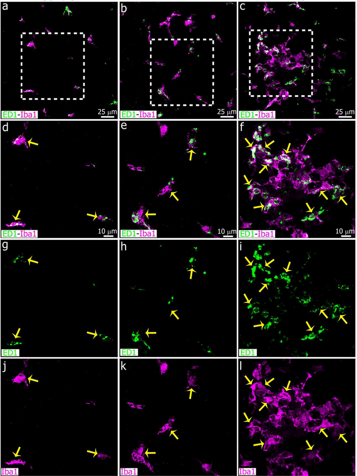
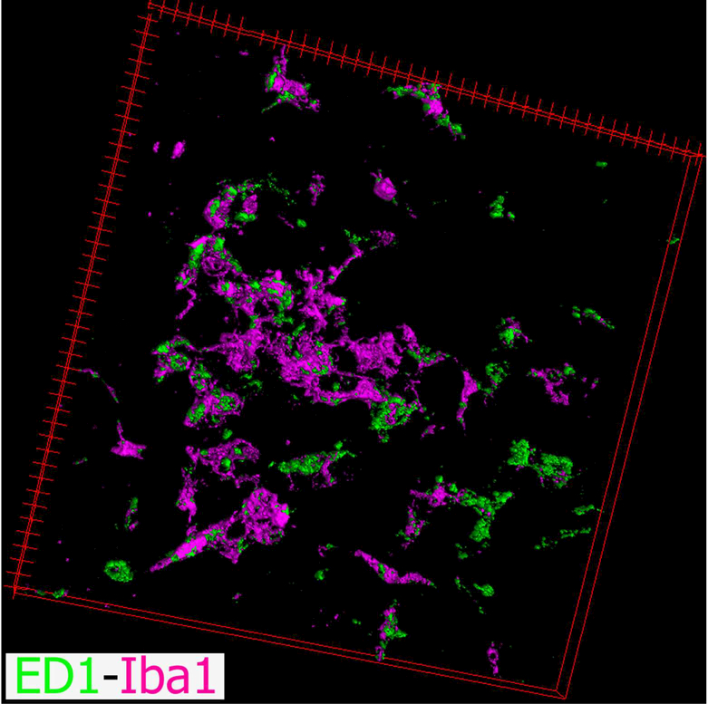
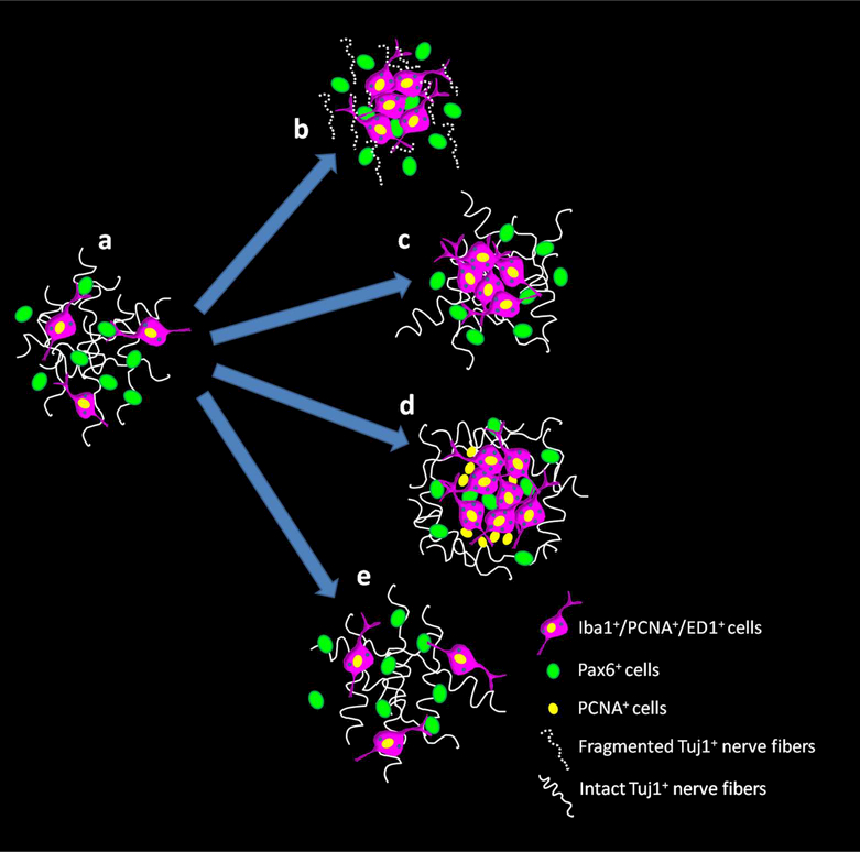
Microglial cells are one of the interstitial elements of the pineal gland (PG). We recently reported the pattern of microglia colonization and activation, and microglia-Pax6+ cell interactions during normal pineal ontogeny. Here, we describe the dynamics of microglia-Pax6+ cell associations and interactions after surgical or pharmacological manipulation. In adult rats, the superior cervical ganglia (SCG) were exposed, and either bilaterally excised (SCGx) or decentralized (SCGd). In the SCGx PGs, the density of Iba1+ microglia increased after surgery and returned to sham baseline levels 13 days later. Pineal microglia also responded to SCGd, a more subtle denervation. The number of clustered Iba1+ /PCNA+ /ED1+ microglia was higher 4 days after both surgeries compared to the sham-operated group. However, the number of Pax6+ /PCNA- cells and the percentage of Pax6+ cells contacted by and/or phagocytosed by microglia increased significantly only after SCGx. Separate groups of rats were treated with either bacterial lipopolysaccharides (LPS) or doxycycline (DOX) to activate or inhibit pineal microglia, respectively. Peripheral LPS administration caused an increase in the number of clustered Iba1+ /PCNA+ /ED1+ microglial cells, and in the percentage of Pax6+ cells associated with and/or engulfed by microglia. In the LPS-treated PGs, we also noted an increase in the number of PCNA+ cells that were Iba1- within the microglial cell clusters. The density of Pax6+ cells did not change after LPS treatment. DOX administration did not influence the parameters analyzed. These data suggest that pineal microglia are highly receptive cells capable of rapidly responding in a differential manner to surgical and pharmacological stimuli.

Keywords: ED1 (RRID: AB_566872); Iba1 (RRID: AB_2224402; RRID: AB_839504); PCNA (RRID: AB_95106); Pax6 (RRID: AB_1566562; RRID: AB_2565003; RRID: AB_291612); Tuj1 (RRID: AB_10063408; RRID: AB_2313773); bacterial lipopolysaccharides; decentralization; differential responses; ganglionectomy; microglia; pineal gland.


Figures

Figure 1.
Figure 1.
Kinetics of microglial cell number in the adult rat pineal gland after bilateral superior cervical ganglionectomy (SCGx). The surgical removal of the SCG increased the number of Iba1+ microglial cells per area (0.05 mm2) in the pineal gland by 3 days (3D) after the surgery, and the density of microglia remained significantly elevated for 9 days. The number of microglial cells returned to the sham baseline level 13 days after SCGx. Three glands (N=3) per group were analyzed at each time point. Data were expressed as mean ± S.E.M. Microglia density was similar among sham-operated rats; the sham bar represents the average of the twelve control animals. Statistics: one-way ANOVA followed by the Tukey post-test; ***P < 0.001; **P < 0.01.
Figure 2.
Figure 2.
Sympathetic nerve fibers degenerate and microglial cell number is increased in the adult pineal gland after bilateral SCGx. (a-f) Sections of adult pineal glands (PG) including the pineal stalk (PS), immunolabeled for the microglial marker Iba1 (magenta) and β- tubulin III (Tuj1, green) at 6 days after sham (left column, a, c, e) and SCGx surgery (right column, b, d, f). Wallerian degeneration of the sympathetic nerve fibers and a higher number of microglial cells, mainly in the proximal and ventral PG and PS, are observed in the SCGx PG compared to the control gland. (a-f) 10x; scale bar: 75 μm.
Figure 3.
Figure 3.
Higher magnification images showing pineal microglial cells interacting and phagocytozing degenerated sympathetic nerve fibers after SCGx. Panels show microglial cells immunoreactive for Iba1 (magenta, yellow arrows) associated with normal or fragmented nerve fibers positive for Tujl (green, white arrowheads) in sham-operated (left column, a, c) and SCGx (right column, b, d) pineal glands at 6 days after surgery. Phagocytosis events were more frequent in the SCGx group. (a, c) 1.5x digital zoom from a 60x image; (b, d) 2x digital zoom from a 40x image; scale bar: 5 μm.
Figure 4.
Figure 4.
Interactions between microglial cells and degenerated sympathetic nerve fibers in the pineal gland after SCGx. Images show sequential confocal planes separated by a distance of 1.5 μm (a-e) and a 3-dimensional reconstruction (f). Immunolabeling for the microglial marker Iba1 (magenta, yellow arrows) and β-tubulin III (Tuj1, green, white arrowheads). The five confocal planes displayed here were used to generate the image shown in the inset of Figure 3b. (a-e) 5x digital zoom from 40x images; scale bar: 5 μm. (f) 3-dimensional reconstruction of the five optical sections in a-e highlights interactions between microglia and fragmented nerve fibers.
Figure 5.
Figure 5.
Sympathetic innervation of the rat pineal gland remains intact after bilateral superior cervical decentralization (SCGd). Panels show sections of pineal glands (PG) from sham-operated (left column, a, d, g), SCGd (center column, b, e, h) and ganglionectomized (SCGx; right column, c, f, i) adult rats four days after surgery. Sections were immunostained for Iba1 (magenta) and Tuj1 (green). Wallerian degeneration was only observed after SCGx, but close association and phagocytosis of nerve fibers (white arrowheads) by microglial cells (yellow arrows) were observed in each group. (a-c) 60x; scale bar: 25 μm. (d-i) 1.8x digital zooms from the insets shown in a-c; scale bar: 10μm.
Figure 6.
Figure 6.
Superior cervical ganglionectomy (SCGx) and decentralization of SCG (SCGd) increase microglial cell number and cellular interactions in the adult rat pineal gland. Panels show images of pineal glands (PG) immunolabeled for Iba1 (magenta, yellow arrows) and the essential transcription factor Pax6 (green, yellow arrowheads) at 4 days after sham (left column, a, d, g, j), SCGd (center column, b, e, h, k), and SCGx (right column, c, f, i, l-o) surgery. The number of Iba1+ microglial cells was higher in both the SCGd and SCGx PGs compared to sham-operated animals, and microglial cell clusters were frequent in the experimental groups. An increase in the number of Pax6+ cells, and associations between microglia and Pax6+ cells were observed in the SCGx PG. Illustrative examples of microglia- Pax6+ cell associations in the SCGx PG are shown in m-o. (a and c) 60x; (b) 1.5x digital zoom from a 40x image; scale bar: 25 μm. (d-l) 1.8x digital zooms from the insets shown in a-c; scale bar: 10 μm. (m-o) 5.3x digital zooms from f; scale bar: 5 μm.
Figure 7.
Figure 7.
Morphometric analysis of microglial cells, Pax6+ cells and cellular interactions between both cell types after SCGx or SCGd. The analysis was performed 4 days (4D) after surgery, and a sham-operated group was included. (a) The number of Iba1+ microglial cells per area (0.05 mm2) increased after surgical disruption of the sympathetic innervation to the pineal gland by bilateral SCGx or SCGd. (b) The density of Pax6+ cells was significantly increased in the SCGx pineal glands. (c) SCGx induced a significant increase in the percentage of Pax6+ cells in contact with and/or engulfed by microglial cells. Data were expressed as mean ± S.E.M. (N=4). Statistics: one-way ANOVA followed by the Tukey posttest; ***P < 0.001; **P < 0.01.
Figure 8.
Figure 8.
The number of proliferative Iba1+ microglial cells is increased in the pineal gland after SCGx or SCGd. Confocal images of adult rat pineal glands (PG) immunostained for Iba1 (magenta) and the mitotic cell marker PCNA (green) show that the majority of microglial cells are positive for both markers (yellow arrows) four days after SCGx (right column, c, f, i, l) or SCGd (center column, b, e, h, k), compared to the sham-operated group (left column, a, d, g, j). White arrows show an Iba1+ cell that does not express PCNA, and white arrowheads point a PCNA+ cell that does not express Iba1. (a-c) 1.5x digital zooms from 40x images; scale bar: 25 μm. (d-l) 1.9x digital zooms from the insets shown in a-c; scale bar: 10 μm.
Figure 9.
Figure 9.
The majority of Pax6+ cells did not express the mitotic cell marker PCNA in the pineal gland at four days after SCGd, SCGx or sham-operation. Panels show confocal images of pineal glands (PG) from control (left column, a, d, g), SCGd (center column, b, e, h) and SCGx (right column, c, f, i) rats, immunolabeled for the essential transcription factor Pax6 (green) and the proliferative marker PCNA (magenta). Pax6+ cells are mostly negative for PCNA (Pax6+/PCNA cells). White arrowheads point Pax6high cells with minimally detectable levels of the mitotic cell marker PCNA (Pax6high/PCNAlow cells). (a-i) 3x digital zooms from 40x images; scale bar: 10 μm.
Figure 10.
Figure 10.
The number of microglial cells that express markers associated with phagocytosis increased in the adult pineal gland four days after SCGx or SCGd. Confocal images show the proportion of Iba1+ microglia (magenta) that co-express the lysosomal marker ED1 (CD68, green). Although microglia in the healthy adult pineal gland (PG) normally exhibit morphological hallmarks of activation (Ibanez Rodriguez et al., 2016), the expression of ED1 was potentiated by both SCGx (right column, c, f, i, l) and SCGd (center column, b, e, h, k), compared to the sham surgery (left column, a, d, g, j). Yellow arrows indicate microglia positive for Iba1 and ED1. (a-c) 1.4x digital zooms from 40x images; scale bar: 25 μm. (d-l) 1.9x digital zooms from the insets shown in a-c; scale bar: 10 μm.
Figure 11.
Figure 11.
An increased number of microglial cells arranged in clusters and associated with Pax6+ cells is observed in the pineal gland after peripheral administration of bacterial lipopolysaccharides (LPS). Panels show confocal images of the adult pineal gland (PG) after intraperitoneal (IP) injections of LPS (right column, c, f, i, l), the antibiotic doxycycline (DOX; center column, b, e, h, k), or vehicle control (left column, a, d, g, j). Close association and phagocytosis of Pax6+ cells (green, yellow arrowheads) by Iba1+ microglia (magenta, yellow arrows) were observed in the three groups. Large microglial cell clusters that encapsulated multiple Pax6+ cells were observed only after LPS administration. (a-c) 60x; scale bar: 25 μm. (d-l) 1.8x digital zooms from the insets shown in a-c; scale bar: 10 μm.
Figure 12.
Figure 12.
The density of Iba1+ microglial cells and the percentage of Pax6+ cells contacted by and/or engulfed by microglia increased in the pineal gland after a peripheral challenge with bacterial lipopolysaccharides (LPS) (a and c). (b) The number of Pax6+ cells in the pineal gland (PG) 24 hours after the second intraperitoneal (IP) injection of LPS was not altered. (a-c) Administration of the antibiotic doxycycline (DOX) did not affect the parameters analyzed. Data were expressed as mean ± S.E.M., in an area of 0.035 mm2 (N=5). Statistics: one-way ANOVA followed by the Tukey post-test; ***P < 0.001.
Figure 13.
Figure 13.
Images showing representative Iba1+ microglial cells and Pax6+ cells in the adult pineal gland after bacterial lipopolysaccharide (LPS) administration. Each microglial cell (magenta, yellow arrows) was in contact with multiple Pax6+ cells (green, yellow arrowheads), and apparently phagocytosed some of them 24 hours after the second intraperitoneal (IP) injection of LPS. (a-c) 2.8x digital zoom from a 60x image; scale bar: 10 μm. (d) 3-dimensional representation of the microglia and Pax6+ cells shown in a-c.
Figure 14.
Figure 14.
Two proliferative cell populations are clearly distinguished within the microglial cell clusters induced by intraperitoneal (IP) administration of bacterial lipopolysaccharides (LPS). Sections of pineal glands (PG) immunolabeled for Iba1 (magenta) and the mitotic cell marker PCNA (green) after treatment with LPS (right column, c, f, i, l), doxycycline (DOX; center column, b, e, h, k) or vehicle (left column, a, d, g, j). In all groups, the majority of Iba1+ microglial cells are positive for PCNA (yellow arrows). In the LPS group, a second cell population immunoreactive only for PCNA (white arrowheads) was observed within the microglial cell clusters. White arrows point an Iba1+ microglial cell negative for PCNA in a control pineal gland. (a-c) 60x; scale bar: 25 μm. (d-l) 1.8x digital zooms from the insets shown in a-c; scale bar: 10 μm.
Figure 15.
Figure 15.
Pax6+ cells appear to be mostly negative for the mitotic cell marker PCNA 24 hours after administration of bacterial lipopolysaccharides (LPS) or the antibiotic doxycycline (DOX). Panels show sections of pineal glands (PG) from LPS (right column, c, f, i), DOX (center column, b, e, h), or vehicle-treated (left column, a, d, g) animals, immunostained for Pax6 (green) and PCNA (magenta). The majority of the Pax6+ cells did not express PCNA, indicating they were not actively proliferating at ZT6. A few Pax6high/PCNAhigh cells (yellow arrowheads) were observed in the LPS-treated PGs. Pax6high/PCNAlow cells (white arrowheads) were present in both DOX and LPS groups. (a-i) 2x digital zooms from 60x images; scale bar: 10 μm.
Figure 16.
Figure 16.
Microglia are highly phagocytic after IP administration of bacterial lipopolysaccharides (LPS). Panels show images of pineal glands (PG) immunostained for Iba1 (magenta) and the lysosomal marker ED1 (CD68, green) after intraperitoneal (IP) injections of LPS (right column, c, f, i, l), doxycycline (DOX; center column, b, e, h, k) or vehicle (left column, a, d, g, j). Microglia in the cell clusters in LPS-treated pineal glands are enriched in cytoplasmic ED1-positive bodies. There were no differences in the ED1 expression pattern between control and DOX-treated PGs. Yellow arrows show microglial cells immunoreactive for both Iba1 and ED1. (a-c) 60x; scale bar: 25 μm. (d-l) 1.8x digital zooms from the insets shown in a-c; scale bar: 10 μm.
Figure 17.
Figure 17.
Three-dimensional projection of the image shown in Figure 16c. Microglial cells (Iba1, magenta) expressing high levels of the lysosomal antigen ED1 (CD68, green) were observed in the pineal gland (PG) following intraperitoneal (IP) administration of bacterial lipopolysaccharides (LPS).
Figure 18.
Figure 18.
Schematic model of the differential response of pineal microglia to surgical and pharmacological stimuli. (a) In the naive adult rat pineal gland (PG), microglial cells are proliferative and display hallmarks of activation and phagocytic activity (Iba1+/PCNA+/ED1+ cells) (Ibanez Rodriguez et al., 2016). Pax6+ cells that appear to derive from Pax6+/Vimentin+ neuroepithelial precursor cells in the developing pineal gland, remain in the adult gland as a quiescent cell reservoir and a preferred target for the microglial cells (Ibanez Rodriguez et al., 2016). (b) Bilateral superior cervical ganglionectomy (SCGx) induces Wallerian degeneration (WD) of the sympathetic nerve fibers that innervate the pineal gland and an increase in the density of microglial cells and Pax6+ cells, and interactions between both cell types. (c) Superior cervical ganglia decentralization (SCGd) is a more subtle disruption of the sympathetic innervation to the pineal gland that increases microglial cell number without significantly altering the Pax6+ cell population, or interactions between microglia and Pax6+ cells. SCGd does not cause WD of the sympathetic nerve fibers. (d) Intraperitoneal (IP) administration of bacterial lipopolysaccharides (LPS) significantly increases the number of microglia in the pineal gland and induces the formation of microglial cell clusters that include at least two cell populations immunoreactive for the mitotic cell marker PCNA. LPS enhanced the association and interaction between pineal microglia and Pax6+ cells, without significantly affecting the number of Pax6+ cells. (e) In the rat pineal gland, doxycycline (DOX) injections did not alter the density of microglial cells or Pax6+ cells, and did not influence interactions between both cell types.

References

    1. Ajmone-Cat MA, Mancini M, De Simone R, Cilli P, & Minghetti L (2013). Microglial polarization and plasticity: evidence from organotypic hippocampal slice cultures. Glia, 61(10), 1698–1711. doi:10.1002/glia.22550 - DOI - PubMed
    1. Ajmone-Cat MA, Nicolini A, & Minghetti L (2003). Prolonged exposure of microglia to lipopolysaccharide modifies the intracellular signaling pathways and selectively promotes prostaglandin E2 synthesis. J Neurochem, 87(5), 1193–1203. - PubMed
    1. Bailey MJ, Coon SL, Carter DA, Humphries A, Kim JS, Shi Q, . . . Klein DC (2009). Night/day changes in pineal expression of >600 genes: central role of adrenergic/cAMP signaling. J Biol Chem, 284(12), 7606–7622. doi:10.1074/jbc.M808394200 - DOI - PMC - PubMed
    1. Benitez SG, Castro AE, Patterson SI, Muñoz EM, & Seltzer AM (2014). Hypoxic preconditioning differentially affects GABAergic and glutamatergic neuronal cells in the injured cerebellum of the neonatal rat. PLoS One, 9(7), e102056. doi:10.1371/journal.pone.0102056 - DOI - PMC - PubMed
    1. Brown GC, & Neher JJ (2014). Microglial phagocytosis of live neurons. Nat Rev Neurosci, 15(4), 209–216. doi:10.1038/nrn3710 - DOI - PubMed

Publication types