. 2018 Sep;84(3):386-400.
doi: 10.1002/ana.25267.

Molecular and clinical relationship between live-attenuated Japanese encephalitis vaccination and childhood onset myasthenia gravis

Affiliations

Molecular and clinical relationship between live-attenuated Japanese encephalitis vaccination and childhood onset myasthenia gravis

Dan He et al. Ann Neurol. 2018 Sep.

Abstract

Objective: The incidence of childhood onset myasthenia gravis (CMG) in China is higher than that in other countries; however, the reasons for this are unclear.

Methods: We investigated the clinical and immunological profiles of CMG, and assessed the potential precipitating factors. For the mouse studies, the possible implication of vaccination in the pathogenesis was explored.

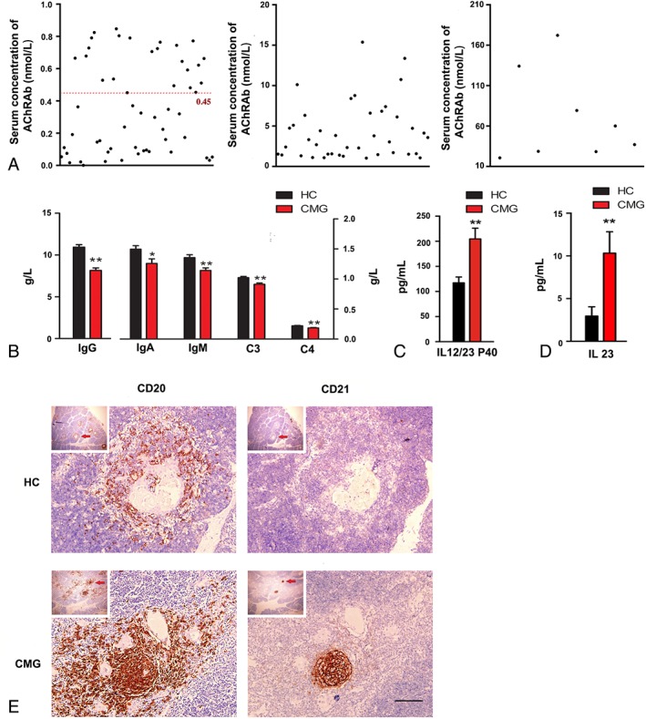
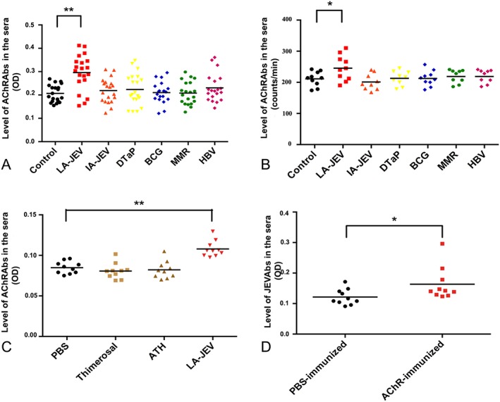
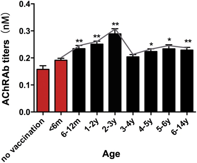
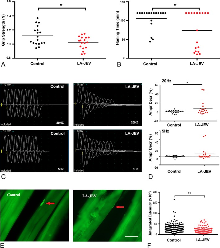
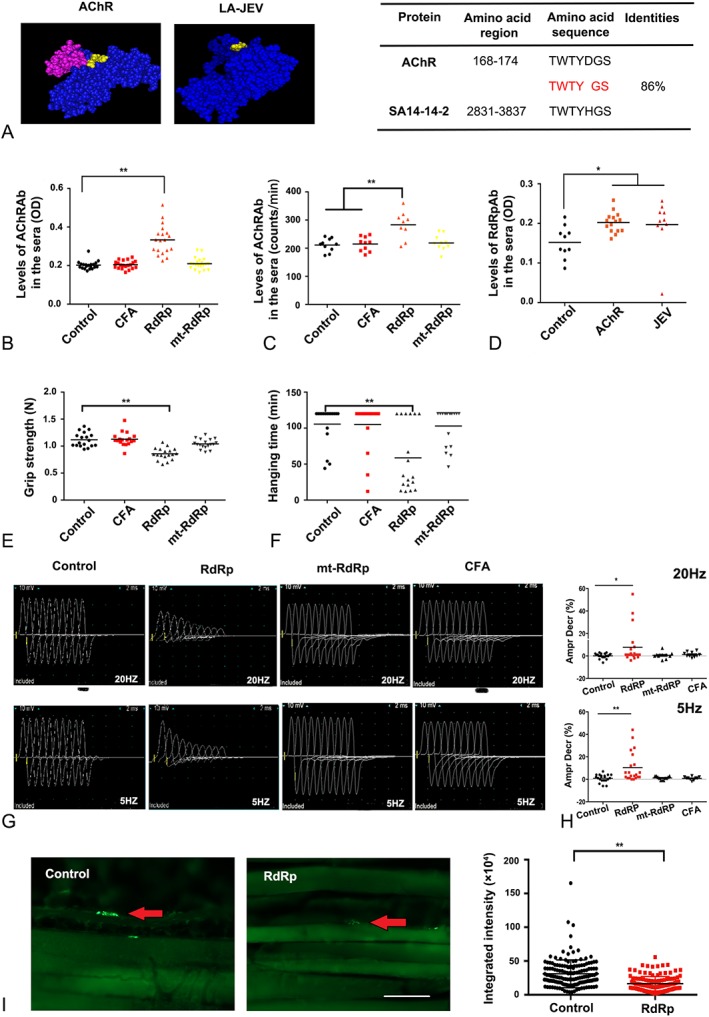
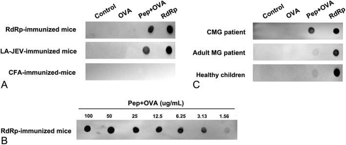
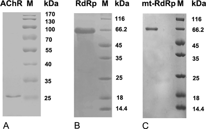
Results: In our retrospective study, 51.22% of the 4,219 cases of myasthenia gravis (MG) were of the childhood onset type. The cohort study uncovered that the pathophysiology of CMG was mediated by immune deviation, rather than through gene mutations or virus infections. The administration of the live-attenuated Japanese encephalitis vaccine (LA-JEV), but not the inactivated vaccine or other vaccines, in mice induced serum acetylcholine receptor (AChR) antibody production, reduced the AChR density at the endplates, and decreased both muscle strength and response to repetitive nerve stimulation. We found a peptide (containing 7 amino acids) of LA-JEV similar to the AChR-α subunit, and immunization with a synthesized protein containing this peptide reproduced the MG-like phenotype in mice.

Interpretation: Our results describe the immunological profile of CMG. Immunization with LA-JEV induced an autoimmune reaction against the AChR through molecular mimicry. These findings might explain the higher occurrence rate of CMG in China, where children are routinely vaccinated with LA-JEV, compared with that in countries, where this vaccination is not as common. Efforts should be made to optimize immunization strategies and reduce the risk for developing autoimmune disorders among children. Ann Neurol 2018;84:386-400.


Figures

Figure 1
Figure 1
Clinical and immunological profiles of patients with childhood onset myasthenia gravis (CMG). (A) Serum concentration of anti–acetylcholine receptor antibodies (AChRAbs) in the CMG patients of our cohort study. Serum titers > 0.45nM were considered positive (red line; n = 104). (B) Serum concentration of IgG, IgM, IgA, C3, and C4 in CMG patients (red columns; n = 104) is significantly lower than that in healthy controls (HC; black columns; n = 100, ); *p < 0.05, **p < 0.01. (C) Serum concentration of IL12/23 P40 in CMG patients (red column; n = 104) is significantly higher than that in HC (black column; n = 100); **p < 0.01. (D) Serum concentration of IL23 in CMG patients (red column; n = 104) is significantly higher in patients than in HC (black column; n = 100); **p < 0.01. (E) Representative images of the thymus in patients with CMG and HC. The CD20+ and CD21+ B cells aggregate to form a germinal center in the thymic medulla. Higher magnification images of the areas indicated by the arrows are shown. Scale bar = 25 μm.
Figure 2
Figure 2
Injection with live‐attenuated Japanese encephalitis vaccine (LA‐JEV) leads to the production of anti–acetylcholine receptor antibodies (AChRAbs). (A) Levels of AChRAbs measured by enzyme‐linked immunosorbent assay (ELISA) in the sera of BALB/c mice injected 4 times at 2‐week intervals with LA‐JEV (red), inactivated Japanese encephalitis vaccine (IA‐JEV; orange), diphtheria/tetanus/pertussis vaccine (DTaP; yellow), bacillus Calmette–Guérin vaccine (BCG; blue), measles/mumps/rubella vaccine (MMR; green), hepatitis B vaccine (HBV; pink), or phosphate‐buffered saline (PBS; control, black). Mice injected with LA‐JEV produced a significantly higher mean level of AChRAbs than mice injected with other vaccines (**p < 0.01, n = 20). (B) The AChRAb levels measured by radioimmunoassay in sera from BALB/c mice that were administered 4 injections (at 2‐week intervals) of one of the following: LA‐JEV (red), IA‐JEV (orange), DTaP (yellow), BCG (blue), MMR (green), HBV (pink), or PBS (control, black). Mice injected with LA‐JEV produced a significantly higher mean level of AChRAbs than mice injected with other vaccines (*p < 0.05, n = 10). (C) Levels of AChRAb measured by ELISA in the sera of BALB/c mice injected 4 times at 2‐week intervals after injection with PBS (black), thimerosal (yellow‐green), aluminum hydroxide (ATH; green‐yellow), or LA‐JEV (red). Adjuvant‐immunized mice and control mice show no significant difference in their AChRAb titers, while mice injected with LA‐JEV produced a significantly higher level of AChRAbs (**p < 0.01). (D) Levels of JEV antibodies (JEVAbs) measured by ELISA in the sera of BALB/c mice injected 4 times at 2‐week intervals with Torpedo californica–derived AChR (red) or PBS control (black). AChRAbs induced by torpedo AChRs reacted with the JEV antigen in the culture supernatants derived from LA‐JEV–transfected cells, but the control did not (*p < 0.05). OD = optical density.
Figure 3
Figure 3
Serological features of children immunized with live‐attenuated Japanese encephalitis vaccine (LA‐JEV). Serum anti–acetylcholine receptor antibody (AChRAb) titers in children vaccinated with LA‐JEV at different ages are shown. The red columns represent serum titers in children who were not inoculated with LA‐JEV (m = months; y = years; *p < 0.05, **p < 0.01). For the no vaccination group, children did not receive any vaccine, including LA‐JEV.
Figure 4
Figure 4
Mice injected with live‐attenuated Japanese encephalitis vaccine (LA‐JEV) develop features of myasthenia gravis. BALB/c mice were given intraperitoneal injections of 5.7 log10 plaque‐forming units/time LA‐JEV or phosphate‐buffered saline (PBS; control) 4 times at 2‐week intervals. (A) The grip strength of control and LA‐JEV–immunized mice, and (B) the lengths of time for which the LA‐JEV–injected and control mice were able to hang from a coat hanger were tested at week 8. Mice vaccinated with LA‐JEV had significantly reduced grip strength and hanging time when compared with the mice injected with the PBS control (*p < 0.05; red dots). (C) Representative image of the compound muscle action potential (CMAP) responses in control and LA‐JEV–injected mice at 8 weeks after first injection upon repetitive nerve stimulation. Mice injected with LA‐JEV had a decreased CMAP response compared to the control mice. (D) When 20Hz stimulation was administered, the percentage decrease in CMAPs was significantly greater in mice injected with LA‐JEV than in control mice (*p < 0.05; n = 20 for the control group; n = 19 for the LA‐JEV group, upper panel). Five‐hertz stimulation did not induce a statistically significant reduction in the CMAP response between the control and LA‐JEV–injected mice (lower panel). Ampr Decr % denotes the percentage decrease in the 5th amplitude of the CMAP compared to the first response. (E) Representative image of the immunofluorescence staining of acetylcholine receptors (AChRs) with biotin‐conjugated α‐bungarotoxin in muscle sections derived from mice injected with PBS (control) or LA‐JEV. In the control mice, AChRs (labeled by green fluorescence) aggregated at the endplates (red arrows), whereas in LA‐JEV–immunized mice, AChR staining was faint (scale bar = 25 μm). (F) The mean fluorescence intensity of AChRs randomly selected from 169 neuromuscular junctions of 5 mice injected with LA‐JEV (red) was significantly lower than that of control mice (black; **p < 0.01).
Figure 5
Figure 5
RdRp induces a myasthenia gravis–like phenotype through molecular mimicry. (A) Structures of the 2 proteins, acetylcholine receptor (AChR) and RNA‐dependent RNA polymerase (RdRp; left panel). The highlighted regions indicate the 7‐amino‐acid‐long region that is 86% identical between AChR and live‐attenuated Japanese encephalitis vaccine (LA‐JEV), based on Basic Local Alignment Search Tool analysis. The table on the right shows the sequences of this region in the 2 proteins. (B) The serum concentration of AChR antibodies (AChRAbs) in mice injected 4 times at 2‐week intervals with RdRp (orange), mutated (mt)‐RdRp (yellow), complete Freund adjuvant (CFA; red), and phosphate‐buffered saline (PBS; control), as measured by enzyme‐linked immunosorbent assay (**p < 0.01, n = 20). Mice injected with RdRp produced a significantly higher mean level of AChRAbs than mice injected with PBS. (C) The serum concentration of AChRAbs in mice injected 4 times at 2‐week intervals with RdRp (orange), mtRdRp (yellow), CFA (red), and PBS (control), as measured by radioimmunoassay (**p < 0.01). (D) AChRAb induced by torpedo AChR reacted strongly to RdRp protein (*p < 0.05). (E) The grip strength and (F) the duration of hanging of control (PBS)‐, CFA‐, RdRp‐, and mtRdRp‐immunized mice as tested at week 8. The grip strength and hanging time were lower in mice injected with RdRp compared to the control mice (**p < 0.01), whereas the mtRdRp‐ and CFA‐immunized groups were not significantly different in their mean grip strength and hanging time. (G) Representative image of the compound muscle action potential (CMAP) responses in control, CFA‐, RdRp‐, and mtRdRp‐injected mice at 8 weeks after first injection upon repetitive nerve stimulation. Mice injected with RdRp showed a decreased CMAP response 8 weeks after the first injection, whereas the mtRdRp‐ and CFA‐immunized groups did not. (H) Compared with the control mice, the percentage reduction in CMAPs after the mice were injected with RdRp was significant greater upon 20Hz (upper panel) and 5Hz (lower panel) stimulation, whereas the mtRdRp‐ and CFA‐immunized groups showed no difference. Ampr Decr % denotes the percentage decrease in the 5th amplitude of the CMAP compared to the first response (*p < 0.05, **p < 0.01). (I) Representative image (left panel) of the immunofluorescence staining of AChRs with biotin‐conjugated α‐bungarotoxin in muscle sections derived from mice injected with PBS (control) or RdRp. In control mice, AChRs (labeled by green fluorescence) aggregated in the endplates (red arrows), whereas in mice injected with RdRp, AChR staining was barely detected (scale bar = 25 μm). In the right panel, the fluorescence intensity of AChRs at 135 neuromuscular junctions from 5 randomly selected mice injected with RdRp (red) were significantly lower than that of AChRs selected from control mice (black), **p < 0.01. OD = optical density.
Figure 6
Figure 6
Injection with live‐attenuated Japanese encephalitis vaccine (LA‐JEV) leads to the production of anti–acetylcholine receptor antibodies (AChRAbs). (A) The reactivity of the serum from LA‐JEV/RNA‐dependent RNA polymerase (RdRp)/complete Freund adjuvant (CFA)‐immunized mice with the TWTYDGS epitope was tested by dot blot. LA‐JEV/RdRp‐immunized mouse serum could react with TWTYDGS conjugated to ovalbumin (Pep+OVA). (B) The samples of Pep+OVA were 2‐fold serial diluted and spotted onto a piece of nitrocellulose membrane. RdRp‐immunized mouse serum could react with Pep+OVA at a concentration of 3.13 µg/ml. (C) The serum of childhood onset myasthenia gravis (CMG) patients could react with the TWTYDGS epitope, whereas that of adult myasthenia gravis (MG) patients and healthy controls were less likely to react with the TWTYDGS epitope. OVA = ovalbumin.
Figure 7
Figure 7
Representative band patterns of the recombinant proteins after sodium dodecyl sulfate–polyacrylamide gel electrophoresis (SDS‐PAGE). Purified proteins were electrophoresed by 12% SDS‐PAGE and stained with Coomassie Brilliant Blue R250. (A) acetylcholine receptor (AChR)‐a1‐210, (B) RNA‐dependent RNA polymerase (RdRp), and (C) mutated (mt)‐RdRp were detected as 25, 66.2, and 66.2kDa bands.

References

    1. Meriggioli MN, Sanders DB. Autoimmune myasthenia gravis: emerging clinical and biological heterogeneity. Lancet Neurol 2009;8:475–490. - PMC - PubMed
    1. Zhu WH, Lu JH, Lin J, et al. HLA‐DQA1*03:02/DQB1*03:03:02 is strongly associated with susceptibility to childhood‐onset ocular myasthenia gravis in Southern Han Chinese. J Neuroimmunol 2012;247:81–85. - PubMed
    1. Zhang X, Yang M, Xu J, et al. Clinical and serological study of myasthenia gravis in HuBei Province, China. J Neurol Neurosurg Psychiatry 2007;78:386–390. - PMC - PubMed
    1. Carr AS, Cardwell CR, McCarron PO, McConville J. A systematic review of population based epidemiological studies in myasthenia gravis. BMC Neurol 2010;10:46. - PMC - PubMed
    1. Lai CH, Tseng HF. Nationwide population‐based epidemiological study of myasthenia gravis in Taiwan. Neuroepidemiology 2010;35:66–71. - PMC - PubMed

Publication types