Multicenter Study
. 2018 Dec;42(12):1625-1635.
doi: 10.1097/PAS.0000000000001146.

Inflammation, Active Fibroplasia, and End-stage Fibrosis in 172 Biliary Atresia Remnants Correlate Poorly With Age at Kasai Portoenterostomy, Visceral Heterotaxy, and Outcome

Affiliations
Multicenter Study

Inflammation, Active Fibroplasia, and End-stage Fibrosis in 172 Biliary Atresia Remnants Correlate Poorly With Age at Kasai Portoenterostomy, Visceral Heterotaxy, and Outcome

Kevin E Bove et al. Am J Surg Pathol. 2018 Dec.

Abstract

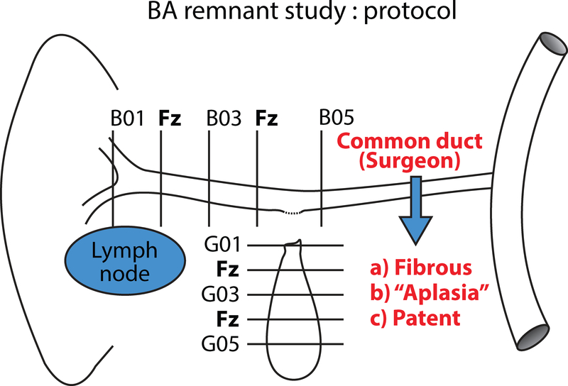
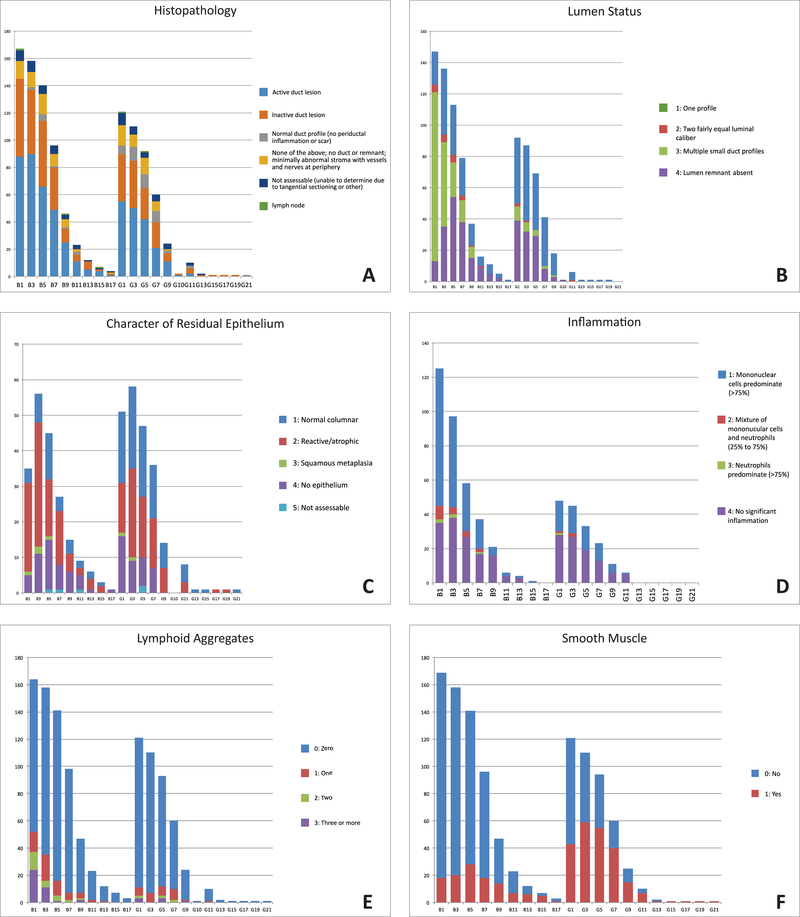
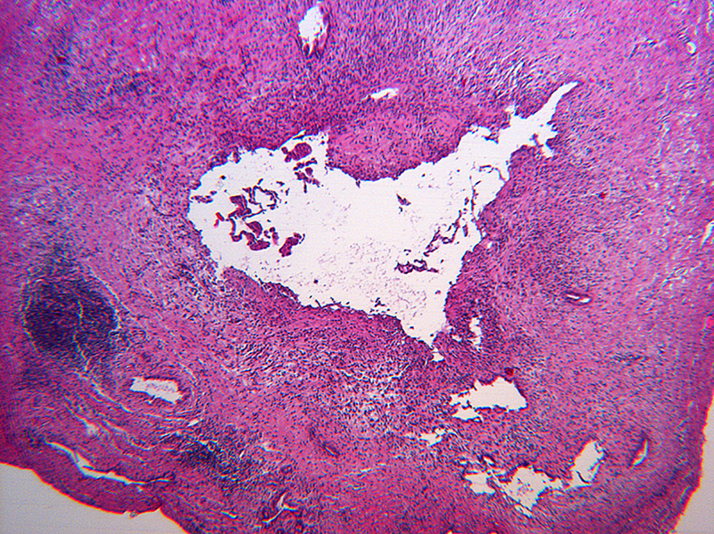
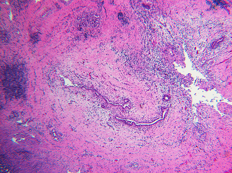
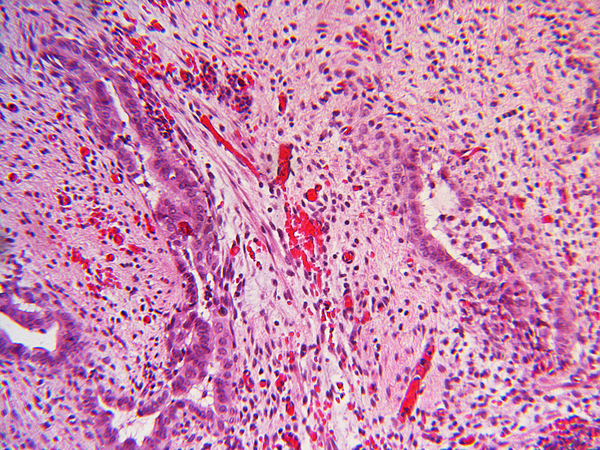
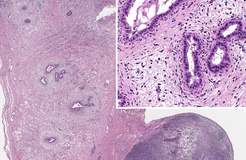
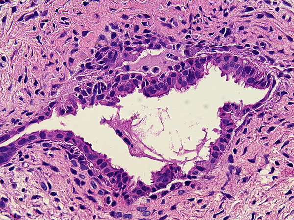
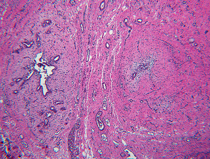
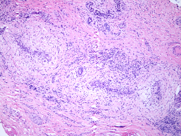
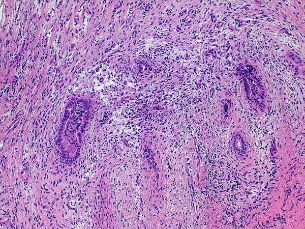
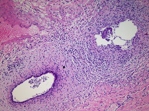
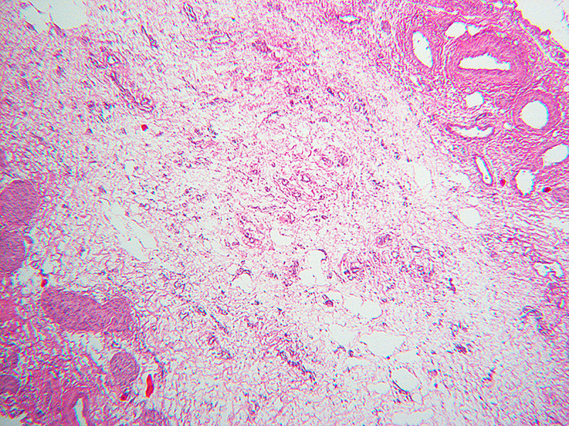
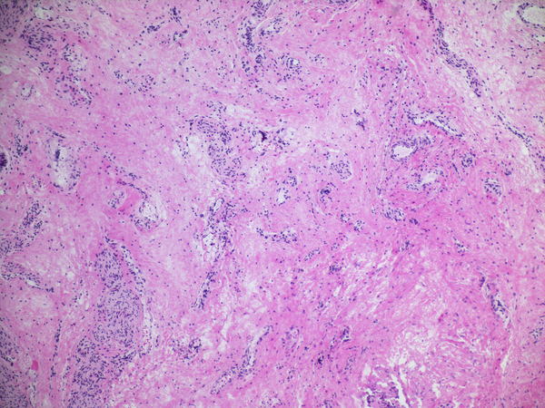
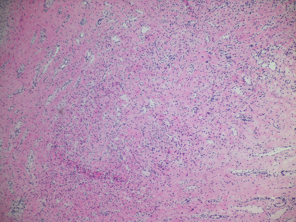
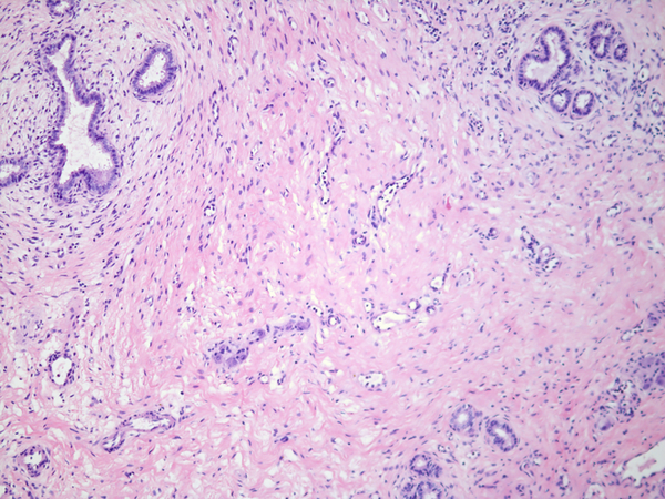
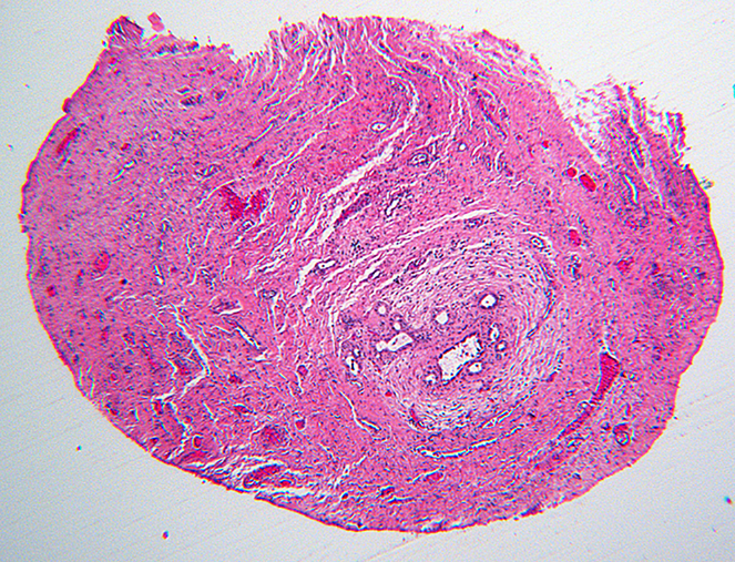
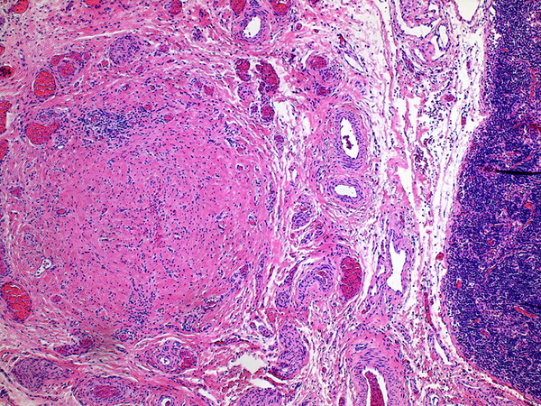
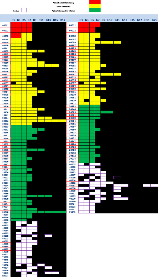
Published histologic studies of the hilar plate or entire biliary remnant at the time of Kasai portoenterostomy (KHPE) have not provided deep insight into the pathogenesis of biliary atresia, relation to age at surgery, prognosis or the basis for successful drainage. We report detailed histologic findings in 172 centrally reviewed biliary remnants with an average of 6 sections per subject. Active lesions were classified as either necroinflammatory (rare/clustered in a few subjects) or active concentric fibroplasia with or without inflammation (common). Inactive lesions showed bland replacement by collagen and fibrous cords with little or no inflammation. Heterogeneity was common within a given remnant; however, relatively homogenous histologic patterns, defined as 3 or more inactive or active levels in the hepatic ducts levels, characterized most remnants. Homogeneity did not correlate with age at KHPE, presence/absence of congenital anomalies at laparotomy indicative of heterotaxy and outcome. Remnants from youngest subjects were more likely than older subjects to be homogenously inactive suggesting significantly earlier onset in the youngest subset. Conversely remnants from the oldest subjects were often homogenously active suggesting later onset or slower progression. More data are needed in remnants from subjects <30 days old at KHPE and in those with visceral anomalies. Prevalence of partially preserved epithelium in active fibroplastic biliary atresia lesions at all ages suggests that epithelial regression or injury may not be a primary event or that reepithelialization is already underway at the time of KHPE. We hypothesize that outcome after KHPE results from competition between active fibroplasia and reepithelialization of retained, collapsed but not obliterated lumens. The driver of active fibroplasia is unknown.


Figures

Figure 1.
Figure 1.
This schematic demonstrates how the biliary remnants in this study were sampled for FFPE with alternate slices frozen for research. The average number of paraffin blocks per case was 6. Additional levels in the B and G series were labelled by expanding the sequences in the schematic as needed*.
Figure 2.
Figure 2.
Topographic bar graphs demonstrating distribution in remnants of histopathologic subsets (a), lumen status (b), residual epithelium character (c), inflammatory status (d), tertiary lymphoid aggregates (e), and smooth muscle (f).
Figure 3.
Figure 3.
Necroinflammatory lesions in BA remnants: All panels show remnant levels B-1 (hilum). A and B: Single lumens with necrotic epithelium are accompanied by intense inflammation, granulation tissue and tertiary lymphoid aggregates (arrows). C. Variably necrotic and retained injured epithelium is accompanied by stromal edema, loose fibroblast proliferation with focal smooth muscle differentiation, minor collagen deposition, reactive epithelium and a mixed mononuclear cell-neutrophil infiltrate.
Figure 3.
Figure 3.
Necroinflammatory lesions in BA remnants: All panels show remnant levels B-1 (hilum). A and B: Single lumens with necrotic epithelium are accompanied by intense inflammation, granulation tissue and tertiary lymphoid aggregates (arrows). C. Variably necrotic and retained injured epithelium is accompanied by stromal edema, loose fibroblast proliferation with focal smooth muscle differentiation, minor collagen deposition, reactive epithelium and a mixed mononuclear cell-neutrophil infiltrate.
Figure 4.
Figure 4.
Active fibroplasia in BA remnants: A, remnant level B-1(hilum) shows active fibroplasia without inflammation related to duct remnants and peribiliary glands with intact epithelium (insert). A non-reactive hilar lymph node at lower right lacked germinal centers. B, remnant level B-1 shows reactive epithelium with focal subepithelial fluid (arrow). C, remnant level B-5 shows two lumen residues, possibly located near junction of cystic and hepatic ducts, with asynchronous incomplete fibrous obliteration. The larger lumen on the left is accompanied by prominent peribiliary glands.
Figure 4.
Figure 4.
Active fibroplasia in BA remnants: A, remnant level B-1(hilum) shows active fibroplasia without inflammation related to duct remnants and peribiliary glands with intact epithelium (insert). A non-reactive hilar lymph node at lower right lacked germinal centers. B, remnant level B-1 shows reactive epithelium with focal subepithelial fluid (arrow). C, remnant level B-5 shows two lumen residues, possibly located near junction of cystic and hepatic ducts, with asynchronous incomplete fibrous obliteration. The larger lumen on the left is accompanied by prominent peribiliary glands.
Figure 5.
Figure 5.
Active fibroplasia classified as mixed with mononuclear cell inflammation. A, remnant level B-1 shows two larger lumens, probably obliquely sectioned surrounded by slightly edematous fibroblastic collars with mild mononuclear cell infiltrates of varied density. Peribiliary glands show mild reactive change of similar nature. B, remnant level B-1 shows mononuclear cells lightly infiltrating zone of circumferential reactive myofibroblasts around epithelial channels of variable size with uncertain distinction between main duct and peribiliary glands. Nuclear debris is prominent (arrow). C, remnant level B-5 shows two lumens, one showing circumferential reactive fibroplasia with preserved epithelium and the other showing mononuclear cells intermixed with reactive fibroblasts and extensive epithelial erosion, more typical of the necroinflammatory subset.
Figure 5.
Figure 5.
Active fibroplasia classified as mixed with mononuclear cell inflammation. A, remnant level B-1 shows two larger lumens, probably obliquely sectioned surrounded by slightly edematous fibroblastic collars with mild mononuclear cell infiltrates of varied density. Peribiliary glands show mild reactive change of similar nature. B, remnant level B-1 shows mononuclear cells lightly infiltrating zone of circumferential reactive myofibroblasts around epithelial channels of variable size with uncertain distinction between main duct and peribiliary glands. Nuclear debris is prominent (arrow). C, remnant level B-5 shows two lumens, one showing circumferential reactive fibroplasia with preserved epithelium and the other showing mononuclear cells intermixed with reactive fibroblasts and extensive epithelial erosion, more typical of the necroinflammatory subset.
Figure 6.
Figure 6.
Paucicollagenous obliteration, a subset of inactive remnants. A, remnant level B-3 shows subtle central obliterative lesion with normal vessels and nerves displaced to periphery. B, remnant level B-7 shows ill-defined central vascularized scar. C, remnant level B-5 shows ill-defined central vascularized scar. Notably absent are peribiliary glands.
Figure 6.
Figure 6.
Paucicollagenous obliteration, a subset of inactive remnants. A, remnant level B-3 shows subtle central obliterative lesion with normal vessels and nerves displaced to periphery. B, remnant level B-7 shows ill-defined central vascularized scar. C, remnant level B-5 shows ill-defined central vascularized scar. Notably absent are peribiliary glands.
Figure 7.
Figure 7.
Inactive fibrous cords. A, remnant level B-5 is the seat of a non-discrete central scar without inflammation. Cicatricial collagen surrounds surviving peribiliary glands at the periphery, one of which communicates with a compressed lumen lined by tall cholangiocytes. B, remnant level B-1 shows discrete fibrous cord with a few retained minute epithelial nests. C, remnant G-1 shows a discrete fibrous cord. An inactive lymph node is adjacent.
Figure 7.
Figure 7.
Inactive fibrous cords. A, remnant level B-5 is the seat of a non-discrete central scar without inflammation. Cicatricial collagen surrounds surviving peribiliary glands at the periphery, one of which communicates with a compressed lumen lined by tall cholangiocytes. B, remnant level B-1 shows discrete fibrous cord with a few retained minute epithelial nests. C, remnant G-1 shows a discrete fibrous cord. An inactive lymph node is adjacent.
Figure 8.
Figure 8.
Color-coded box plot illustrates the number and location of levels in homogeneous remnants. Left, B series; right, G series. Red, necroinflammatory; Yellow, active fibroplasia; Green, active fibroplasia with mixed inflammation; White, Inactive; Black, indeterminate or non-assessable. Inclusion criteria were at least three levels in the B or G series that were either active or inactive. The only exception in the boxplot is one remnant that had only two levels classed as necroinflammatory, included here to emphasize the clustering of necroinflammatory lesions in 4 subjects accounted for 17 of 19 levels so classified (Table 1). Twenty-two of 23 remnants shown as black boxes were in the homogeneously inactive hepatic duct sets.

Similar articles

Cited by

References

    1. Mack CL, Feldman AG, Sokol RJ. Clues to the etiology of bile duct injury in biliary atresia. Semin Liver Dis. 2012;32:307–16 - PMC - PubMed
    1. Feldman AG, Mack CL. Biliary atresia: cellular dynamics and immune dysregulation. Semin Pediatr Surg. 2012: 21;192–200 - PMC - PubMed
    1. Petersen C, Davenport M. Aetiology of biliary atresia: what is actually known? Orphanet J Rare Dis. 2013: 8; 128- - PMC - PubMed
    1. Gautier M, Jehan P, Odievre M. Histologic Study of biliary fibrous remnants in 48 cases of extrahepatic biliary atresia: correlation with postoperative bile flow restoration. J Pediatr. 1976;89:704–9. - PubMed
    1. Gautier M Atresia of the extrahepatic bile ducts. Etiologic hypothesis founded on a histological study of 130 fibrous remnants. Arch Fr Pediatr. 1979;36(9 suppl):III–XII. - PubMed

Publication types

MeSH terms