. 2018 Oct;95(10):959-970.
doi: 10.1097/OPX.0000000000001286.

Neutralizing Peripheral Refraction Eliminates Refractive Scotomata in Tilted Disc Syndrome

Affiliations

Neutralizing Peripheral Refraction Eliminates Refractive Scotomata in Tilted Disc Syndrome

Jack Phu et al. Optom Vis Sci. 2018 Oct.

Abstract

Significance: We demonstrate that the visual field defects in patients with tilted disc syndrome can be reduced or eliminated by neutralizing the peripheral scotoma in the area of posterior retinal bowing, which may allow differentiation between a congenital anomaly and acquired pathology.

Purpose: Tilted disc syndrome is a congenital and unchanging condition that may present with visual field defects mimicking loss seen in neurological diseases, such as transsynaptic retrograde degeneration. Our purpose was to systematically investigate the ability of a neutralized peripheral refraction to eliminate refractive visual field defects seen in tilted disc syndrome. This was compared with the same technique performed on patients with neurological deficits.

Methods: The Humphrey Field Analyzer was used to measure sensitivities across the 30-2 test grid in 14 patients with tilted disc syndrome using four refractive corrections: habitual near correction and with an additional -1.00, -2.00 or -3.00 D negative lens added as correction lenses. Peripheral refractive errors along the horizontal meridian were determined using peripheral retinoscopy and thus allowed calculation of residual peripheral refraction with different levels of refractive correction. Visual field defects were assessed qualitatively and quantitatively using sensitivities and probability scores in both patient groups.

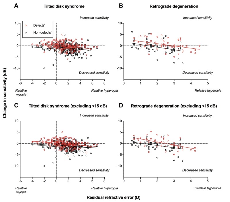
Results: A smaller residual refractive error after the application of negative addition lenses correlated with improvement in visual field defects in terms of sensitivity and probability scores in patients with tilted disc syndrome. Patients with established neurological deficits (retrograde degeneration) showed improvement in sensitivities but not in probability scores.

Conclusions: Neutralizing the refractive error at the region of posterior retinal bowing due to tilted disc syndrome reduces the apparent visual field defect. This may be a useful and rapid test to help differentiate between tilted disc syndrome and other pathological causes of visual field defects such as neurological deficits.


Conflict of interest statement

Conflict of Interest Disclosure: None of the authors have reported a financial conflict of interest.

Figures

FIGURE 1
FIGURE 1
Theoretical framework behind the use of a negative addition lens in tilted disc syndrome. The yellow dashed lines indicate a circular arc. In (A), a patient with tilted disc syndrome has posterior bowing of the retina (blue arrow). A negative addition lens can focus rays of light more appropriately at the region of posterior retinal bowing by diverging rays of light. A patient with generally regular retinal curvature is shown in (B). The green arrows indicate regions with near normal curvature and/or slight anterior shift (relative hyperopic defocus). The fundus photographs of the corresponding representative patients are shown directly below. In (C), the optic nerve head displays tilt and torsion, with situs inversus of the adjacent retinal blood vessels typical of tilted disc syndrome. In (D), the optic nerve head is fairly oval in appearance, without tilt or torsion.
FIGURE 2
FIGURE 2
(A) Visual field results for a patient with tilted disc syndrome. Baseline visual field results show a cluster of visual field depressions primarily located temporally around the blind spot. Sequential negative addition lenses (−1.00, −2.00, and −3.00 D NAL) reduce the significance of the defects. (B) Visual field results for a patient with retrograde degeneration due to a neurological deficit. Baseline visual field shows a primarily inferotemporal quadrantonopia. Note the presence of defects that were flagged by the Humphrey Field Analyzer as highly significant (P < .005). Use of negative addition lenses did not appear to change the appearance of the visual field defect.
FIGURE 3
FIGURE 3
Difference in sensitivity (dB) as a function of visual field eccentricity (°) for a patient with tilted disc syndrome (A) and a patient with a neurological deficit and retrograde degeneration (B). A positive difference in sensitivity indicated improvement, and a negative difference indicated worsening. A positive visual field eccentricity was considered temporal, and negative eccentricity was considered nasal. Each color denotes a different negative addition lens. Datum points indicate mean, and the error bars indicate 1 standard error of the mean.
FIGURE 4
FIGURE 4
Change in sensitivity (dB) as a function of residual refractive error (D) for patients with tilted disc syndrome (A) and retrograde degeneration due to a neurological deficit (B). A positive change in sensitivity indicates improvement in sensitivity, and a negative change indicates worsening. A positive residual refractive error indicates relative hyperopia, and a negative refractive error indicates myopia. Defects (locations with a baseline pattern deviation P < .05) are shown in red, and nondefects (all other points with a pattern deviation result of P > .05) are shown in black. Note that all patients with retrograde degeneration only exhibited relative hyperopia, so the x axis begins at 0. Each datum point represents an individual test location along the horizontal meridian (i.e., one point from the four lines about the midline). The solid lines indicate the linear regression results, analyzed separately for relative hyperopia and relative myopia. (C) and (D) represent the results of patients with tilted disc syndrome and retrograde degeneration as per (A) and (B), respectively, but with results with a baseline sensitivity of less than 19 dB excluded from analysis. Note the similarity between (A) and (C) owing to the small number of censored points.
FIGURE 5
FIGURE 5
Individual differences in P score as a function of residual relative refraction (D), with color codes and x axis as per Fig. 4 (results for patients with tilted disc syndrome in (A) and patients with retrograde degeneration due to neurological deficits in (B)). A positive difference in P score indicates a decreased probability of abnormality on the Humphrey Field Analyzer deviation map (i.e., more normal), and a negative difference indicates an increased probability of abnormality (i.e., more abnormal). (C) and (D) represent the results of patients with tilted disc syndrome and retrograde degeneration as per (A) and (B), respectively, but with results with a baseline sensitivity of less than 19 dB excluded from analysis. Note the similarity between (A) and (C) owing to the small number of censored points.
FIGURE 6
FIGURE 6
Spectralis optical coherence tomography results showing the structural correlate of visual field defects in three representative patients. In (A), the patient with tilted disc syndrome has clear posterior bowing with intact inner and outer retinal layers nasally (orange arrow), corresponding to the refractive scotoma in the temporal visual field (orange box). These defects tend to be shallow. In (B), the patient with a neurological deficit and manifest retrograde degeneration shows clear thinning of the inner retinal layers (retinal nerve fiber layer and ganglion cell layer, green arrows) temporal to the fovea corresponding the clear, deep inferonasal visual field defect (green box). In (C), a patient with pathological myopia (−11.00 D) has a posterior staphyloma within the central retina, with thinning of the inner retinal layers, similar to (B), temporal to the fovea (red arrows). This corresponds to the inferonasal visual field defect (red box), which appears deeper than the defect caused by posterior retinal bowing in tilted disc syndrome (A).
None

Similar articles

Cited by

References

    1. Witmer MT, Margo CE, Drucker M. Tilted Optic Disks. Surv Ophthalmol 2010;55:403–28. - PubMed
    1. How AC, Tan GS, Chan YH, et al. Population Prevalence of Tilted and Torted Optic Discs among an Adult Chinese Population in Singapore: The Tanjong Pagar Study. Arch Ophthalmol 2009;127:894–9. - PubMed
    1. Vongphanit J, Mitchell P, Wang JJ. Population Prevalence of Tilted Optic Disks and the Relationship of This Sign to Refractive Error. Am J Ophthalmol 2002;133:679–85. - PubMed
    1. You QS, Xu L, Jonas JB. Tilted Optic Discs: The Beijing Eye Study. Eye (Lond) 2008;22:728–9. - PubMed
    1. Apple DJ, Rabb MF, Walsh PM. Congenital Anomalies of the Optic Disc. Surv Ophthalmol 1982;27:3–41. - PubMed

Publication types