. 2019 Feb;160(2):375-384.
doi: 10.1097/j.pain.0000000000001405.

Contribution of dorsal root ganglion octamer transcription factor 1 to neuropathic pain after peripheral nerve injury

Affiliations

Contribution of dorsal root ganglion octamer transcription factor 1 to neuropathic pain after peripheral nerve injury

Jingjing Yuan et al. Pain. 2019 Feb.

Abstract

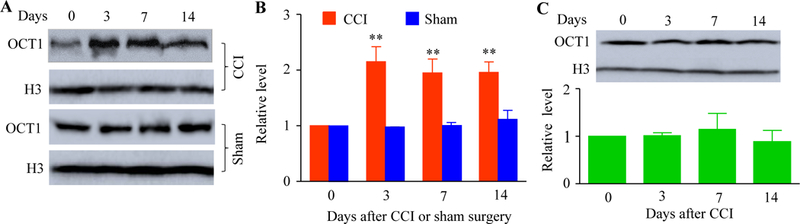
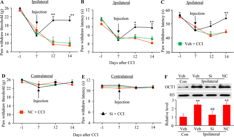
Neuropathic pain genesis is related to gene alterations in the dorsal root ganglion (DRG) after peripheral nerve injury. Transcription factors control gene expression. In this study, we investigated whether octamer transcription factor 1 (OCT1), a transcription factor, contributed to neuropathic pain caused by chronic constriction injury (CCI) of the sciatic nerve. Chronic constriction injury produced a time-dependent increase in the level of OCT1 protein in the ipsilateral L4/5 DRG, but not in the spinal cord. Blocking this increase through microinjection of OCT1 siRNA into the ipsilateral L4/5 DRG attenuated the initiation and maintenance of CCI-induced mechanical allodynia, heat hyperalgesia, and cold allodynia and improved morphine analgesia after CCI, without affecting basal responses to acute mechanical, heat, and cold stimuli as well as locomotor functions. Mimicking this increase through microinjection of recombinant adeno-associated virus 5 harboring full-length OCT1 into the unilateral L4/5 DRG led to marked mechanical allodynia, heat hyperalgesia, and cold allodynia in naive rats. Mechanistically, OCT1 participated in CCI-induced increases in Dnmt3a mRNA and its protein and DNMT3a-mediated decreases in Oprm1 and Kcna2 mRNAs and their proteins in the injured DRG. These findings indicate that OCT1 may participate in neuropathic pain at least in part by transcriptionally activating Dnmt3a and subsequently epigenetic silencing of Oprm1 and Kcan2 in the DRG. OCT1 may serve as a potential target for therapeutic treatments against neuropathic pain.


Conflict of interest statement

Conflict of interest statement

The authors have no conflicts of interest to declare.

Figures

Fig. 1.
Fig. 1.
Peripheral nerve injury-caused increase in the OCT1 expression in the injured DRG. (A and B) The amounts of OCT1 protein in the ipsilateral L4/5 DRG at the different days post-CCI or sham surgery. A: Representative Western blots. B: A statistical summry of the densitometric analysis. n = 3 biological replicates (6 rats) per time point. Two-way ANOVA followed by post hoc Tukey test. **P < 0.01 versus the corresponding naïve group (0 day). (C) OCT1 protein expression in the ipsilateral L4/5 spinal cord at the different days after CCI. n = 3 biological replicates (6 rats) per time point. One-way ANOVA followed by post hoc Tukey test.
Fig. 2.
Fig. 2.
Immunohistochemistry for OCT1 in the DRG. (A abd B) OCT1 is colocalized with NeuN (A) and glutamine synthetase (GS; Arrows; B) in the individual neurons of naïve DRG. Scale bar: 50 μm. (C) Distribution of OCT1-labeled neuronal somata in naïve DRG. Large, 40%; medium: 44.8%; small: 21.2%. (D-F) OCT1 is co-expressed with NF200 (D), IB4 (E), and CGRP (F) in the individual neurons of naïve DRG. Scale bar: 25 μm. (G and H) The cells labeled by OCT1 in the ipsilateral and contralalteral L5 DRG on day 7 post-CCI. G: Representative immunostaining images. H: A statistical summary of the analysi in number of OCT1-labeled cells. n = 3 rats. *P < 0.05 versus the contralateral side by two-tailed unpaired Student’s t-test.
Fig. 3.
Fig. 3.
Effect of DRG pre-microinjection of OCT1 siRNA on CCI-caused induction of pain hypersensitivities and dorsal horn central sensitization. (A and B) Effect of pre-microinjection of OCT1 siRNA (Si), vehicle (Veh), or negative control siRNA (NC) into the ipsilateral L4/5 DRG on the expression of Oct1 mRNA (A) and OCT1 protein (B) in the ipsilateral L4/5 DRG on day 5 after CCI or sham surgery. n = 3 biological replicates (6 rats) per group. One-way ANOVA followed by post hoc Tukey test. *P < 0.05 or **P < 0.01 versus the vehicle (Veh) plus sham group. #P < 0.05 versus the vehicle plus CCI group. (C-G) Effect of pre-micoinjection of OCT1 siRNA (Si), vehicle (Veh), or negative control siRNA (NC) into the ipsilateral L4/5 DRG on paw withdrawal threshold to mechanical stimulation (C and F), paw withdrawal latency to heat stimulation (D and G) and paw withdrawal latency to cold stimulation (E) on the ipsilateral (C-E) and contralateral (F and G) sides at the different days after CCI or sham surgery. n = 5 rats per group. Two-way ANOVA followed by post hoc Tukey test. **P < 0.01 versus the vehicle plus CCI group at the corresponding time point. (H) Effect of pre-microinjection of OCT1 siRNA (Si) or vehicle (Veh) into the ipsilateral L4/5 DRG on CCI-caused increases in the phosphorylation of ERK1/2 and expression of GFAP in the ipsilateral L4/5 dorsal horn on day 5 after CCI or sham surgery. Left: Represetaitive Western blots; Right: A statistical summary of densitometric analysis. n = 3 biological replicates (6 rats) per group. One-way ANOVA followed by post hoc Tukey test. *P < 0.05 or **P < 0.01 versus the corresponding vehicle plus sham group. ##P < 0.01 versus the corresponding vehicle plus CCI group.
Fig. 4.
Fig. 4.
Effect of DRG post-microinjection of OCT1 siRNA on CCI-caused maintenance of pain hypersensitivities. (A-E) Effect of DRG microinjection of OCT1 siRNA (Si), vehicle (Veh) or negative control siRNA (NC) into the ipsilateral L4/5 DRG starting on day 7 after CCI on paw withdrawal threshold to mechanical stimulation (A and D), paw withdrawal latency to heat stimulatuon (B and E), and paw withdrawal latency to cold stimulation (C) on the ipsilateral (A-C) and contralateral (D and E) sides. n = 5 rats per group. Two-way ANOVA followed by post hoc Tukey test. *P < 0.05 or **P < 0.01 versus the vehicle plus CCI group at the corresponding time point. (F) Effect of DRG microinjection of OCT1 siRNA (Si), vehicle (Veh) or negative control siRNA (NC) into the ipsilateral L4/5 DRG starting on day 7 after CCI on the expression of OCT1 protein in the ipsilateral and contralaterl (Con) L4/5 DRG on day 14 after CCI. n = 3 biological replicates (6 rats) per group. One-way ANOVA followed by post hoc Tukey test. **P < 0.01 versus the vehicle (Veh) plus CCI group on the contralateral side. ##P < 0.01 versus the vehicle plus CCI group on the ipsilateral side.
Fig.5.
Fig.5.
Effect of DRG OCT1 overexpression on nocicpetive thereshold in naive rats. (A and B) Oct1 mRNA (A) and OCT1 protein (B) expression in the injected L4/5 DRG 8 weeks after microinjection of AAV5-OCT1 or control AAV5-EGFP into unilateral L4/5 DRG. n = 3 biological replicates (3 rats) per group. **P < 0.01 versus the AAV5-EGFP group by two-tailed unpaired Student’s t-test. (C-G) Effect of micorinjection of AAV5-OCT1 or AAV5-EGFP into the unilateral L4/5 DRG on paw withdrawal threshold to mechanical stimulation (C and F), paw withdrawal latency to heat stimulation (D and G), and paw withdrawal latency to cold stimulation (E) on the ipsilateral (C-E) and controlateral (F and G) sides at the differenct weeks after microinjection. n = 5 rats/group. Two-way ANOVA followed by post hoc Tukey test. *P < 0.05 or **P < 0.01 versus the AAV5-EGFP group at the corrsponding time point.
Fig. 6.
Fig. 6.
OCT1 transcriptionally activates Dnmt3a gene and represses DNMT3a-mediated MOR and Kv1.2 expression in the DRG. (A and B) The expression of Dnmt3a, Oprm1, and Kcna2 mRNA (A) and protein (B) in the ipsilateral L4/5 DRG on day 5 after sham surgery or CCI in the rats pre-microinjected with vehicle (Veh), negative control siRNA (NC), or OCT1 siRNA into the unilateral L4/5 DRG. n = 3–6 biological replicates (3–6 rats) per group. One-way ANOVA followed by post hoc Tukey test. *P < 0.05 or **P < 0.01 versus the corresponding vehicle plus sham group. #P < 0.05 or ##P < 0.01 versus the corresponding vehicle plus CCI group. (C and D) The expression of Dnmt3a, Oprm1 and Kcna2 mRNA (C) and protein (D) in the injected L4/5 DRG 8 weeks after microinjection of AAV5-EGFP or AAV5-OCT1 into the unilateral L4/5 DRG. n = 3–5 biological replicates (3–5 rats) per group. *P < 0.05 or **P < 0.01 versus the corresponding AAV5-EGFP group by two-tailed unpaired Student’s t-test. (E) Colocalization of OCT1 with MOR and Kv1.2 Double-label immunofluorescent staining of OCT1 (red) with MOR (green) or Kv1.2 (green) in the DRG neurons of naïve rats. Scale bar: 50 μm.
Fig. 7.
Fig. 7.
Blocking CCI-caused increase in OCT1 in the injured DRG improves MOR-mediated analgesia under CCI-caused neuropathic pain conditions. (A) Effect of pre-microinjection of OCT1 siRNA or vehicle (Veh) into the unilateral L4/5 DRG on morphine analgesia on the ipsilateral and contralateral sides 3 days after CCI or sham surgery. n = 5 rats/group. Two-way ANOVA followed by post hoc Tukey test. **P < 0.01 versus the correspondung vehicle plus sham group. #P < 0.05 versus the correspondung vehicle plus CCI group. (B and C) Effect of intraperitoneal injection of methylnatrexone (M) on the OCT1 siRNA-produced antinociceptive effect 4 days post-CCI or sham surgery on the ipsilateral side. n = 5 rats/group. Two-way ANOVA followed by post hoc Tukey test. **P < 0.01 versus the vehicle plus sham group at the corresponding time point. #P < 0.05 versus the vehicle plus CCI group at the corresponding time point.

Similar articles

Cited by

References

    1. Campbell JN, Meyer RA. Mechanisms of neuropathic pain. Neuron 2006;52:77–92. - PMC - PubMed
    1. Costigan M, Scholz J, Woolf CJ. Neuropathic pain: a maladaptive response of the nervous system to damage. Annu Rev Neurosci 2009;32:1–32. - PMC - PubMed
    1. Dixon WJ. Efficient analysis of experimental observations. Annu Rev Pharmacol Toxicol 1980;20:441–62. - PubMed
    1. Fan L, Guan X, Wang W, Zhao JY, Zhang H, Tiwari V, Hoffman PN, Li M, Tao YX. Impaired neuropathic pain and preserved acute pain in rats overexpressing voltage-gated potassium channel subunit Kv1.2 in primary afferent neurons. Mol Pain 2014;10:8. - PMC - PubMed
    1. Kehlet H, Jensen TS, Woolf CJ. Persistent postsurgical pain: risk factors and prevention. Lancet 2006;367:1618–25. - PubMed

MeSH terms