. 2019 Feb 1;100(2):547-560.
doi: 10.1093/biolre/ioy200.

Sources of all-trans retinal oxidation independent of the aldehyde dehydrogenase 1A isozymes exist in the postnatal testis†

Affiliations

Sources of all-trans retinal oxidation independent of the aldehyde dehydrogenase 1A isozymes exist in the postnatal testis†

My-Thanh Beedle et al. Biol Reprod. .

Abstract

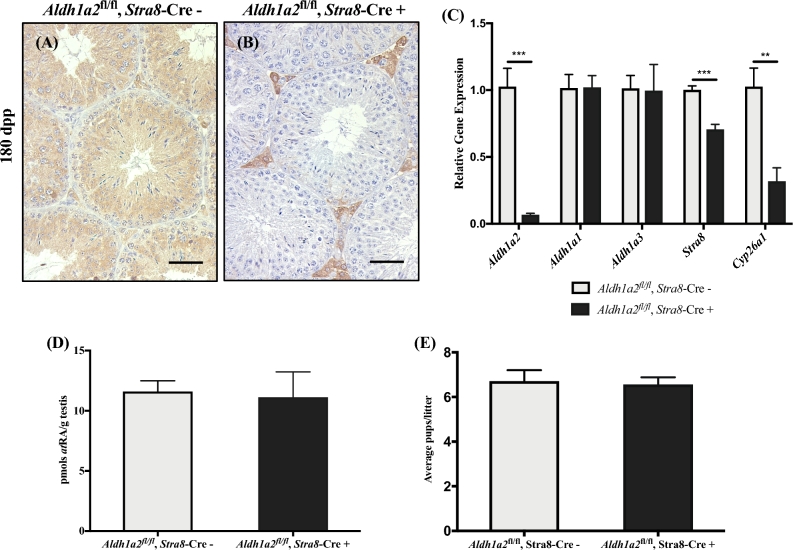
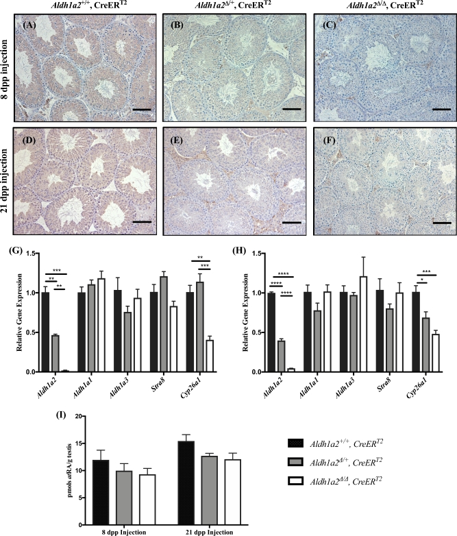
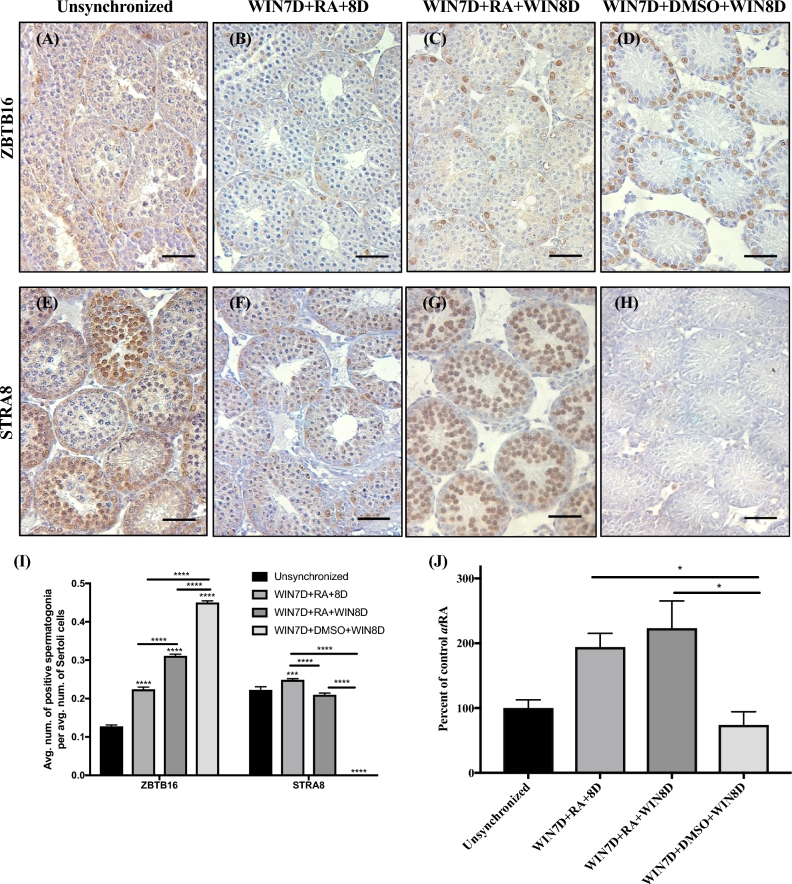
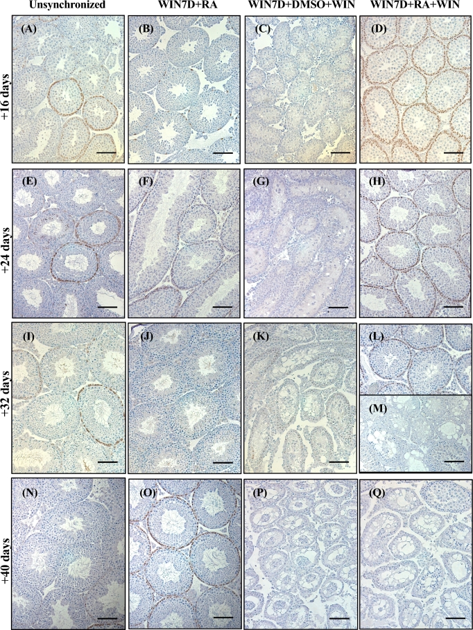
Despite the essential role of the active metabolite of vitamin A, all-trans retinoic acid (atRA) in spermatogenesis, the enzymes, and cellular populations responsible for its synthesis in the postnatal testis remain largely unknown. The aldehyde dehydrogenase 1A (ALDH1A) family of enzymes residing within Sertoli cells is responsible for the synthesis of atRA, driving the first round of spermatogenesis. Those studies also revealed that the atRA required to drive subsequent rounds of spermatogenesis is possibly derived from the ALDH1A enzymes residing within the meiotic and post-meiotic germ cells. Three ALDH1A isozymes (ALDH1A1, ALDH1A2, and ALDH1A3) are present in the testis. Although, ALDH1A1 is expressed in adult Sertoli cells and is suggested to contribute to the atRA required for the pre-meiotic transitions, ALDH1A2 is proposed to be the essential isomer involved in testicular atRA biosynthesis. In this report, we first examine the requirement for ALDH1A2 via the generation and analysis of a conditional Aldh1a2 germ cell knockout and a tamoxifen-induced Aldh1a2 knockout model. We then utilized the pan-ALDH1A inhibitor (WIN 18446) to test the collective contribution of the ALDH1A enzymes to atRA biosynthesis following the first round of spermatogenesis. Collectively, our data provide the first in vivo evidence demonstrating that animals severely deficient in ALDH1A2 postnatally proceed normally through spermatogenesis. Our studies with a pan-ALDH1A inhibitor (WIN 18446) also suggest that an alternative source of atRA biosynthesis independent of the ALDH1A enzymes becomes available to maintain atRA levels for several spermatogenic cycles following an initial atRA injection.

Keywords: Aldh1A; Aldh1a2; WIN 18,446; retinoic acid; spermatogenesis; spermatogonia; testis.


Figures

Figure 1.
Figure 1.
Elimination of Aldh1a2 using the Stra8-Cre. Control (Aldh1a2fl/fl, Stra8-Cre–) and cKO (Aldh1a2fl/fl, Stra8-Cre+) animals analyzed at 180 dpp. Representative control (A) and cKO (B) cross-section stained for ALDH1A2. Immunopositive cells are indicated by brown precipitate. (C) qRT-PCR analysis of Aldh1a2, Aldh1a1, Aldh1a3, Stra8, and Cyp26a1. (D) Graphical representation of atRA measurements. (E) Graphical representation of the average number of pups per litter. Scale bars = 100 μM. n = 3–4. * P < 0.05, ** P < 0.01, and *** P < 0.001.
Figure 2.
Figure 2.
Elimination of Aldh1a2 using the inducible CreERT2. Analysis of Aldh1a2+/+, CreERT2; Aldh1a2/+, CreERT2; and Aldh1a2▵/▵, CreERT2 animals injected with tamoxifen. Representative immunohistochemistry from animals injected at 8 (A, B, and C; 68 dpp at euthanasia) and 21 dpp (D, E and F; 81 dpp at euthanasia). Cross-sections are stained for ALDH1A2 protein and immunopositive cells are indicated by brown precipitate. qRT-PCR analysis was performed to determine the relative expression of Aldh1a2, Aldh1a1, Aldh1a3, Stra8, and Cyp26a1 in animals injected with tamoxifen at 8 (G) and 21 dpp (H). (I) Graphical representation of atRA measurements. Scale bars = 100 μm. n = 3–7. *P < 0.05, **P < 0.01, ***P < 0.001, and ****P < 0.0001.
Figure 3.
Figure 3.
Maintenance on WIN7D + RA + WIN8D increases the number of ZBTB16-positive spermatogonia but does not decrease the number of STRA8-positive spermatogonia or atRA levels. Immunostaining of testis sections from unsynchronized (A and E), WIN7D + RA + 8D (B and F), WIN7D + RA + WIN8D (C and G), and WIN7D + DMSO + WIN8D treated mice (D and H). Testis sections were immunostained for ZBTB16 (AD) and STRA8 (EH) protein. For each treatment group, immmunopositive cells are denoted by brown precipitate. (I) Graphical representation of ZBTB16 and STRA8 counts. (J) Graphical representation of atRA measurements reported as percent of control. Asterisks represent statistical difference of a treatment group compared to control. Black lines represent statistical significance between treatment groups. Animals were 17 dpp at euthanasia. Scale bar = 100 μM. n = 3–4. *P < 0.05, ***P < 0.001, ****P < 0.0001.
Figure 4.
Figure 4.
Maintenance on WIN for several spermatogenic cycles eventually results in degenerative seminiferous tubules. Immunostaining of testis sections from unsynchronized (A, E, I, and N), WIN7D + RA (B, F, J, and O), WIN7D + DMSO + WIN (C, G, K, and P), and WIN7D + RA + WIN (D, H, L, M, and Q) treated mice for 16, 24, 32, and 40 additional maintenance days. Testis sections were immunostained for STRA8. Immunopositive cells are indicated by brown precipitate. Scale bar = 100 μM. n = 3–4.
Figure 5.
Figure 5.
WIN7D + RA + HYD8D animals do not show any spermatogenic defects, although WIN7D + RA + WIN/HYD8D animals show an increased number of ZBTB16-positive spermatogonia. Immunostaining of testis sections for ZBTB16 and STRA8 for WIN7D + RA + HYD8D (A and B) and WIN7D + RA + WIN/HYD8D treated mice (C and D). Graphical representation of the average number of ZBTB16- (E) and STRA8-positive (F) spermatogonia per tubule within each treatment group. Asterisks represent statistical difference of a treatment group compared to control. Black lines indicate statistical significance between treatment groups. Animals were 17 dpp at euthanasia. Scale bar = 100 μM. n = 3. *P < 0.05, **P < 0.01, ****P < 0.0001.
Figure 6.
Figure 6.
WIN7D + RA + WIN/HYD8D animals have an increased number of tubules with abnormal or missing layers of GCNA-positive germ cells. Representative testis sections from WIN7D + RA + 8D (A), WIN7D + RA + HYD8D (B), WIN7D + RA + WIN8D (C), and WIN7D + RA + WIN/HYD8D (D) treated animals immunostained for GCNA. Immunopositive cells are indicated by brown precipitate. (E) Graphical representation of the number of tubules from WIN7D + RA + 8D, WIN7D + RA + HYD8D, WIN7D + RA + WIN8D, and WIN7D + RA + WIN/HYD8D treated testes containing normal GCNA-positive germ cell layers. Asterisks represent statistical difference of a treatment group compared to control. Black lines represent statistical significance between treatment groups. Animals were 17 dpp at euthanasia. n = 3–4. *P < 0.05.

References

    1. Yang QE, Oatley JM. Spermatogonial stem cell functions in physiological and pathological conditions. Curr Top Dev Biol 2014; 107:235–267. - PubMed
    1. Busada JT, Geyer CB. The role of retinoic acid (RA) in spermatogonial differentiation. Biol Reprod 2016; 94:10. - PMC - PubMed
    1. Drumond AL, Meistrich ML, Chiarini-Garcia H. Spermatogonial morphology and kinetics during testis development in mice: a high-resolution light microscopy approach. Reproduction 2011; 142:145–155. - PubMed
    1. Niedenberger BA, Busada JT, Geyer CB. Marker expression reveals heterogeneity of spermatogonia in the neonatal mouse testis. Reproduction 2015; 149:329–338. - PMC - PubMed
    1. Oakberg EF. A description of spermiogenesis in the mouse and its use in analysis of the cycle of the seminiferous epithelium and germ cell renewal. Am J Anat 1956; 99:391–413. - PubMed

Publication types