Review
. 2018 Sep 22;218(suppl_2):S68-S74.
doi: 10.1093/infdis/jiy403.

Immunobiology of Varicella-Zoster Virus Infection

Affiliations
Review

Immunobiology of Varicella-Zoster Virus Infection

Kerry J Laing et al. J Infect Dis. .

Erratum in

  • Erratum.
    [No authors listed] [No authors listed] J Infect Dis. 2019 Apr 16;219(9):1514. doi: 10.1093/infdis/jiy693. J Infect Dis. 2019. PMID: 30535082 Free PMC article. No abstract available.

Abstract

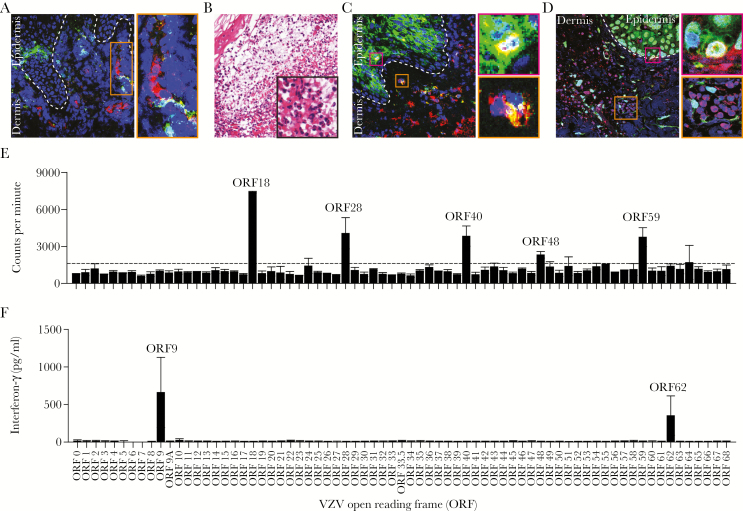
Varicella-zoster virus (VZV) causes clinically significant illness during acute and recurrent infection accompanied by robust innate and acquired immune responses. Innate immune cells in skin and ganglion secrete type I interferon (IFN-I) and proinflammatory cytokines to control VZV. Varicella-zoster virus subverts pattern recognition receptor sensing to modulate antigen presentation and IFN-I production. During primary infection, VZV hijacks T cells to disseminate to the skin and establishes latency in ganglia. Durable T- and B-cell memory formed within a few weeks of infection is boosted by reactivation or re-exposure. Antigen-specific T cells are recruited and potentially retained in VZV-infected skin to counteract reactivation. In latently VZV-infected ganglia, however, virus-specific T cells have not been recovered, suggesting that local innate immune responses control VZV latency. Antibodies prevent primary VZV infection, whereas T cells are fundamental to resolving disease, limiting severity, and preventing reactivation. In this study, we review current knowledge on the interactions between VZV and the human immune system.


Figures

Figure 1.
Figure 1.
Immunopathology of herpes zoster lesions and systemic virus-specific T-cell immune responses in latently varicella-zoster virus (VZV)-infected adults. (A–D) Sections of formalin-fixed and paraffin-embedded skin biopsy obtained from a herpes zoster patient were analyzed by immunofluorescence ([IF] A, C, and D) or stained with hematoxylin and eosin (B). (A) Dual IF staining for VZV glycoprotein E ([gE] green) and CD123 (plasmacytoid dendritic cell [pDC]; red) reveals dermal pDC adjacent to VZV-infected cells. Inset: magnification of indicated area. (B) Hematoxylin and eosin staining showing polymorphonuclear cell infiltrates in a zoster vesicle. Magnification: ×100; inset: ×400. (C) Dual IF staining for VZV gE (green) and CD68 (macrophages; red) showing high numbers of dermal macrophages adjacent to VZV-infected epidermis. Insets: magnification of the indicated areas showing VZV gE-positive epidermal (pink inset) and dermal (orange inset) macrophages. (D) Dual IF staining for VZV open reading frame (ORF)63 (green) and CD3 (T cells; red) showing dermal T-cell infiltrates in proximity to VZV-infected cells. Pink inset: magnification showing VZV ORF63-positive T cell in epidermis. Orange inset: magnification showing dermal T cells adjacent to VZV ORF63-positive cells. (A, C, and D) Nuclei were counterstained with 4’,6-diamidino-2-phenylindole (blue), and dashed lines indicate epidermal—dermal junction and images are shown at ×200 magnification. (E–F) Bulk in vitro VZV-enriched CD4 (E) and CD8 (F) T-cell lines (TCLs) obtained from different representative latently VZV-infected healthy adults were assayed for reactivity to proteins encoded by individual VZV ORFs (indicated on the x-axis). (E) CD4 T-cell reactivity was determined using [3H]-thymidine incorporation, as described previously (Supplementary Data Ref. 77). Bars and error bars indicate mean ± standard deviation of duplicate measurements. Dashed horizontal line indicates threshold for positivity [(median of all negative control antigens) + (2.33 times median absolute deviation of negative control antigens)]. Both duplicates needed to exceed the threshold for the ORF to be determined as positive. (F) Interferon-γ secretion levels determined by enzyme-linked immunosorbent assay in conditioned medium of CD8 TCL cocultured with Cos-7 cells cotransfected with the responsive human leukocyte antigen (HLA)-I allele (ie, HLA-B*0802) and the individual VZV ORFs, as described previously (Supplementary Data Ref. 76). The VZV ORFs specifically recognized by T cells in the individuals analyzed are indicated.

References

    1. Gershon AA, Breuer J, Cohen JI, et al. . Varicella zoster virus infection. Nat Rev Dis Primers 2015; 1:15016. - PMC - PubMed
    1. Gilden DH, Kleinschmidt-DeMasters BK, LaGuardia JJ, Mahalingam R, Cohrs RJ. Neurologic complications of the reactivation of varicella-zoster virus. N Engl J Med 2000; 342:635–45. - PubMed
    1. Liesegang TJ. Herpes zoster ophthalmicus natural history, risk factors, clinical presentation, and morbidity. Ophthalmology 2008; 115:S3–12. - PubMed
    1. Zerboni L, Sen N, Oliver SL, Arvin AM. Molecular mechanisms of varicella zoster virus pathogenesis. Nat Rev Microbiol 2014; 12:197–210. - PMC - PubMed
    1. Ouwendijk WJ, Mahalingam R, de Swart RL, et al. . T-cell tropism of simian varicella virus during primary infection. PLoS Pathog 2013; 9:e1003368. - PMC - PubMed

Publication types

MeSH terms