. 2018 Sep 24;14(9):e1007308.
doi: 10.1371/journal.ppat.1007308. eCollection 2018 Sep.

NLRP6 negatively regulates pulmonary host defense in Gram-positive bacterial infection through modulating neutrophil recruitment and function

Affiliations

NLRP6 negatively regulates pulmonary host defense in Gram-positive bacterial infection through modulating neutrophil recruitment and function

Laxman Ghimire et al. PLoS Pathog. .

Abstract

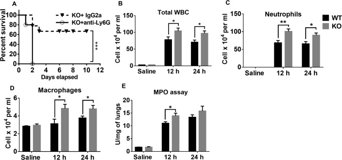
Gram-positive bacteria, including Staphylococcus aureus are endemic in the U.S., which cause life-threatening necrotizing pneumonia. Neutrophils are known to be critical for clearance of S. aureus infection from the lungs and extrapulmonary organs. Therefore, we investigated whether the NLRP6 inflammasome regulates neutrophil-dependent host immunity during pulmonary S. aureus infection. Unlike their wild-type (WT) counterparts, NLRP6 knockout (KO) mice were protected against pulmonary S. aureus infection as evidenced by their higher survival rate and lower bacterial burden in the lungs and extrapulmonary organs. In addition, NLRP6 KO mice displayed increased neutrophil recruitment following infection, and when neutrophils were depleted the protective effect was lost. Furthermore, neutrophils from the KO mice demonstrated enhanced intracellular bacterial killing and increased NADPH oxidase-dependent ROS production. Intriguingly, we found higher NK cell-mediated IFN-γ production in KO mouse lungs, and treatment with IFN-γ was found to enhance the bactericidal ability of WT and KO neutrophils. The NLRP6 KO mice also displayed decreased pyroptosis and necroptosis in the lungs following infection. Blocking of pyroptosis and necroptosis in WT mice resulted in increased survival, reduced bacterial burden in the lungs, and attenuated cytokine production. Taken together, these novel findings show that NLRP6 serves as a negative regulator of neutrophil-mediated host defense during Gram-positive bacterial infection in the lungs through regulating both neutrophil influx and function. These results also suggest that blocking NLRP6 to augment neutrophil-associated bacterial clearance should be considered as a potential therapeutic intervention strategy for treatment of S. aureus pneumonia.


Conflict of interest statement

The authors have declared that no competing interests exist.

Figures

Fig 1
Fig 1. Upregulation and activation of NLRP6.
(A) Lungs tissue sections from healthy controls and pneumonic patients were stained with antibodies against neutrophils (lipocalin-2+), macrophages (CD68+), and epithelial (CD326+) cells. NLRP6 is shown in red, neutrophils, macrophages, and epithelial cell markers are in green, and DAPI is blue. NLRP6-positive cells are indicated by white arrowheads. (B) Immunofluorescence microscopy was performed on lung sections from healthy controls and S. aureus-infected mice to assess expression of NLRP6. Neutrophils (Ly6G+), macrophages (F4/80+), and epithelial cells (CD 326+) are stained green and NLRP6 is red. NLRP6-positive cells are indicated by white arrowheads. (C) NLRP6 expression was analyzed by western blot in lysates of human healthy control tissue, pneumonic lung tissue, and HL-60 (human neutrophil-like) cells infected with S. aureus (MOI10) for 4 hours. (D) THP-1 (human monocytic) cells were infected with S. aureus (MOI 20) or purified α-hemolysin (hla) for 8 hours and expression of NLRP6 was assessed by western blotting. (E, F) Bone marrow-derived macrophages (BMDMs) were infected with S. aureus (MOI: 20) or hla from S. aureus (50 μg/ml) for 8 hours (E). Expression of NLRP6 protein was analyzed by immunoblotting. (F) Caspase-1 processing by the NLRP6 inflammasome in response to S. aureus or hla in BMDMs. (G) IL-1β secretion by WT and NLRP6 KO BMDMs following S. aureus infection. (H) IL-1β secretion by WT and NLRP6 KO mice after S. aureus (5 X 10⁷ CFU/mouse) infection (N = 6-8/group). (I) BMDMs from WT mice were infected with S. aureus (MOI: 50) and stained for NLRP6 (red) and ASC (green). The white arrowheads demonstrate co-localization of NRP6 and ASC. (J) BMDMs from WT mice were infected with S. aureus (MOI: 50) and stained for NLRP6 (red) and caspase-1 (green). The white arrowheads show co-localization of NRP6 and caspase-1. The graphs show the mean ± SEM of three independent experiments. The images shown are the representative of five different fields from three independent experiments. Magnification: 40X. SA: S. aureus, hla: α-hemolysin. *, p<0.05, **, p<0.01, and *** p<0.001.
Fig 2
Fig 2. NLRP6-/- mice are resistant to pulmonary S. aureus infection.
(A) WT, NLRP6 KO, and ASC KO mice (N = 13-15/group) were infected intratracheally with a lethal inoculum of S. aureus (USA 300) (2X10⁸ CFU/mouse) and survival was then observed for 10 days. WT and NLRP6 KO mice (N = 5-8/group) were infected intratracheally with a sublethal inoculum of S. aureus (5 X 10⁷ CFU/mouse) and then were euthanized 12 or 24 h post-infection to quantitate the bacterial burden in (B) lungs, (C) BALF, and (D) liver. Total protein in BALF was measured (E). WT and KO mice (N = 6-8/group) were infected intratracheally with 5X10⁷ CFU/mouse of S. aureus (MSSA strain). Twenty-four-hours post-infection, the bacterial burden was measured in lungs (F) and BALF (G). WT and KO mice (N = 5/group) were co-housed for 4 weeks and infected with a sublethal dose of S. aureus. Mice were euthanized at 24 hours post-infection to estimate bacterial burden in the lungs (H) and BALF (I). Bone marrow chimeras were generated as described in the methods (N = 6-8/group) and then were infected with a sublethal dose of S. aureus. At 24-hours post-infection, mice were euthanized to estimate bacterial burden in the lungs (J) and BALF (K). *, p<0.05, **, p<0.01, and *** p<0.001.
Fig 3
Fig 3. Neutrophils confer host protection in NLRP6 KO mice.
(A) NLRP6 KO mice (N = 10-12/group) were treated either with anti-Ly6G antibody or IgG2a, 24 and 2 hours prior to infection with a lethal dose of S. aureus (USA300). Survival was monitored for 10 days. NLRP6 KO mice (N = 5-8/group) were infected intratracheally with a sublethal inoculum of S. aureus (5 X 10⁷ CFU/mouse). Mice were euthanized 12- or 24-hours post-infection to estimate total leukocytes (B), neutrophils (C), and macrophages (D) in the BALF. (E) Myeloperoxidase assay was performed in the lungs. *, p<0.05, **, p<0.01, and *** p<0.001.
Fig 4
Fig 4. NLRP6-/- neutrophils exhibit enhanced intracellular killing through increased IFN-γ and NADPH oxidase-dependent ROS production.
(A) BMDNs from WT and KO mice were infected with S. aureus (MOI 10) and the intracellular killing ability of neutrophils was measured at the indicated time points. (B) ROS production by BMDNs from WT and KO mice were compared after infection with S. aureus (MOI 1). (C) WT and KO mice (N = 6-8/group) were infected with a sublethal dose of S. aureus (5 X 10⁷ CFU/mouse). At designated time points, lungs were collected and processed for immunoblotting to compare the expression of NADPH oxidase enzyme components between WT and KO mice. (D) IFN-γ in BALF obtained from mice in 3C was measured. (E) Lungs obtained from S. aureus-infected WT and KO mice were homogenized and expressions of phospho-38, phospho-JNK, and phospho-STAT3 were measured through immunoblotting. (F) BMDNs from WT and KO mice were isolated and pre-treated with either IFN-γ or PBS for 30 minutes before infection with S. aureus (MOI 10). Two-hours post-infection, WT neutrophils were lysed and immunoblotted to measure the expression of NADPH oxidase enzyme components. (G) ROS production by neutrophils in 3F were measured as described in the methods. All figures are representative of three independent experiments. ROS: reactive oxygen species, RFU: relative florescence unit. WT: Wild-type, KO: NLRP6 KO. *, p<0.05, **, p<0.01, and *** p<0.001.
Fig 5
Fig 5. IFN-γ mediates bacterial clearance in NLRP6 KO mice during S. aureus-induced pneumonia.
KO mice (n = 6-8/group) were treated with either anti-IFN-γ antibody (100 μg/mouse, i.p.) or isotype control antibody 12 hours prior to infection with S. aureus (5 X 10⁷ CFU/mouse). WT mice were treated with equal volume of isotype control antibody 12 hours prior to infection with S. aureus (5 X 10⁷ CFU/mouse). Twelve-hours post-infection, mice were euthanized to collect lungs and BALF. Bacterial burden in the (A) lungs, and (B) BALF were compared. All figures are representative of three independent experiments. *, p<0.05, **, p<0.01, *** p<0.001.
Fig 6
Fig 6. NK cells and CD4+ T cells are the major sources of IFN-γ production during pulmonary S. aureus infection.
WT and KO mice (N = 6-8/group) were infected with S. aureus (5 X 10⁷ CFU/mouse). Twelve-hours post-infection, mice were euthanized to harvest lungs. Single cell suspensions obtained through digestion of lungs were treated with PMA/Ionomycin and GolgiStop for 5 hours and then stained for flow cytometry. (A) Representative zebra plot showing CD3- IFN-γ +NK 1.1+ cells. (B) Quantification of A. (C) Representative zebra plot showing CD3+ IFN-γ+ CD4+T cells. (D) Quantification of C. (E) WT and KO mice (N = 6-8/group) were infected with S. aureus (5 X 10⁷ CFU/mouse). Twenty-four hours post-infection, mice were euthanized to collect lungs tissues. Single cell suspensions obtained after lung digestion were stained to estimate NK and CD4 T cells using flowcytometry. The figures shown above are representatives of three independent experiments. *, p<0.05, **, p<0.01.
Fig 7
Fig 7. NLRP6-/- mice display reduced pyroptosis and necroptosis during S. aureus induced pneumonia.
WT and KO (N = 6-8/group/time point) mice were infected with a sublethal dose of S. aureus (5 X 10⁷ CFU/mouse). (A) LDH released in BALF, (B) IL-1α in BALF, and (C) HMGB1 in serum were measured. (D) BMDNs from WT and KO mice were isolated and pre-treated with either Ac-YVAD-CMK (100μg/ml), Nec-1s (300 μM), or an equivalent amount of DMSO for 30 minutes prior to infection with S. aureus (MOI 50). Cytotoxicity was measured 2 hours post-infection. (E) Expression of cleaved gasdermin-D in BMDMs obtained from WT and KO mice after infection with S. aureus for 8 hours. (F) BMDMs from WT and NLRP6 KO mice were infected with S. aureus (MOI 50) for 8 hours. Caspase-1 activation was observed through fluorescence microscopy. The upper panel shows WT and the lower panel shows KO macrophages. Caspase-1 positive cells are shown by white arrowheads. (G) BMDMs from WT and NLRP6 KO mice were infected with S. aureus as in F. Gasdermin-D positive cells were observed through fluorescence microscopy. The upper panel indicates WT and lower panel indicates KO macrophages after infection with S. aureus. (H) Western blots to show expression of phospho-MLKL, RIP3, RIP1, and cleaved caspase-8 in lungs homogenates obtained from S. aureus-infected mice. WT and KO mice (N = 6-8/group/time-point) were infected with a sublethal dose of S. aureus (5 X 10⁷ CFU/mouse, i.t). 12 and 24 h post-infection, mice were euthanized to collect lungs and processed for western blotting. (I) Fluorescent microscopy showing expression of molecules involved in necroptosis in macrophages after infection with S. aureus. BMDMs obtained from WT and KO mice were infected with S. aureus (MOI 50) for 8 hours and then stained for phospho-MLKL (red) and RIP3 (green) antibody. Necroptotic cells are represented as orange (indicated by white arrowheads). (J) Human pneumonic lungs showing necroptosis. Lungs tissue sections from healthy and pneumonic patients were deparaffinized and stained with phospho-MLKL, RIP3, and DAPI. Tissue undergoing necroptosis is shown by white arrowheads. Immunofluorescence pictures were taken at 40X. All figures are representative of three independent experiments. Ac-YVAD-CMK: caspase-1 inhibitor, Nec-1s: Necrostatin-1s, CL-GSMD: Cleaved Gasdermin-D, FL-GSMD: Full-length Gasdermin-D, WT: *, p<0.05, **, p<0.01, and *** p<0.001.
Fig 8
Fig 8. NLRP6-mediated pyroptosis is detrimental during pulmonary S. aureus infection.
WT and KO mice (N = 6-8/group) were administered either Ac-YVAD-CMK (100 μg/mouse) or an equal amount of DMSO 12 hours prior to infection with a sublethal dose of S. aureus. Caspase-1/11 double knock out mice (N = 6-8/group) received an equal amount of DMSO 12 hours prior to infection with a sublethal dose of S. aureus. Mice were euthanized 24 hours post-infection to collect BALF and lungs. (A) Total protein and (B) LDH release in the lungs, bacterial burden in (C) lungs and (D) BALF were measured. (E) IL-1β, (F) TNF-α, and (G) MCP-1 in the BALF were measured through standard ELISA procedure. (H) WT mice (N = 15/group) were treated with either Ac-YVAD-CMK (100 μg/mouse 12 hours prior to infection with S. aureus), or an equivalent amount of DMSO prior to infection with a lethal dose of S. aureus (2 X 10⁸ CFU/mouse, i.t.). Survival was monitored up to 80 hours post infection. Each figure is a representative of at least three independent experiments. Ac-YVAD-CMK: caspase-1 inhibitor, TNF-α: Tumor necrosis factor-α, MCP-1: Monocyte chemoattractant protein-1, *, p<0.05, **, p<0.01, *** p<0.001, and **** p<0.0001.
Fig 9
Fig 9. Blocking NLRP6-mediated necroptosis in WT mice improves host defense during S. aureus infection.
WT and KO mice (N = 6-8/group) were treated with either GW806742X (100 μl of 100 μM solution, i.p., 12 hours prior to infection) or an equivalent amount of DMSO prior to infection with a sublethal dose of S. aureus. Mice were euthanized 24 hours post infection to collect lungs and BALF. (A) Total protein and (B) LDH release in the BALF, bacterial burden in (C) lungs and (D) BALF were measured. (E) IL-1β, (F) TNF-α, and (G) MCP-1 in the cell-free BALF were measured. (H) Total leukocytes, (I) neutrophils, and (J) macrophages in the BALF was determined. (K) WT mice (N = 15/group) were treated with either Nec-1s (300 μg/mouse) or an equivalent amount of DMSO, 18 hours before and at the time of infection with S. aureus (2X10⁸ CFU/mouse). Survival was monitored for up to 80 hours. Each figure is representative of three independent experiments. GW806742X: MLKL inhibitor, Nec-1s: Necrostatin-1s. *, p<0.05, **, p<0.01, and *** p<0.001.

References

    1. World Health Organization. Pneumonia fact sheet [Updated 2016 Sep; accessed 2017 Dec]. Available from http://www.who.int/mediacentre/factsheets/fs331/en/.
    1. Moran GJ, Krishnadasan A, Gorwitz RJ, Fosheim GE, McDougal LK, Carey RB, et al. Methicillin-resistant S. aureus infections among patients in the emergency department. N Engl J Med. 2006;355(7):666–74. 10.1056/NEJMoa055356 . - DOI - PubMed
    1. Shahangian A, Chow EK, Tian X, Kang JR, Ghaffari A, Liu SY, et al. Type I IFNs mediate development of postinfluenza bacterial pneumonia in mice. J Clin Invest. 2009;119(7):1910–20. 10.1172/JCI35412 ; PubMed Central PMCID: PMCPMC2701856. - DOI - PMC - PubMed
    1. Rodvold KA, McConeghy KW. Methicillin-resistant Staphylococcus aureus therapy: past, present, and future. Clin Infect Dis. 2014;58 Suppl 1:S20–7. Epub 2013/12/18. 10.1093/cid/cit614 . - DOI - PubMed
    1. Parker D, Prince A. Immunopathogenesis of Staphylococcus aureus pulmonary infection. Semin Immunopathol. 2012;34(2):281–97. 10.1007/s00281-011-0291-7 ; PubMed Central PMCID: PMCPMC3577067. - DOI - PMC - PubMed

Publication types

MeSH terms

Substances