. 2018 Sep 21;19(10):2873.
doi: 10.3390/ijms19102873.

Melatonin Can Strengthen the Effect of Retinoic Acid in HL-60 Cells

Affiliations

Melatonin Can Strengthen the Effect of Retinoic Acid in HL-60 Cells

Olga Krestinina et al. Int J Mol Sci. .

Abstract

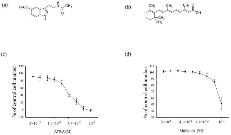
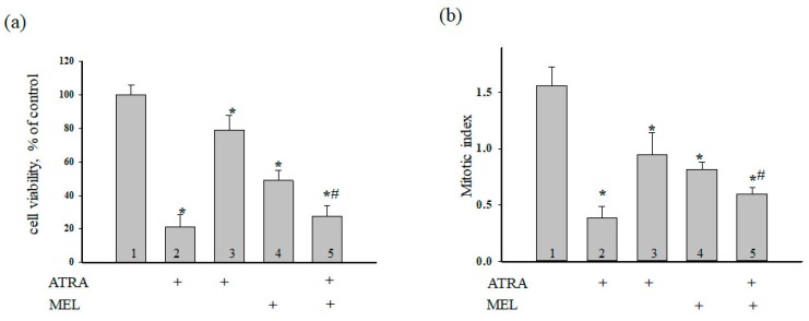
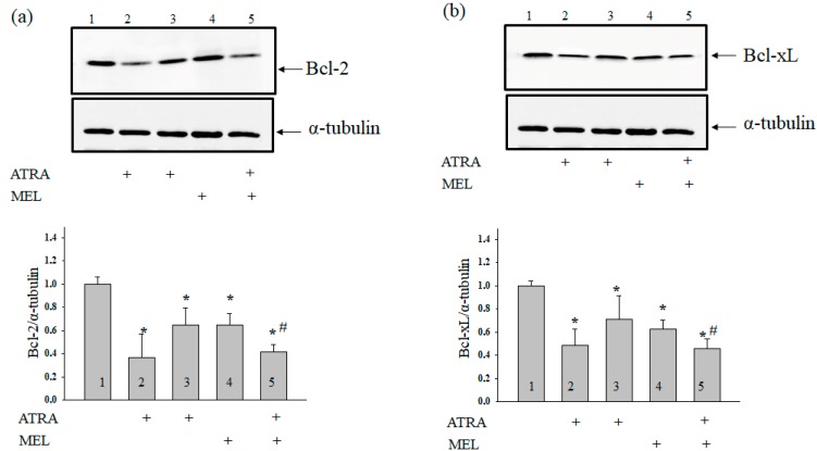
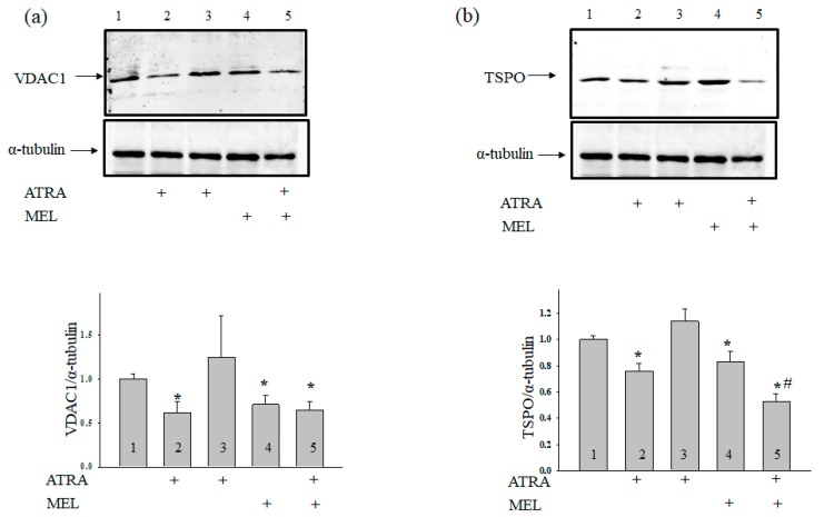
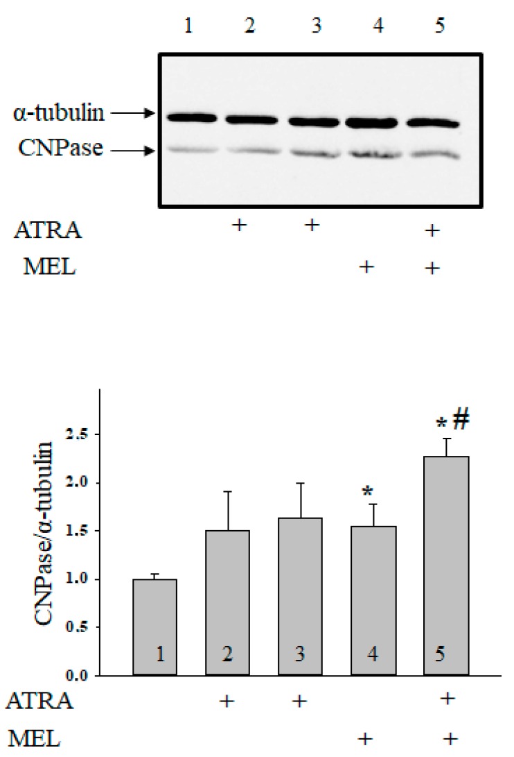
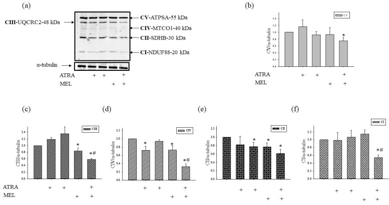
Melatonin is produced by the pineal gland. It can be regarded as an anticancer agent and used for combined therapy, owing to its oncostatic, antioxidant, and immunoregulatory activities. Retinoic acid is widely used for the treatment of acute promyelocytic leukemia; however, it has adverse effects on the human organism. We investigated the effect of melatonin and reduced concentrations of retinoic acid on the activation of proliferation in acute promyelocytic leukemiaon a cell model HL-60. The combined effect of these compounds leads to a reduction in the number of cells by 70% and the index of mitotic activity by 64%. Combined treatment with melatonin and retinoic acid decreased the expression of the Bcl-2. The mitochondrial isoform VDAC1 can be a target in the treatment of different tumors. The combined effect of and retinoic acid at a low concentration (10 nM) decreased VDAC1 expression. Melatonin in combination with retinoic acid produced a similar effect on the expression of the translocator protein. The coprecipitation of VDAC with 2',3'-cyclonucleotide-3'-phosphodiesterase implies a possible role of its in cancer development. The combined effect of retinoic acid and melatonin decreased the activity of the electron transport chain complexes. The changes in the activation of proliferation in HL-60 cells, the mitotic index, and Bcl-2 expression under combined effect of retinoic acid (10 nM) with melatonin (1 mM) are similar to changes that are induced by 1 μM retinoic acid. Our results suggest that MEL is able to improve the action the other chemotherapeutic agent.

Keywords: 2′,3′-cyclonucleotide-3′-phosphodiesterase; HL-60 cells; acute promyelocytic leukemia; apoptosis; melatonin; retinoic acid; translocator protein; voltage dependent anion channel-1.


Conflict of interest statement

The authors declare no conflict of interest.

Figures

Figure 1
Figure 1
Upper part (a) and (b). Chemical structures of melatonin (MEL) and all-trans retinoic acid (ATRA). (a) MEL, (b) ATRA. Lower part (c) and (d). Concentration dependence of the cytotoxic effects of MEL and ATRA. Cells were seeded in a 96-well plate at a density of 5 × 10³ cells per well and treated with indicated concentrations of (a) MEL and (b) ATRA for 96 h. The data are presented as means ± S.D. of ten separate experiments.
Figure 2
Figure 2
Combined effect of MEL and ATRA on the viability and proliferation status of HL-60 cells. Cells were seeded in a 96-well plate at a density of 5 × 10³ cells per well and treated with 1 µM ATRA (column 2), and 10 nM ATRA (columns 3) and 1 mM MEL (column 4), and MEL in combination with 10 nM ATRA (column 5); untreated cells (control, column 1). (a) Cell viability in % relative to the control; (b) The mitotic index (MI) is defined as the ratio between the number of cells in mitosis and the total number of cells. The data are presented as the means ± S.D. of ten separate experiments. * p < 0.05 significant difference in values in comparison with the corresponding control, # p < 0.05 significant difference in values compared to the value obtained after the addition of MEL alone.
Figure 3
Figure 3
Combined effect of MEL and ATRA on the level of Bcl-2 (a) and Bcl-xL (b) proteins in HL-60 cells. Cells were seeded in a 96-well plate at a density of 5 × 10³ cells per well and treated with 1 µM ATRA (column 2), and 10 nM ATRA (columns 3) and 1 mM MEL (column 4), and MEL in combination with 10 nM ATRA (column 5); untreated cells (control, column 1). Protein samples were extracted and subjected to Western blot for Bcl-2 and Bcl-xL detection. Immunodetection of α-tubulin was used as a loading control. Upper parts-immunostaining of Bcl-2, Bcl-xL and α-tubulin. Lower part-quantitation of immunostaining using computer-assisted densitometry. Bar graphs represent the proteins levels in relative units. The protein level in a cell lysate without any addition was taken to be unity and served as a control. The data are presented as the means ± S.D. of five separate experiments. * p < 0.05 significant difference in the protein level compared with the corresponding control, # p < 0.05 significant difference in the protein level compared to the value that was obtained in the presence of MEL alone.
Figure 4
Figure 4
Combined effect of MEL and ATRA on the level of TSPO and VDAC1 in HL-60 cells. Cells were seeded in a 96-well plate at a density of 5 × 10³ cells per well and treated with 1 µM ATRA (column 2), and 10 nM ATRA (columns 3) and 1 mM MEL (column 4), and MEL in combination with 10 nM ATRA (column 5); untreated cells (control, column 1). Immunodetection of α-tubulin was used as a loading control. (a) Immunostaining (upper part) and quantitation (lower part) of the protein level of VDAC1; (b)-of TSPO. Bar graphs represent the levels of appropriate proteins in relative units. The protein level in a cell lysate without any addition was taken to be unity and served as a control. The data are presented as means ± S.D. of five separate experiments. * p < 0.05 significant difference in protein level in comparison with corresponding control, # p < 0.05 significant difference in the protein level compared to the value obtained in the presence of MEL alone.
Figure 5
Figure 5
Combined effect of MEL and ATRA on the level of CNPase in HL-60 cells. Cells were seeded in a 96-well plate at a density of 5 × 10³ cells per well and treated with 1 µM ATRA (column 2), and 10 nM ATRA (columns 3) and 1 mM MEL (column 4), and MEL in combination with 10 nM ATRA (column 5); untreated cells (control, column 1). Protein samples were extracted and subjected to Western blot for CNPase detection. The immunodetection of α-tubulin was used as a loading control. Upper part-immunostaining of CNPase and α-tubulin. Lower part-quantitation of immunostaining using computer-assisted densitometry. Bar graphs represent the Bcl-2 level in relative units. The protein level in a cell lysate without any addition was taken to be unity and served as a control. The data are presented as means ± S.D. of five separate experiments. * p < 0.05 significant difference in protein level in comparison with corresponding control, # p < 0.05 significant difference in the protein level compared to the value obtained in the presence of MEL alone.
Figure 6
Figure 6
The effect of MEL and ATRA on the mitochondrial respiratory chain complexes in HL-60 cells. Cells were seeded in a 96-well plate at a density of 5 × 10³ cells per well and treated with 1 µM ATRA (column 2), and 10 nM ATRA (columns 3) and 1 mM MEL (column 4), and MEL in combination with 10 nM ATRA (column 5); untreated cells (control, column 1). Protein samples were extracted and subjected to Western blotting. Changes in mitochondrial complexes were detected using, the Total OXPHOS Rodent WB Antibody Cocktail. The immunodetection of α-tubulin was used as a loading control. (a) Immunostaining with OXPHOS antibody cocktail and α-tubulin; (bf)-quantification of immunostaining using computer-assisted densitometry. Bar graphs represent the levels of appropriate complexes (I–V) in relative units. The protein level in a cell lysate without any addition was taken to be unity and served as a control. The data are presented as means ± S.D. of five separate experiments. * p < 0.05 significant difference in protein level in comparison with the corresponding control, # p < 0.05 significant difference in the protein level compared to the value obtained in the presence of MEL alone.

Similar articles

Cited by

References

    1. Stehle J.H., Saade A., Rawashdeh O., Ackermann K., Jilg A., Sebesteny T., Maronde E. A survey of molecular details in the human pineal gland in the light of phylogeny, structure, function and chronobiological diseases. J. Pineal Res. 2011;51:17–43. doi: 10.1111/j.1600-079X.2011.00856.x. - DOI - PubMed
    1. Carlberg C. Gene regulation by melatonin. Ann. N. Y. Acad. Sci. 2000;917:387–396. doi: 10.1111/j.1749-6632.2000.tb05403.x. - DOI - PubMed
    1. Bizzarri M., Proietti S., Cucina A., Reiter R.J. Molecular mechanisms of the pro-apoptotic actions of melatonin in cancer: A review. Expert Opin. Ther. Targets. 2013;17:1483–1496. doi: 10.1517/14728222.2013.834890. - DOI - PubMed
    1. Vijayalaxmi, Thomas C.R., Jr., Reiter R.J., Herman T.S. Melatonin: From basic research to cancer treatment clinics. J. Clin. Oncol. 2002;20:2575–2601. doi: 10.1200/JCO.2002.11.004. - DOI - PubMed
    1. Shiu S.Y. Towards rational and evidence-based use of melatonin in prostate cancer prevention and treatment. J. Pineal Res. 2007;43:1–9. doi: 10.1111/j.1600-079X.2007.00451.x. - DOI - PubMed

MeSH terms

LinkOut - more resources