Review
. 2018 Sep 21;19(10):2878.
doi: 10.3390/ijms19102878.

The Multifaceted Role of Pectin Methylesterase Inhibitors (PMEIs)

Affiliations
Review

The Multifaceted Role of Pectin Methylesterase Inhibitors (PMEIs)

Alexandra Wormit et al. Int J Mol Sci. .

Abstract

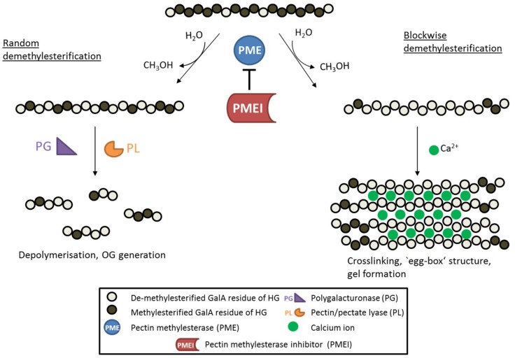
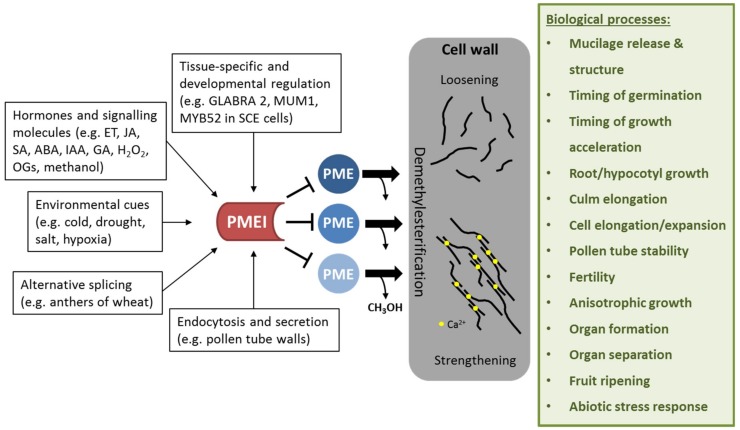
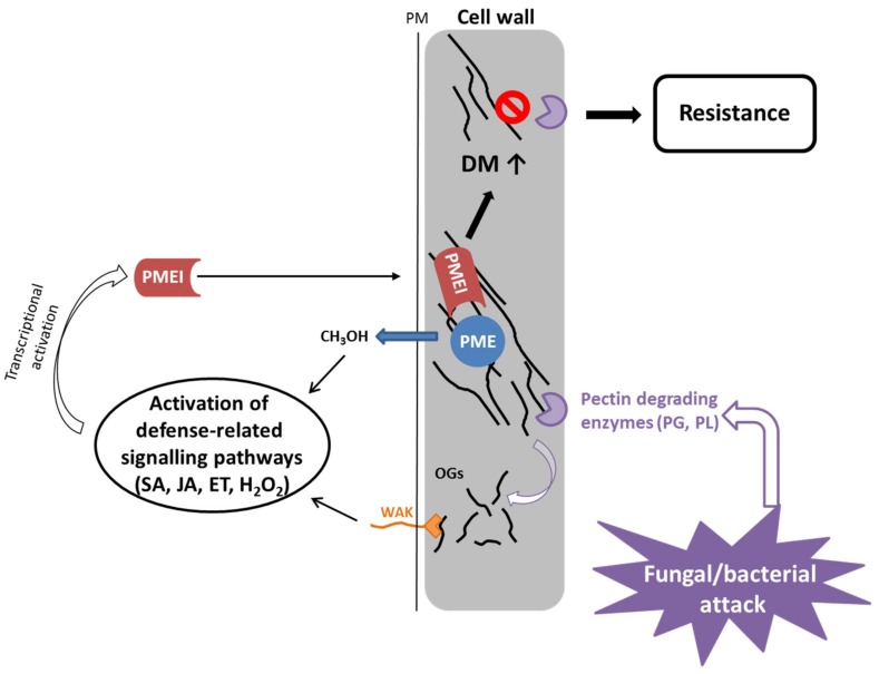
Plant cell walls are complex and dynamic structures that play important roles in growth and development, as well as in response to stresses. Pectin is a major polysaccharide of cell walls rich in galacturonic acid (GalA). Homogalacturonan (HG) is considered the most abundant pectic polymer in plant cell walls and is partially methylesterified at the C6 atom of galacturonic acid. Its degree (and pattern) of methylation (DM) has been shown to affect biomechanical properties of the cell wall by making pectin susceptible for enzymatic de-polymerization and enabling gel formation. Pectin methylesterases (PMEs) catalyze the removal of methyl-groups from the HG backbone and their activity is modulated by a family of proteinaceous inhibitors known as pectin methylesterase inhibitors (PMEIs). As such, the interplay between PME and PMEI can be considered as a determinant of cell adhesion, cell wall porosity and elasticity, as well as a source of signaling molecules released upon cell wall stress. This review aims to highlight recent updates in our understanding of the PMEI gene family, their regulation and structure, interaction with PMEs, as well as their function in response to stress and during development.

Keywords: applications; cell wall properties; degree of methylesterification (DM); development; homogalacturonan (HG); pectin methylesterase inhibitor (PMEI), pectin; stress.


Conflict of interest statement

The authors declare no conflict of interest

Figures

Figure 1
Figure 1
Schematic diagram showing the de-methylesterification of HG and the effects on its structure. HG is highly methylesterified when deposited into the cell wall. PMEs can de-methylesterify HG in a block-wise fashion, leading to several consecutive GalA residues without methylester groups. These HG backbones are negatively charged and can therefore form crosslinks with cations like calcium ions, leading to so called ‘egg-box‘ structures responsible for gel formation. On the other hand, PMEs can de-methylesterify single GalA residues leading to a random methylesterification pattern. Low-methylesterified HG is depolymerized by pectin-degrading enzymes such as polygalacturonases (PG) and pectin/pectate lyases (PL), which leads to the formation of oligogalacturonides (OG). PME activity is inhibited by its proteinaceous inhibitor PMEI.
Figure 2
Figure 2
Effect of PMEI regulation on cell wall properties and biological processes affected by PMEI manipulation. PMEIs are transcriptionally regulated in a tissue-specific and development-dependent manner. Several plant hormones and signaling molecules as well as environmental stresses can activate PMEI gene expression. Alternative splicing and directed endocytosis and secretion regulate the level of active PMEIs in the cell wall. PMEI can inhibit several PME enzymes, thereby regulating de-methylesterification of HG. This in turn modulates cell wall properties such as loosening or strengthening, which is required for several biological processes.
Figure 3
Figure 3
PMEIs modulate PME activity and DM of pectin in response to fungal and bacterial attack. Bacteria and fungi secrete pectin degrading enzymes, like polygalacturonases and pectate/pectin lyases, upon infection. PME activity leads to release of methanol, which serves as an alarm signal, activating the expression of pathogen-related PMEIs. Similarly, OGs, which are generated by degradation of pectin by fungal/bacterial PGs and PLs, serve as DAMPs, leading also to activation of defense related signaling pathways, which have been shown to activate PMEI gene expression. PMEIs inhibit PME activity leading to a higher degree of methylesterification of pectin, which contributes to resistance to fungal/bacterial enzymes resulting in less disease symptoms.

Similar articles

Cited by

References

    1. Wolf S., Mouille G., Pelloux J. Homogalacturonan methyl-esterification and plant development. Mol. Plant. 2009;2:851–860. doi: 10.1093/mp/ssp066. - DOI - PubMed
    1. Mohnen D. Pectin structure and biosynthesis. Curr. Opin. Plant Biol. 2008;11:266–277. doi: 10.1016/j.pbi.2008.03.006. - DOI - PubMed
    1. Braybrook S.A., Hofte H., Peaucelle A. Probing the mechanical contributions of the pectin matrix. Plant Signal. Behav. 2012;7:1037–1041. doi: 10.4161/psb.20768. - DOI - PMC - PubMed
    1. Palin R., Geitmann A. The role of pectin in plant morphogenesis. Biosystems. 2012;109:397–402. doi: 10.1016/J.BIOSYSTEMS.2012.04.006. - DOI - PubMed
    1. Daher F.B., Braybrook S.A. How to let go: Pectin and plant cell adhesion. Front. Plant Sci. 2015;6:523. doi: 10.3389/fpls.2015.00523. - DOI - PMC - PubMed

MeSH terms

LinkOut - more resources