Review
. 2018 Sep 24;15(1):276.
doi: 10.1186/s12974-018-1313-3.

Inflammation: the link between comorbidities, genetics, and Alzheimer's disease

Affiliations
Review

Inflammation: the link between comorbidities, genetics, and Alzheimer's disease

Estella A Newcombe et al. J Neuroinflammation. .

Abstract

Alzheimer's disease (AD) is a neurodegenerative disorder, most cases of which lack a clear causative event. This has made the disease difficult to characterize and, thus, diagnose. Although some cases are genetically linked, there are many diseases and lifestyle factors that can lead to an increased risk of developing AD, including traumatic brain injury, diabetes, hypertension, obesity, and other metabolic syndromes, in addition to aging. Identifying common factors and trends between these conditions could enhance our understanding of AD and lead to the development of more effective treatments. Although the immune system is one of the body's key defense mechanisms, chronic inflammation has been increasingly linked with several age-related diseases. Moreover, it is now well accepted that chronic inflammation has an important role in the onset and progression of AD. In this review, the different inflammatory signals associated with AD and its risk factors will be outlined to demonstrate how chronic inflammation may be influencing individual susceptibility to AD. Our goal is to bring attention to potential shared signals presented by the immune system during different conditions that could lead to the development of successful treatments.

Keywords: APOE4; Aging; Alzheimer’s disease; CD33; Diabetes; Microglia; Neuroinflammation; Obesity; TBI; TREM2.


Conflict of interest statement

Ethics approval and consent to participate

Not applicable.

Consent for publication

Not applicable.

Competing interests

The authors declare that they have no competing interests.

Publisher’s Note

Springer Nature remains neutral with regard to jurisdictional claims in published maps and institutional affiliations.

Figures

Fig. 1
Fig. 1
The pathogenic hallmarks of AD in the human brain over time. In the earliest stages of AD, the formation of Aβ occurs due to abnormal cleavage of amyloid precursor protein (APP) by β- and γ-secretases, whereas it is normally cleaved by α- and γ-secretases. Aβ monomers are intrinsically disordered and have a propensity to oligomerize and aggregate into Aβ plaques. Aβ activates microglia and astrocytes, causing them to clear Aβ via phagocytosis and proteolysis. The presence of Aβ has also been linked to the hyperphosphorylation and destabilization of tau and the subsequent formation of tau tangles. Inflammatory activation and signaling can also cause further production of Aβ. Tau pathology is observed approximately 10 years after the initiation of Aβ aggregation. Tau is a microtubule-associated protein that is predominately found in neurons, where it is regulated by phosphorylation and other post-translational modifications (i.e., acetylation, ubiquitylation) to stabilize microtubules, regulate axonal stability, and maintain cell function. While tau contains 2–3 mol of phosphate in the healthy brain, it can accumulate up to three times more phosphate in AD. This hyperphosphorylation lowers the affinity of tau for the microtubules, increases its resistance to degradation by proteases and the proteasome, and leads to its fibrillization and aggregation into neurofibrillary tangles, which ultimately causes neuronal loss and cognitive decline. The increase in Aβ aggregation, inflammation, and tau hyperphosphorylation leads to a variety of downstream effects on neuronal synapses, including inhibition of LTP, impaired dendritic trafficking, increased excitotoxicity, and a reduction in synaptic density. This leads to synaptic loss and eventual neuronal loss approximately 20 years following initial disease pathogenesis, which is followed by the symptomatic cognitive decline
Fig. 2
Fig. 2
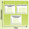
Time-course of pro- and anti-inflammatory responses. The accepted model of acute inflammation shows the activation and resolution of inflammation that occurs due to pro- and then anti-inflammatory modulators. These processes become dysfunctional during chronic inflammation where the resolution phase is not achieved due to excessive pro-inflammatory signaling. Considering more recent developments in the studies of inflammation, we propose a hypothetical model of inflammation during aging, in which an event is not resolved to a baseline inflammatory level, but leaves some residual pro-inflammatory effects (i.e., microglial priming)
Fig. 3
Fig. 3
Inflammatory responses in the young and old brain. The BBB is intact in the young brain, reinforced by tight junctions, endothelial cells, pericytes, and astrocytes (blue). During homeostasis, microglia (orange) perform a surveillance role with extended processes. Microglia and astrocytes are activated by an inflammatory event, during which the microglia prepare for phagocytosis to eliminate the inflammatory stimulus, such as during a bacterial infection following an injury. Peripheral immune cells such as monocytes (green) are also able to cross the BBB to enhance phagocytosis and debris clearance in the brain. After the inflammatory event, microglia in the young brain are able to return to their surveillance state. After a lifetime of such events, however, microglia in the aged brain acquire a primed state, where they exhibit persistent low-level inflammation during homeostasis. During aging, the BBB also becomes leaky. Primed microglia are fast to react to inflammatory stimuli, but have a high risk of becoming senescent and unable to perform phagocytosis and clear infection. If the microglia are no longer able to remove the infection, they continue to release inflammatory mediators, leading to increased inflammatory cell migration past the BBB, which is further permeabilized during disease
Fig. 4
Fig. 4
Proposed model of immunosenescence over time. We suggest that immunosenescence is related to the cumulative effects of many inflammatory events throughout life, including infection, metabolic disorders, or genetic risks. It may be possible to reduce immunosenescence by modifying lifestyle, leading to a reduced cumulative effect, which may subsequently decrease the risk of developing AD
Fig. 5
Fig. 5
Inflammation uniquely affects each of the hallmarks of AD. Each hallmark of AD has a unique relationship with inflammation, whereby it can both be triggered by and trigger inflammation. Aβ plaques activate microglia and trigger their pro-inflammatory signaling pathways, which can cause increased Aβ production. Tau phosphorylation can be caused by the production of Aβ plaques, as well as by increased inflammatory signaling. Tau hyperphosphorylation and tangles can also lead to increased inflammation. Finally, neurodegeneration can be caused by an excessive microglial response, while neuronal debris from neurodegeneration can lead to further stimulation of microglia

References

    1. Cummings J, Lee G, Mortsdorf T, Ritter A, Zhong K. Alzheimer’s disease drug development pipeline: 2017. Alzheimers Dement (N Y) 2017;3(3):367–384. - PMC - PubMed
    1. Fullerton JN, Gilroy DW. Resolution of inflammation: a new therapeutic frontier. Nat Rev Drug Discov. 2016;15(8):551–567. doi: 10.1038/nrd.2016.39. - DOI - PubMed
    1. Tay TL, Mai D, Dautzenberg J, Fernandez-Klett F, Lin G, Sagar, et al. A new fate mapping system reveals context-dependent random or clonal expansion of microglia. Nat Neurosci. 2017;20(6):793–803. doi: 10.1038/nn.4547. - DOI - PubMed
    1. Grabert K, Michoel T, Karavolos MH, Clohisey S, Baillie JK, Stevens MP, et al. Microglial brain region-dependent diversity and selective regional sensitivities to aging. Nat Neurosci. 2016;19(3):504–516. doi: 10.1038/nn.4222. - DOI - PMC - PubMed
    1. Askew K, Li K, Olmos-Alonso A, Garcia-Moreno F, Liang Y, Richardson P, et al. Coupled proliferation and apoptosis maintain the rapid turnover of microglia in the adult brain. Cell Rep. 2017;18(2):391–405. doi: 10.1016/j.celrep.2016.12.041. - DOI - PMC - PubMed