. 2018 Sep-Oct:103:74-85.
doi: 10.1016/j.diff.2018.08.001. Epub 2018 Aug 23.

Development of the human penis and clitoris

Affiliations

Development of the human penis and clitoris

Laurence Baskin et al. Differentiation. 2018 Sep-Oct.

Abstract

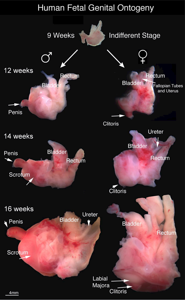
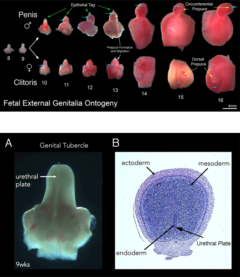
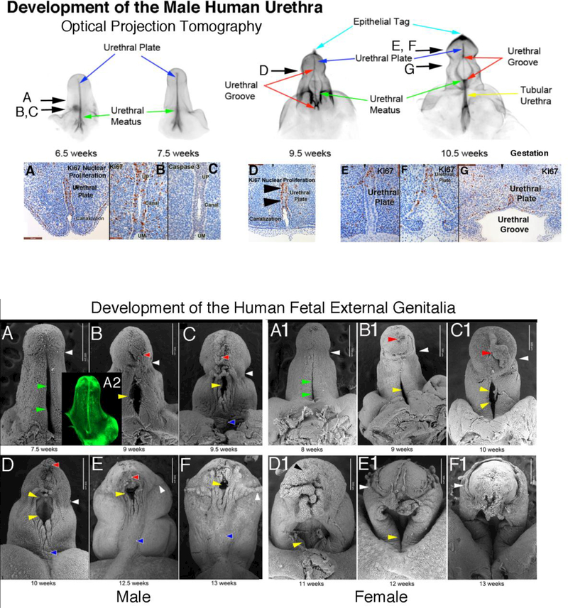
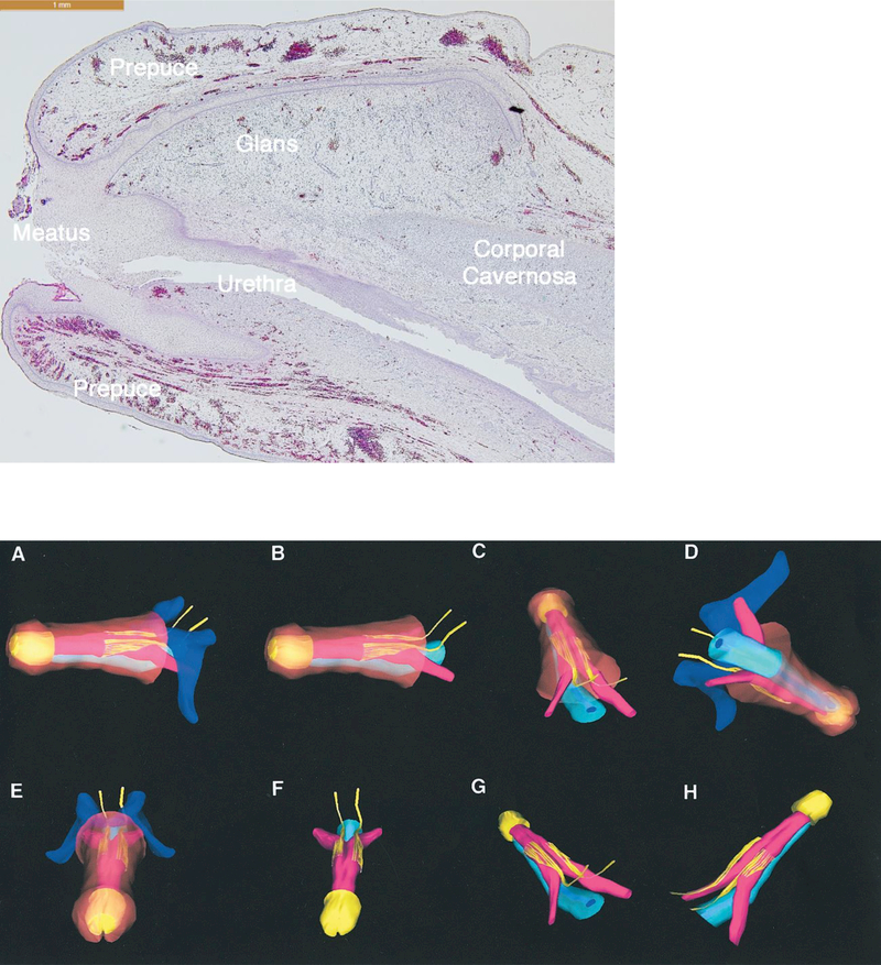
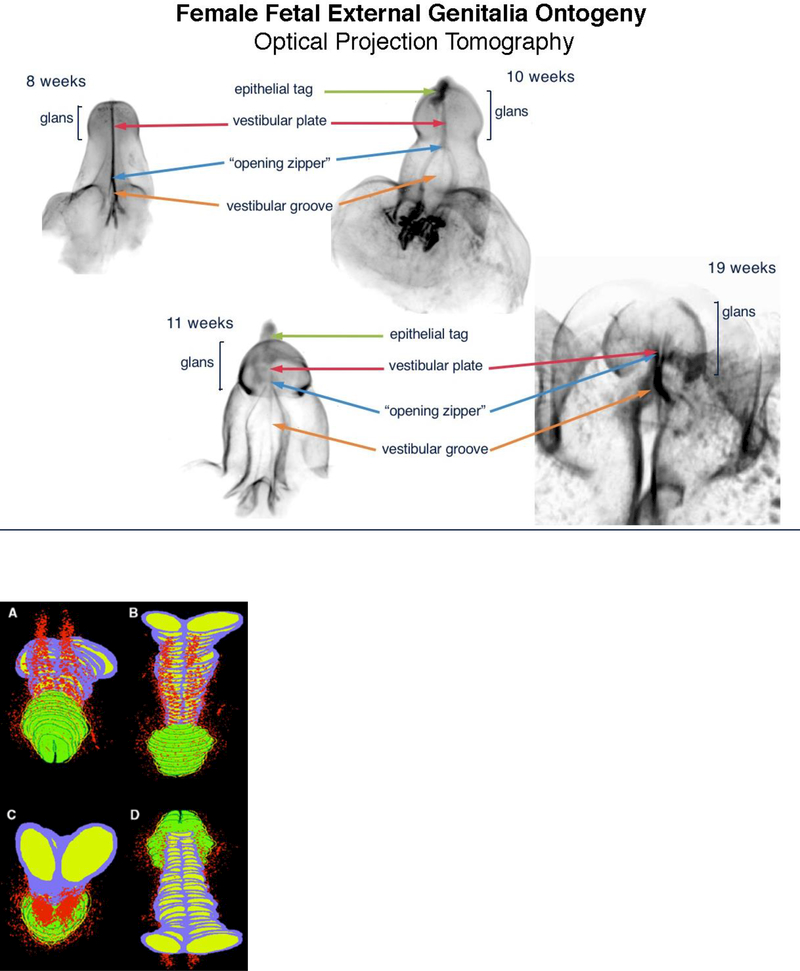
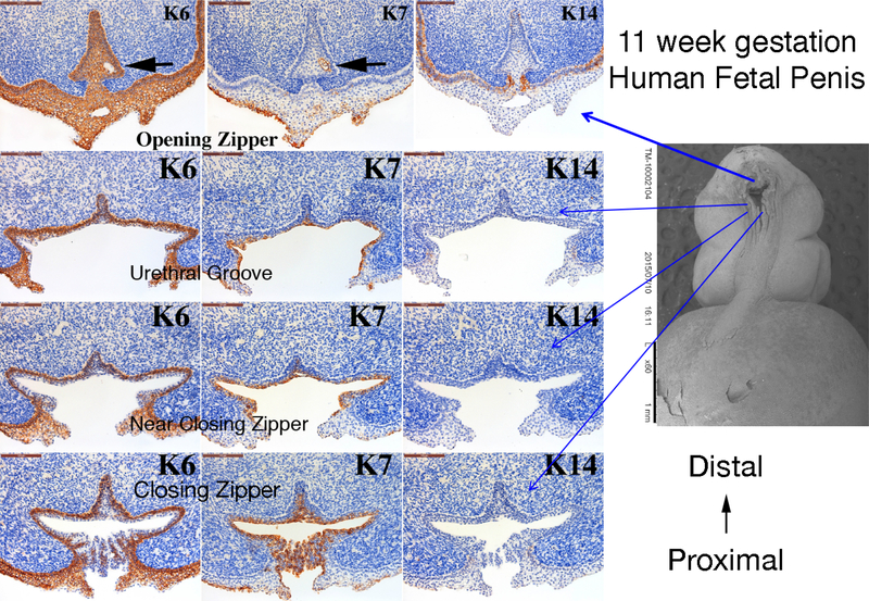
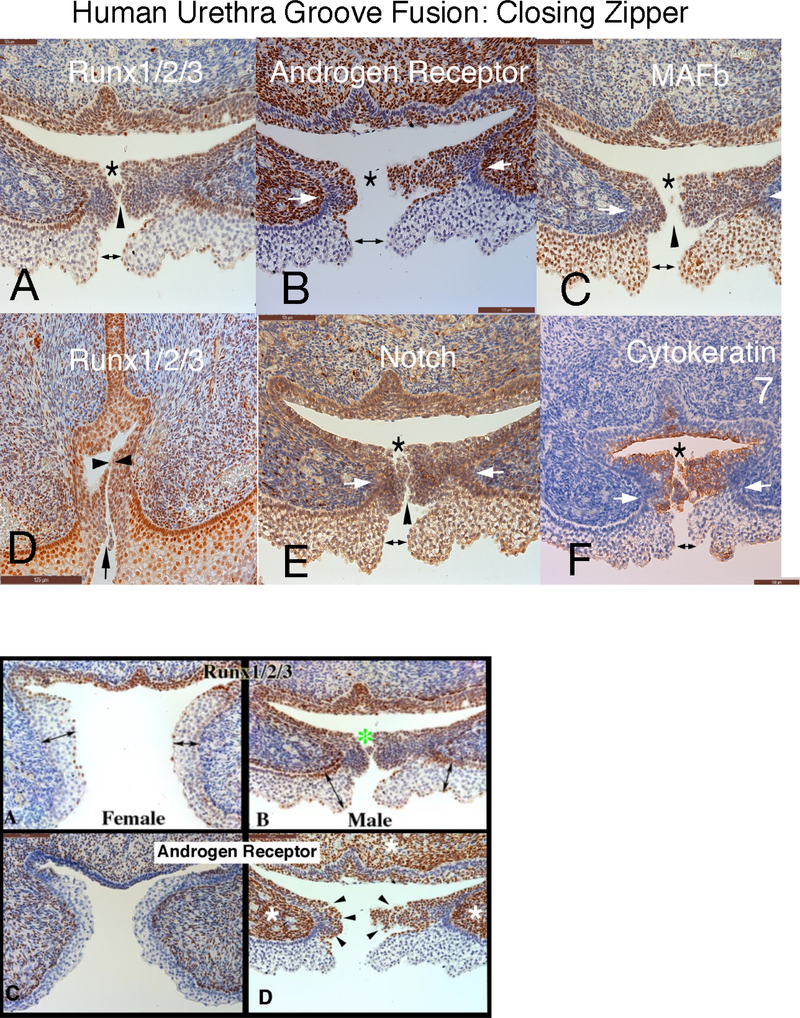
The human penis and clitoris develop from the ambisexual genital tubercle. To compare and contrast the development of human penis and clitoris, we used macroscopic photography, optical projection tomography, light sheet microscopy, scanning electron microscopy, histology and immunohistochemistry. The human genital tubercle differentiates into a penis under the influence of androgens forming a tubular urethra that develops by canalization of the urethral plate to form a wide diamond-shaped urethral groove (opening zipper) whose edges (urethral folds) fuse in the midline (closing zipper). In contrast, in females, without the influence of androgens, the vestibular plate (homologue of the urethral plate) undergoes canalization to form a wide vestibular groove whose edges (vestibular folds) remain unfused, ultimately forming the labia minora defining the vaginal vestibule. The neurovascular anatomy is similar in both the developing human penis and clitoris and is the key to successful surgical reconstructions.

Keywords: Canalization and fusion; Clitoris; Development; Human; Penis.


Figures

Figure 1.
Figure 1.
Gross Human Fetal Pelvic Ontogeny: Gross ontogeny of the human fetal pelvis at 9 weeks of gestation (end of indifferent stage), 12 weeks, 14 weeks and 16 weeks of gestation. Note the divergent development after 9 weeks of gestation especially in respect to the orientation of the external genitalia with the penis clearly visible at ~ 90-degree angle from the body and the clitoris recessed close to the body wall.
Figure 2.
Figure 2.
Fetal External Genitalia Ontogeny: Representative ventral views of external genitalia of the human males (top row) and females (bottom row) (8–16 weeks of gestation). Note the morphologic differences between male and female specimens after the indifferent stage (8–9 weeks of gestation) with formation of the penile urethra within the shaft due to urethral fold fusion in the penis and lack of urethral (vestibular) fold fusion in female specimens (light blue arrows depict the location of the urethra meatus in both the male and female specimens). Note the divergent evolution of the male and female prepuce (yellow arrows) with complete circumferential formation of the prepuce at 14–16 weeks of gestation in the male and a resulting dorsal prepuce in the female. The epithelial tag is seen in both male (green arrows) and female (clearly visible without arrows)) specimens from 10–13 weeks of gestation disappearing after this time point.
Figure 3:
Figure 3:
Human male genital tubercle/ future human penis at 9 weeks of gestation. Note the urethral plate in the gross specimen (A). In the corresponding histologic cross section (B), the three embryonic layers of external genitalia development are labeled.
Figure 4:
Figure 4:
Optical projection tomography of male urethral development from 6.5 to 10.5 weeks fetal age. Note the urethral plate (blue arrow) that ends within the glans. The wide-open urethral groove (red arrows) is best seen from 9.5 to 10.5 weeks with clear progression of proximal to distal fusion of the edges of the urethral groove to form the tubular urethra (yellow arrow). The epithelial tag is marked by the light blue arrow. The proliferation marker Ki67 are labeled with arrows in the OPT specimens A-G, with the exception of C which illustrates no staining for the apoptotic marker caspase 3. Canalization of the urethral plate is visible in histologic section A-D. Reproduced from Li, Y., A. Sinclair, M. Cao, et al., Canalization of the urethral plate precedes fusion of the urethral folds during male penile urethral development: the double zipper hypothesis. J Urol, 2015. 193(4): p. 1353–59, with permission.
Figure 5:
Figure 5:
Scanning electron microscopy ontogeny of the developing human fetal penis (A-F) from 7.5 −13 weeks and developing human clitoris (A1-F1) 8–13 weeks of gestation. White arrowheads indicate the junction of the penile and clitoral shaft with the glans (ages 7.5–11 weeks A-D & A1-D1). At 12 weeks of gestation in both males and females note advancing of the prepuce over the glans (white arrowheads) (E, E1, F & F1). The red arrowheads denote the epithelial tag. The blue arrowheads indicate the median penile raphe. Yellow arrowheads indicate the open urethral groove in males and vestibular groove in females. The green arrows in A & A1 denote the urethral plate in males and vestibular plate in females which is not open based on histologic analysis (see figure 3) and lightsheet microscopy staining with E-cadherin (A2). Modified from Shen, J., M. Overland, A. Sinclair, et al., Complex epithelial remodeling underlie the fusion event in early fetal development of the human penile urethra. Differentiation, 2016. 92(4): p. 169–182, with permission.
Figure 6:
Figure 6:
Sagittal section of an 18-weeks gestation human fetal glans penis. Note that the penile epidermis meets the distal aspect of the urethral epithelium at the meatus.
Figure 7.
Figure 7.
3-D Neuroanatomy of the fully formed fetal penis at 17.5 weeks of gestation. A to H, multiple views of 3-D reconstruction of human fetal penis dorsal nerve neuroanatomy with respective layers removed for selective visualization. Dark blue areas represent pubic arch and symphysis. Yellow areas represent nerves and glans.Light blue areas represent urethra. Pink areas represent corporal bodies. Brown areas represent penile skin. (Used with permission Akman, Y et al., Penile anatomy under the pubic arch: reconstructive implications. J Urol, 2001. 166: p. 229.) with permission.
Figure 8.
Figure 8.
Optical Projection Tomography: Fetal ontogeny of human clitoris from 8 to 19 weeks of fetal age. Note the opening zipper, which facilitates vestibular plate opening to form vestibular groove, and lack of closing zipper (seen in the male) with the vestibular groove remaining open. Epithelial tag (green arrows), vestibular plate (red arrows) opening zipper (blue arrows) and vestibular groove (orange arrows).
Figure 9.
Figure 9.
Computer generated 3-D reconstruction of normal human fetal clitoris at 24 weeks of gestation. Red areas represent nerve pathway with paucity of nerves at bottom and top midline of clitori., Purple= tunica of corporal bodies, yellow= interior of corporal bodies, green= glans clitoris, and dark green and yellow= clitoral hood. A, dorsal front view. B, dorsal back view. C, back ventral view. D, ventral view. (Used with permission Baskin, L.S., et al., Anatomical studies of the human clitoris. J Urol, 1999. 162(3 Pt 2): p. 1017.) with permission.
Figure 10.
Figure 10.
Cytokeratin (K) 6, 7 and 14 expression in an 11-week human fetal penis. Representative sections from the opening zipper, urethral groove, near the closing zipper and the closing zipper. Approximate section location is depicted by the blue arrows in the scanning electron microscopic image. Note the canalization (black arrows) in the opening zipper. Note the K6 localization to basal epithelial cells and K7 apical epithelial cells, and the complex arrangement of epithelial cells fusing in the closing zipper.
Figure 11.
Figure 11.
Human fetal penis at 11 weeks of gestation: Closing zipper - fusion of the urethral folds. Immunohistochemical localization of: A) Runx 1/2/3. B) Androgen Receptor. C) MAFb. D) Runx 1/2/3. E) Notch. and F) Cytokeratin 7. This figure illustrates that formation of the penile urethra occurs as a result of 3 separate fusion events of all three germ layers. Note that in all the panels the cells that ultimately undergo fusion to form the tubular urethra appear to fuse in three areas. 1) ectoderm (double headed arrows) 2) mesenchymal confluence (white arrows) and 3) endoderm (*). Furthermore, epithelial cells appear to have a “delayed cellular fusion”, with clear spaces visible between epithelial cells (black arrowheads).
Figure 12.
Figure 12.
Runx1/2/3 and androgen receptor Immunohistochemistry of 11-week female and male human fetal clitoris and penis at the level of the closing zipper. RUNX1/2/3-positive epithelial cells are seen in males (B) in the floor of the urethral groove and at the point of epithelial fusion (green asterisk). The corresponding epithelium during female (A) external genitalia development has a paucity of RUNX1/2/3 expression. Note the different location of the maturing epidermis (A & B, double-headed arrows). Note the increased amount of androgen receptor expression in the male (D) in both the epithelium (black arrowheads) and mesenchyme compared to the female (C) (white asterisk).

References

    1. Agras K, Willingham E, Liu B, and Baskin LS (2006) Ontogeny of androgen receptor and disruption of its mRNA expression by exogenous estrogens during morphogenesis of the genital tubercle. J Urol 176:1883–1888. - PubMed
    1. Akman Y, Liu W, Li YW, and Baskin LS (2001) Penile anatomy under the pubic arch: reconstructive implications. J Urol 166:225–230. - PubMed
    1. Altemus AR, and Hutchins GM (1991) Development of the human anterior urethra. J Urol 146:1085–1093. - PubMed
    1. Baskin L (2017a) What Is Hypospadias? Clin Pediatr (Phila) 56:409–418. - PubMed
    1. Baskin L, Duckett J, and Lue T (1996) Penile Curvature. Urology 48:347–356. - PubMed

Publication types