Review
. 2019 Apr 1;9(4):a033183.
doi: 10.1101/cshperspect.a033183.

Stem Cells and the Bird Cochlea-Where Is Everybody?

Affiliations
Review

Stem Cells and the Bird Cochlea-Where Is Everybody?

Amanda S Janesick et al. Cold Spring Harb Perspect Med. .

Abstract

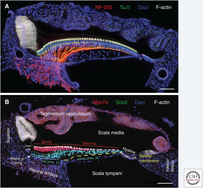
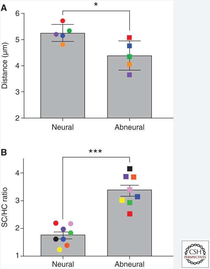
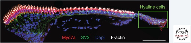
In sharp contrast to the adult mammalian cochlea, which lacks regenerative ability, the mature avian cochlea, or basilar papilla (BP) is capable of complete recovery from hearing loss after damage. Avian sensory hair cell regeneration relies on rousing quiescent supporting cells to proliferate or transdifferentiate after hair cell death. Unlike mammalian cochlear supporting cells, which have clearly defined subtypes, avian BP supporting cells are deceptively indistinguishable and molecular markers have yet to be identified. Despite the importance of supporting cells as the putative stem cells in avian regeneration, it is unknown whether all supporting cells possess equal capability to give rise to a hair cell or if a specialized subpopulation exists. In this perspective, we reinvigorate the concept of a stem cell in the BP, and form comparisons to other regenerating tissues that show cell-cycle reentry after damage. Special emphasis is given to the structure of the BP and how anatomy informs both the potential, intrinsic heterogeneity of the supporting cell layer as well as the choice between mitotic and nonmitotic regenerative strategies.


Figures

Figure 1.
Figure 1.
Cross-sectional anatomy of the chicken basilar papilla (BP). Shown are transverse vibratome sections taken from the middle tonotopic region of the chicken BP, roughly equidistant from proximal and distal ends. Embryonic day 20 (A) and posthatch day 8 (B) were immunostained for (A) neurofilament-200 (NF-200, Sigma) and tubulin β III (TuJ1, EMD Millipore) or (B) myosin-VIIa (Myo7a, Proteus) and Sox2 (Santa Cruz Biotech). Phalloidin labels filamentous (F)-actin and Dapi stains nuclei. Sections were imaged with a Zeiss LSM880 confocal microscope at 40× magnification using Zen Black acquisition software. SC, Supporting cell; HC, hair cell. Scale bars, 50 µm.
Figure 2.
Figure 2.
Abneural supporting cells (SCs) in the chicken basilar papilla (BP) are more tightly packed (A) and more numerous than hair cells (HCs) (B) compared with the neural side. Transverse vibratome sections taken from the middle tonotopic region of the chicken BP (posthatch day 8–10) were immunostained for myosin-VIIa (Proteus) and Sox2 (Santa Cruz Biotech). Sections were imaged as in Figure 1. Using syGlass software (see syglass.io), SC and HC nuclei number and location were recorded, standardizing voxel size and Z-depth across sections. Counts and XYZ coordinates were exported. Distance measurements (A) between the nearest neighboring SC nuclei was conducted in MATLAB vR2017b. Data visualization and statistics (paired t-test) were performed in GraphPad Prism v7. Each data point of the same color represents one chicken. Error bars, SEM. *p ≤ 0.05, ***p ≤ 0.001.
Figure 3.
Figure 3.
Chicken basilar papilla (BP) hyaline cells are innervated. Shown is a transverse vibratome section taken from the middle tonotopic region of the posthatch day 7 chicken BP immunostained for synaptic vesicles (SV2, Developmental Studies Hybridoma Bank) and myosin-VIIa (Myo7a, Proteus). Phalloidin labels F-actin and Dapi stains nuclei. Sections were imaged as in Figure 1. Scale bar, 50 µm.

Similar articles

Cited by

References

    1. Adler HJ, Raphael Y. 1996. New hair cells arise from supporting cell conversion in the acoustically damaged chick inner ear. Neurosci Lett 205: 17–20. - PubMed
    1. Adler HJ, Komeda M, Raphael Y. 1997. Further evidence for supporting cell conversion in the damaged avian basilar papilla. Int J Dev Neurosci 15: 375–385. - PubMed
    1. Alvarado DM, Hawkins RD, Bashiardes S, Veile RA, Ku YC, Powder KE, Spriggs MK, Speck JD, Warchol ME, Lovett M. 2011. An RNA interference-based screen of transcription factor genes identifies pathways necessary for sensory regeneration in the avian inner ear. J Neurosci 31: 4535–4543. - PMC - PubMed
    1. Baird RA, Steyger PS, Schuff NR. 1996. Mitotic and nonmitotic hair cell regeneration in the bullfrog vestibular otolith organs. Ann NY Acad Sci 781: 59–70. - PubMed
    1. Baird RA, Burton MD, Lysakowski A, Fashena DS, Naeger RA. 2000. Hair cell recovery in mitotically blocked cultures of the bullfrog saccule. Proc Natl Acad Sci 97: 11722–11729. - PMC - PubMed

Publication types

LinkOut - more resources