. 2018 Nov 16;293(46):17838-17852.
doi: 10.1074/jbc.RA118.005649. Epub 2018 Sep 24.

Molecular determinants of α-conotoxin potency for inhibition of human and rat α6β4 nicotinic acetylcholine receptors

Affiliations

Molecular determinants of α-conotoxin potency for inhibition of human and rat α6β4 nicotinic acetylcholine receptors

Arik J Hone et al. J Biol Chem. .

Abstract

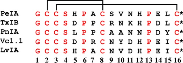
Nicotinic acetylcholine receptors (nAChRs) containing α6 and β4 subunits are expressed by dorsal root ganglion neurons and have been implicated in neuropathic pain. Rodent models are often used to evaluate the efficacy of analgesic compounds, but species differences may affect the activity of some nAChR ligands. A previous candidate α-conotoxin-based therapeutic yielded promising results in rodent models, but failed in human clinical trials, emphasizing the importance of understanding species differences in ligand activity. Here, we show that human and rat α6/α3β4 nAChRs expressed in Xenopus laevis oocytes exhibit differential sensitivity to α-conotoxins. Sequence homology comparisons of human and rat α6β4 nAChR subunits indicated that α6 residues forming the ligand-binding pocket are highly conserved between the two species, but several residues of β4 differed, including a Leu-Gln difference at position 119. X-ray crystallography of α-conotoxin PeIA complexed with the Aplysia californica acetylcholine-binding protein (AChBP) revealed that binding of PeIA orients Pro13 in close proximity to residue 119 of the AChBP complementary subunit. Site-directed mutagenesis studies revealed that Leu119 of human β4 contributes to higher sensitivity of human α6/α3β4 nAChRs to α-conotoxins, and structure-activity studies indicated that PeIA Pro13 is critical for high potency. Human and rat α6/α3β4 nAChRs displayed differential sensitivities to perturbations of the interaction between PeIA Pro13 and residue 119 of the β4 subunit. These results highlight the potential significance of species differences in α6β4 nAChR pharmacology that should be taken into consideration when evaluating the activity of candidate human therapeutics in rodent models.

Keywords: X-ray crystallography; analgesic; neuroinflammation; neuropathic pain; neuroscience; neurotoxin; neurotransmitter receptor; nicotinic acetylcholine receptors (nAChR); nicotinic subunit α6; nicotinic subunit β4; nuclear magnetic resonance spectroscopy; small peptide; α1;-conotoxin.


Conflict of interest statement

The authors declare that they have no conflicts of interest with the contents of this article

Figures

Figure 1.
Figure 1.
Sequence comparison of select 4/7 framework α-Ctxs. Conotoxins are classified into various subclasses based on the number of cysteine residues present in the sequence and the number of amino acid residues between them (63). Peptides of the α-Ctx subclass contain two disulfide-connected pairs of cysteines. Thus, for example, α-Ctxs with four residues between Cys2 and Cys3 and seven between Cys8 and Cys16 belong to the 4/7 framework subclass. Residues in black are variable, and those in red are conserved among this α-Ctx set. The disulfide bonds between Cys residues are depicted with lines. *, C-terminal amidation.
Figure 2.
Figure 2.
α-Ctxs distinguish between human and rat α6/α3β4 nAChRs. X. laevis oocytes expressing human or rat α6/α3β4 nAChRs were subjected to TEVC electrophysiology, and the IC50 values were determined for inhibition of ACh-evoked currents by PeIA (A), PnIA (B), and TxIB (C). Values are provided in Table 1. Error bars, S.D. from at least four oocytes for each IC50 determination.
Figure 3.
Figure 3.
Potency of α-Ctx PeIA on α6/α3β4 nAChRs formed by switching species subunits. Different combinations of H and R α6/α3 and β4 subunits were expressed in X. laevis oocytes and subjected to TEVC electrophysiology as described under “Experimental procedures.” A–C, inhibition curves for ACh-evoked currents by PeIA, PnIA, and TxIB were obtained from oocytes expressing human α6/α3 with rat β4 subunits (closed circles) or rat α6/α3 with human β4 subunits (open circles). Data for inhibition of human (dashed red) and rat (dashed green) α6/α3β4 nAChRs by PeIA, PnIA, and TxIB were previously presented and shown for ease of comparison. Values are provided in Table 2. Error bars, S.D. from at least four oocytes for each IC50 determination.
Figure 4.
Figure 4.
α-Ctxs PeIA, PnIA, and TxIB do not distinguish between human and rat α6/α3β2β3 nAChRs. X. laevis oocytes expressing human or rat α6/α3β2β3 nAChRs were subjected to TEVC electrophysiology as described under “Experimental procedures,” and the IC50 values were determined for inhibition of ACh-evoked currents by PeIA (A), PnIA (B), and TxIB (C). Values are provided in Table 3. Error bars, S.D. from at least four oocytes for each IC50 determination.
Figure 5.
Figure 5.
Residue Leu119 of human β4 is an important determinant of PeIA potency for human α6/α3β4 nAChRs. X. laevis oocytes expressing mutant α6/α3β4 nAChRs were subjected to TEVC electrophysiology as described under “Experimental procedures,” and the IC50 values for inhibition of ACh-evoked currents by PeIA were determined. A, concentration–response curves for inhibition of human α6I177V/α3β4, α6/α3β4L110V, α6/α3β4V118I, α6/α3β4L119F α6/α3β4L119Q (blue), and α6/α3β4V118I,L119Q nAChRs by PeIA. B, concentration–response curves for inhibition of rat α6V177I/α3β4, α6/α3β4V110L, α6/α3β4I118V, α6/α3β4Q119L (blue), α6/α3β4Q119F, and α6/α3β4I118V,Q119L nAChRs by PeIA. Values are provided in Table 4. Data for inhibition of human (dashed red) and rat (dashed green) α6/α3β4 nAChRs by PeIA were previously presented and shown for ease of visual comparison. Error bars, S.D. from at least four oocytes for each IC50 determination.
Figure 6.
Figure 6.
X-ray crystallography of PeIA complexed with the A. californica AChBP. A and B, cartoon rendition of the AChBP–PeIA structure shown from the side (A) and from the top or extracellular view (B). The AChBP is shown in green, and PeIA is shown in red. Residues 1–10 of PeIA are recognized as forming two α-helices split between residues His5 and Pro6. An electron density for a fifth PeIA molecule was observed but omitted because the clarity of the electron density was such that only the peptide backbone structure could be determined accurately, not the side-chain positions. C, cartoon rendition of two subunits showing the location of residues Ala110, Val118, and Met119 (yellow) in the complementary (−) subunit (cyan) and Leu177 (yellow) of the principal subunit (green). Note that the side chain of Met119 is oriented toward PeIA, whereas the side chains of Ala110 and Val118 are oriented away from PeIA. Pro13 of PeIA is depicted as a stick model. The distances between the γ-carbon of PeIA Pro13 and the δ-sulfur and ϵ-carbon of Met119 were 3.6 and 5.4 Å, respectively. D, cartoon rendition of the AChBP–PeIA structure with positions 177 of the principal (+) subunit and 110, 118, and 119 of the complementary (−) subunit mutated to the residues found in the homologous positions of human α6 and β4 subunits, respectively. The distance between the γ-carbon of PeIA Pro13 and the γ-carbon of Leu119 of the complementary subunit was 4.0 Å. Note that position 177 is located outside the ligand-binding pocket and therefore unlikely to directly interact with PeIA.
Figure 7.
Figure 7.
PeIA residue Pro13 is an important determinant of PeIA potency for α6/α3β4 nAChRs. X. laevis oocytes expressing human or rat α6/α3β4 nAChRs were subjected to TEVC electrophysiology as described under “Experimental procedures,” and the IC50 values were determined for inhibition of ACh-evoked currents by analogs of PeIA. A, concentration–response curves for inhibition of human α6/α3β4 nAChRs by analogs of PeIA. B, concentration–response curves for inhibition of rat α6/α3β4 nAChRs by analogs of PeIA. Values are provided in Table 5. Error bars, S.D. from at least four individual oocytes for each IC50 determination. Data for inhibition of human α6/α3β4 (dashed red) and rat α6/α3β4 nAChRs (dashed green) by PeIA were presented previously and shown for ease of visual comparison.
Figure 8.
Figure 8.
NMR indicates that Ala substitution of Pro13 has minimal impact on peptide structure. A, secondary αH shifts of native PeIA and the [P13A]PeIA analog in aqueous solution at 290 K. The horizontal axis represents the sequence of PeIA. B, backbone superposition of the 20 lowest-energy structures of [P13A]PeIA. C, ribbon diagram of [P13A]PeIA (blue) overlaid with PeIA (gray, PDB obtained from ConoServer (25)). An α-helix is present from residue 7 to 10, and the side chains of residue 13 are shown as sticks.
Figure 9.
Figure 9.
Structure–activity studies identify a critical interaction between PeIA Pro13 and β4Leu-119 for high potency on human α6/α3β4 nAChRs. X. laevis oocytes expressing human nAChRs were subjected to TEVC electrophysiology as described under “Experimental procedures,” and the IC50 values were determined for inhibition of ACh-evoked currents by α-Ctxs. A–C, concentration–response curves for inhibition of α6/α3β4L119Q mutant nAChRs by [P13A]PeIA, [P13R]PeIA, and [P13Q]PeIA. The IC50 values are provided in Table 6. Error bars, S.D. from at least four individual oocytes for each IC50 determination. Dashed lines, data previously presented and shown for ease of visual comparison.
Figure 10.
Figure 10.
α-Ctx potency for human α6/α3β4 nAChRs is not affected by mutation of Leu119 to Phe. X. laevis oocytes expressing human nAChRs were subjected to TEVC electrophysiology as described under “Experimental procedures,” and the IC50 values were determined for inhibition of ACh-evoked currents by α-Ctxs. A, concentration–response curves for inhibition of α6/α3β4L119F mutant nAChRs by [P13A]PeIA and [P13R]PeIA. B, concentration–response curves for inhibition of α6/α3β2β3 nAChRs by [P13A]PeIA and [P13R]PeIA. Note that P13A and P13R substitutions of PeIA have very little effect on PeIA potency for inhibition of the α6/α3β2β3 subtype. C, concentration–response curves for inhibition of α6/α3β4L119F mutant and α6/α3β2β3 nAChRs by PnIA. Note that mutating Leu119 of the β4 subunit to Phe has no effect on PnIA potency for α6/α3β4 nAChRs. The IC50 values are provided in Table 6. Error bars, S.D. from at least four individual oocytes for each IC50 determination. The ACh concentrations used were 300 μm for α6/α3β4, α6/α3β4L119Q, and α6/α3β4L119F nAChRs and 100 μm for the α6/α3β2β3 subtype. Dashed lines, data previously presented and shown for ease of visual comparison.
Figure 11.
Figure 11.
Structure–activity studies of PnIA and PeIA demonstrate interaction between position 119 of rat β4 and α-Ctxs. X. laevis oocytes expressing rat nAChRs were subjected to TEVC electrophysiology as described under “Experimental procedures,” and the IC50 values were determined for inhibition of ACh-evoked currents by α-Ctxs. A, concentration–response curves for inhibition of α6/α3β4Q119L and α6/α3β4Q119F mutant nAChRs by PnIA. The IC50 values are provided in Table 7. Error bars, S.D. from at least four individual oocytes for each IC50 determination. Dashed lines, data previously presented for PnIA on rat α6/α3β2β3 and α6/α3β4 nAChRs and shown for ease of visual comparison. B, representative current traces showing inhibition of α6/α3β4Q119F and α6/α3β4Q119L by 10 μm PnIA. Traces in black are control responses, and those in red are responses in the presence of PnIA. C, response analysis of [P13Q]PeIA and [P13R]PeIA inhibition of ACh-evoked currents mediated by mutant α6/α3β4Q119L and α6/α3β4Q119F nAChRs. The currents mediated by α6/α3β4Q119L and α6/α3β4Q119F nAChRs in the presence of 10 μm [P13Q]PeIA were 82 ± 5% (n = 4) and 69 ± 5% (n = 5), respectively, of control responses and were significantly larger than those obtained for α6/α3β4 nAChRs (47 ± 5% (n = 4)). The responses of α6/α3β4Q119L and α6/α3β4Q119F nAChRs after exposure to 10 μm [P13R]PeIA were also significantly larger than those obtained with α6/α3β4 nAChRs under the same conditions (98 ± 2% (n = 4) and 86 ± 4% (n = 4), respectively, of control responses compared with 58 ± 3% (n = 4) for α6/α3β4 nAChRs). Statistical significance was determined using an analysis of variance and Fisher's least significant difference test (***, p ≤ 0.001; ****, p ≤ 0.0001). Error bars, S.D. for the indicated number of individual replicates. D, representative current traces for inhibition of mutant α6/α3β4Q119L and α6/α3β4Q119F nAChRs by the indicated PeIA analogs. Traces in black are control responses, and those in red are responses in the presence of the α-Ctxs.

Similar articles

Cited by

References

    1. Albuquerque E. X., Pereira E. F., Alkondon M., and Rogers S. W. (2009) Mammalian nicotinic acetylcholine receptors: from structure to function. Physiol. Rev. 89, 73–120 10.1152/physrev.00015.2008 - DOI - PMC - PubMed
    1. Dani J. A., and Bertrand D. (2007) Nicotinic acetylcholine receptors and nicotinic cholinergic mechanisms of the central nervous system. Annu. Rev. Pharmacol. Toxicol. 47, 699–729 10.1146/annurev.pharmtox.47.120505.105214 - DOI - PubMed
    1. Bradaïa A., and Trouslard J. (2002) Fast synaptic transmission mediated by α-bungarotoxin-sensitive nicotinic acetylcholine receptors in lamina X neurones of neonatal rat spinal cord. J. Physiol. 544, 727–739 10.1113/jphysiol.2002.028894 - DOI - PMC - PubMed
    1. Exley R., McIntosh J. M., Marks M. J., Maskos U., and Cragg S. J. (2012) Striatal α5 nicotinic receptor subunit regulates dopamine transmission in dorsal striatum. J. Neurosci. 32, 2352–2356 10.1523/JNEUROSCI.4985-11.2012 - DOI - PMC - PubMed
    1. Salminen O., Murphy K. L., McIntosh J. M., Drago J., Marks M. J., Collins A. C., and Grady S. R. (2004) Subunit composition and pharmacology of two classes of striatal presynaptic nicotinic acetylcholine receptors mediating dopamine release in mice. Mol. Pharmacol. 65, 1526–1535 10.1124/mol.65.6.1526 - DOI - PubMed

Publication types

MeSH terms

LinkOut - more resources