. 2018 Nov 23;293(47):18138-18150.
doi: 10.1074/jbc.RA118.003302. Epub 2018 Sep 24.

Functional metagenomics identifies an exosialidase with an inverting catalytic mechanism that defines a new glycoside hydrolase family (GH156)

Affiliations

Functional metagenomics identifies an exosialidase with an inverting catalytic mechanism that defines a new glycoside hydrolase family (GH156)

Léa Chuzel et al. J Biol Chem. .

Abstract

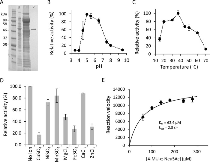
Exosialidases are glycoside hydrolases that remove a single terminal sialic acid residue from oligosaccharides. They are widely distributed in biology, having been found in prokaryotes, eukaryotes, and certain viruses. Most characterized prokaryotic sialidases are from organisms that are pathogenic or commensal with mammals. However, in this study, we used functional metagenomic screening to seek microbial sialidases encoded by environmental DNA isolated from an extreme ecological niche, a thermal spring. Using recombinant expression of potential exosialidase candidates and a fluorogenic sialidase substrate, we discovered an exosialidase having no homology to known sialidases. Phylogenetic analysis indicated that this protein is a member of a small family of bacterial proteins of previously unknown function. Proton NMR revealed that this enzyme functions via an inverting catalytic mechanism, a biochemical property that is distinct from those of known exosialidases. This unique inverting exosialidase defines a new CAZy glycoside hydrolase family we have designated GH156.

Keywords: extremophile; functional metagenomics; glycoside hydrolase; glycosylation; high-throughput screening (HTS); inverting mechanism; neuraminidase; sialic acid; sialidase.


Conflict of interest statement

The authors declare that they have no conflicts of interest with the contents of this article.

Figures

Figure 1.
Figure 1.
Screening for sialidase activity from a hot spring metagenomic library. A, restriction fragment analysis of 12 randomly selected clones from the hot spring metagenomic library were isolated and digested with the rare-cutting endonuclease SbfI. Digested fosmids were separated overnight on a 1% agarose gel along with a λHindIII size marker and a linearized pSMART FOS empty vector control (lane 14; contains one SbfI site). Each clone showed a unique banding pattern, with fragments whose combined sizes indicated the presence of an insert of at least 30–40 kb. B, E. coli cells harboring individual fosmid clones were assayed for sialidase activity with X-Neu5Ac incorporated into agar medium. A single positive clone forming a blue colony is denoted with an arrow. C, lysate from microcultures of E. coli cells harboring individual fosmid clones were assayed for sialidase activity with 4MU-α-Neu5Ac. A single positive clone is denoted with an arrow.
Figure 2.
Figure 2.
Predicted ORFs encoded in the fosmid G7 nucleotide sequence. A, fosmid G7 was sequenced on the PacBio RSII platform. ORFs were predicted with MetaGeneMark and classified into three categories based on their homology to proteins from the nonredundant protein database (NCBI). Black, proteins with a known annotated function; gray, “hypothetical proteins” with no annotated function; orange, proteins involved in saccharide utilization. B, database annotations for all 40 ORFs.
Figure 3.
Figure 3.
Identification of the sialidase-encoding ORF on fosmid G7 and its in vitro expression. A, a map of fosmid G7 transposon insertion sites (red lines) in mutants with abolished sialidase activity. B, SDS–PAGE of ORF9 and ORF12 proteins expressed in vitro using the PURExpress system. C, sialidase activity produced in PURExpress reaction mixtures was assessed using the substrate 4MU-α-Neu5Ac as described under “Experimental procedures.” D, the deduced amino acid sequence of ORF12p. The nucleotide sequence and the deduced protein sequence for ORF12 are annotated in the fosmid G7 sequence record (GenBankTM accession number MH016668).
Figure 4.
Figure 4.
Purification and biochemical characterization of recombinant ORF12p. A, His-tagged ORF12p sialidase was expressed in E. coli and purified using a His-trap column as described under “Experimental procedures.” Shown is SDS–PAGE separation of lysates from uninduced cells (U), induced cells (I), and nickel-purified ORF12p–His (P). B–D, purified ORF12p–His was used to determine the pH (B) and temperature (C) optima of ORF12p–His and the effect of metal ions on its catalysis (D). In these experiments, reactions were performed in triplicate using the substrate 3′-sialyl-N-acetyllactosamine-2AB. Reaction products were analyzed by UPLC–HILIC–FLR and quantitated by peak integration. E, Michaelis–Menten plot of ORF12p catalyzed hydrolysis of 4MU-α-Neu5Ac. The initial velocity was determined in triplicate for each 4MU-α-Neu5Ac concentration.
Figure 5.
Figure 5.
Specificity of ORF12p on sialic acid containing substrates using UPLC–HILIC–FLR analysis. A and B, the ability of ORF12p to cleave the fluorescently labeled substrates 3′- or 6′-sialyl-N-acetyllactosamine-2AB. Undigested substrates 3′- or 6′-sialyllactosamine-2AB run at ∼10.6- or 12.3-min retention times, respectively (A and B, top panels). Control digestion with the NeuA sialidase shifted both substrate peaks to ∼5.5-min retention time (A and B, bottom panels). Digestion of these substrates with 1 unit of ORF12p–His resulted in the same peak shift (A and B, middle panels). C, ORF12p's ability to hydrolyze α2–8 Neu5Ac was assessed using a 2AB-labeled GD3 ganglioside headgroup substrate that contains two sialic acid residues linked via an α2–8 bond. Undigested substrate ran at ∼16.5-min retention time with a very minor peak at ∼11-min retention time corresponding to partially degraded substrate comprised of a single α2–6 terminal sialic acid (C, top panel). NeuA-treated substrate shifted at ∼5.5 min retention time (C, bottom panel). Treatment with 1 unit of ORF12p did not shift the major substrate peak (C, middle panel). D and E, activity of ORF12p on biantennary complex N-glycans with terminal sialic acid residues (Neu5Ac, D; or Neu5Gc, E). Undigested substrates run at ∼26.6- and 27.9-min retention time, respectively (D and E, top panels). NeuA treatment shifted both substrate peaks at ∼23-min retention time (D and E, bottom panels). Incubation of the substrates with 1 or 10 units of ORF12p resulted in the same peak shift, but incomplete substrate desialylation was observed resulting in another smaller peak shift at ∼24.9- and 25.6-min retention time, respectively (D and E, middle panels). Symbolic representation of glycan structures was drawn following the guidelines of the Consortium for Functional Glycomics (50). EU, emission units.
Figure 6.
Figure 6.
Stereochemical course of the hydrolysis reaction catalyzed by ORF12p (A) compared with NeuA (B). One-dimensional proton NMR was used to monitor the reaction products formed over time upon hydrolysis of 4MU-α-Neu5Ac by ORF12p and NeuA. A, the up-field region of the NMR spectra showed two groups of peaks at 1.99 and 2.86 ppm corresponding to the substrate 4MU-α-Neu5Ac (black triangles). Over time, groups of peaks appeared at 1.81 and 2.20 ppm corresponding to released β-Neu5Ac (red triangles). After 1 h, two sets of signals at 1.61 and 2.72 ppm appear as a result of spontaneous mutarotation of β-Neu5Ac to α-Neu5Ac (blue triangles). B, the spectra showed the substrate 4MU-α-Neu5Ac groups of peaks at 2.03 and 2.89 ppm (black triangles). After 30 min, H3a and H3e peaks corresponding to the α-anomer appeared at 1.64 and 2.76 ppm (blue triangles). These were quickly converted by mutarotation to the β-anomer at 1.85 and 2.23 ppm (red triangles).
Figure 7.
Figure 7.
ORF12 protein family. A, ORF12p homologs were identified with the BLASTP algorithm and aligned with Muscle. A phylogenetic tree was then generated with the BLOSSUM62 matrix and the neighbor-joining method. ORF12p and Armatimonadetes OIO94155 are shown in red and blue text, respectively. B, the Armatimonadetes OIO94155 protein homolog was expressed in E. coli and crude lysate was tested with 4MU-α-Neu5Ac. C, protein sequence alignment (Muscle) of ORF12p and its closest homologs (A, yellow box), including Armatimonadetes OIO94155. Conserved residues are highlighted in yellow, and those potentially involved in catalysis are shown with red triangles.

Similar articles

Cited by

References

    1. Leis B., Angelov A., and Liebl W. (2013) Screening and expression of genes from metagenomes. Adv. Appl. Microbiol. 83, 1–68 10.1016/B978-0-12-407678-5.00001-5 - DOI - PubMed
    1. Cheng J., Romantsov T., Engel K., Doxey A. C., Rose D. R., Neufeld J. D., and Charles T. C. (2017) Functional metagenomics reveals novel β-galactosidases not predictable from gene sequences. PLoS One 12, e0172545 10.1371/journal.pone.0172545 - DOI - PMC - PubMed
    1. Mewis K., Armstrong Z., Song Y. C., Baldwin S. A., Withers S. G., and Hallam S. J. (2013) Biomining active cellulases from a mining bioremediation system. J. Biotechnol. 167, 462–471 10.1016/j.jbiotec.2013.07.015 - DOI - PubMed
    1. Coughlan L. M., Cotter P. D., Hill C., and Alvarez-Ordóñez A. (2015) Biotechnological applications of functional metagenomics in the food and pharmaceutical industries. Front. Microbiol. 6, 672 - PMC - PubMed
    1. Maruthamuthu M., Jiménez D. J., Stevens P., and van Elsas J. D. (2016) A multi-substrate approach for functional metagenomics-based screening for (hemi)cellulases in two wheat straw-degrading microbial consortia unveilsnovel thermoalkaliphilic enzymes. BMC Genomics. 17, 86 10.1186/s12864-016-2404-0 - DOI - PMC - PubMed

Publication types

MeSH terms

LinkOut - more resources