Review
. 2018 Oct 9;115(41):10209-10217.
doi: 10.1073/pnas.1804586115. Epub 2018 Sep 24.

Identity and novelty in the avian syrinx

Affiliations
Review

Identity and novelty in the avian syrinx

Evan P Kingsley et al. Proc Natl Acad Sci U S A. .

Abstract

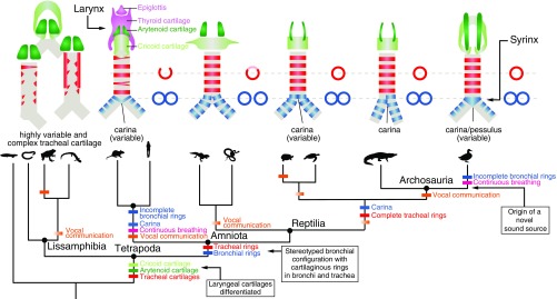
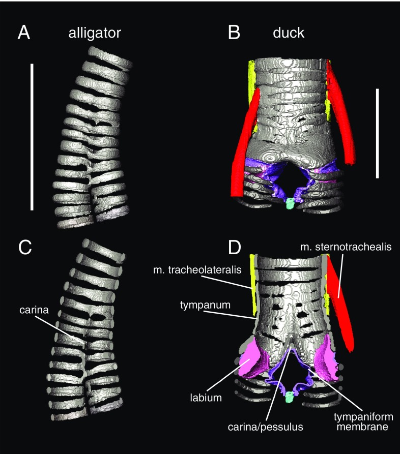
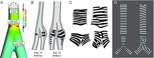
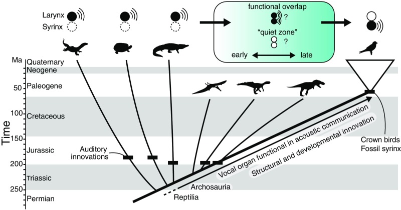
In its most basic conception, a novelty is simply something new. However, when many previously proposed evolutionary novelties have been illuminated by genetic, developmental, and fossil data, they have refined and narrowed our concept of biological "newness." For example, they show that these novelties can occur at one or multiple levels of biological organization. Here, we review the identity of structures in the avian vocal organ, the syrinx, and bring together developmental data on airway patterning, structural data from across tetrapods, and mathematical modeling to assess what is novel. In contrast with laryngeal cartilages that support vocal folds in other vertebrates, we find no evidence that individual cartilage rings anchoring vocal folds in the syrinx have homology with any specific elements in outgroups. Further, unlike all other vertebrate vocal organs, the syrinx is not derived from a known valve precursor, and its origin involves a transition from an evolutionary "spandrel" in the respiratory tract, the site where the trachea meets the bronchi, to a target for novel selective regimes. We find that the syrinx falls into an unusual category of novel structures: those having significant functional overlap with the structures they replace. The syrinx, along with other evolutionary novelties in sensory and signaling modalities, may more commonly involve structural changes that contribute to or modify an existing function rather than those that enable new functions.

Keywords: bioacoustics; birds; tetrapods; tracheal rings; vocal communication.


Conflict of interest statement

The authors declare no conflict of interest.

Figures

Fig. 1.
Fig. 1.
Tetrapod phylogeny showing the sequence of the acquisition of different airway traits through evolutionary time. (Upper, Left to Right) Schematic diagrams depict airway morphology in caecilians, frog, salamander, rat, gecko, tortoise, alligator, and a duck. Paired arytenoid cartilages (dark green) are present in most tetrapods (26). Cricoid cartilage (light green) is absent in some lissamphibians. Tracheal cartilage morphologies (red) are highly irregular (i.e., not ring-shaped) in lissamphibians (29), and irregular, forked rings are also sometimes observed in mammals (88). Stereotyped configuration of trachea and paired bronchi is common to all amniotes, and cartilaginous rings are observed in both the bronchi and trachea (49). Fusion of bronchial rings (blue) at the tracheobronchial juncture forms a carina, or a pessulus in birds (26). (Lower) Colored dashes indicate the branches along which distinct morphological and behavioral innovations (15, 61) may have evolved, with uncertainty (i.e., variability among species in a group) indicated by color gradients. Boxes describe major transitions leading to a syrinx in modern birds.
Fig. 2.
Fig. 2.
3D morphology of airway cartilage in archosaurs visible with diffusible iodine-based contrast-enhanced computed tomography (diceCT). 3D models of tracheal cartilage structure in the alligator (Alligator mississippiensis) (A and C) and Muscovy duck (Cairina moschata) (B and D) in gray. Panels show both external (A and B) and cross-sectional views of the tracheobronchial juncture (C and D). Soft tissue anatomy is clearly visible in B and D, including both intrinsic syringeal muscles (red and yellow), membranes (purple), and labia/vocal folds (pink). Specimens were dissected out, stained following ref. , and scanned at The University of Texas High-Resolution Computed Tomography Facility. Image segmentation was done in Avizo 6.3 (FEI Visualization Sciences Group). See ref. for further details on staining and scanning parameters. (Scale bars: 2 cm.)
Fig. 3.
Fig. 3.
Models inform hypotheses of airway cartilage evolution and development. (A) Airflow through tubes exposes tube walls to a wall shear stress (WSS) that is proportional to the flow velocity gradient (shown schematically at the top of the airway). Steep velocity gradients at the wall give rise to high WSS levels. Flow velocity throughout an airway bifurcation can be predicted by computational fluid dynamics simulations (SI Appendix, Methods S1). Spatial distribution of WSS derived from computed velocity profiles during exhalation is shown for a bifurcation, with regions of elevated WSS denoted. Regions of elevated WSS near the tracheobronchial juncture, such as those shown here, suggest different structural requirements than the rest of the airway, which may have influenced the evolution of vibrating sound sources and/or cartilage structure. (B) Earliest stages of syrinx cartilage formation in the male duck (adapted from ref. 77). The derived (larger, left–right asymmetric) morphology of the syrinx cartilage is present from its initiation and does not form by later shaping or differential growth of the airway cartilage. (C) Examples of human airway cartilage patterns (redrawn from ref. 49) in the trachea and the tracheobronchial juncture. (D) Simulation of a Turing system (with a gradient to orient stripes). Diverse cartilage patterns are predicted at the tracheobronchial juncture. Each panel in D corresponds to a different parameter value (SI Appendix, Methods S2). For computational ease, we solve on a flat, 2D domain, which we argue is a reasonable assumption given the low Gaussian curvature of the system.
Fig. 4.
Fig. 4.
Evolution of sound-producing mechanisms in archosaurs. There are two primary hypotheses for the evolutionary transition from a laryngeal sound source to a syringeal sound source (blue-shaded box). Auditory innovations shown as black dashes (15) suggest a sustained role for acoustic communication in archosaurs. Understanding whether the shift to a syringeal sound source occurred early or late in bird-lineage archosaurs will require further comparative genomic and paleontological work.

References

    1. Xu X, et al. An integrative approach to understanding bird origins. Science. 2014;346:1253293. - PubMed
    1. Muller GB, Wagner GP. Novelty in evolution: Restructuring the concept. Annu Rev Ecol Syst. 1991;22:229–256.
    1. Shubin N, Tabin C, Carroll S. Fossils, genes and the evolution of animal limbs. Nature. 1997;388:639–648. - PubMed
    1. Hall BK, Kerney R. Levels of biological organization and the origin of novelty. J Exp Zoolog B Mol Dev Evol. 2012;318:428–437. - PubMed
    1. Riede T, Li Z, Tokuda IT, Farmer CG. Functional morphology of the Alligator mississippiensis larynx with implications for vocal production. J Exp Biol. 2015;218:991–998. - PubMed

Publication types

LinkOut - more resources