. 2018 Oct 9;115(41):10481-10486.
doi: 10.1073/pnas.1804198115. Epub 2018 Sep 24.

Small molecule inhibits α-synuclein aggregation, disrupts amyloid fibrils, and prevents degeneration of dopaminergic neurons

Affiliations

Small molecule inhibits α-synuclein aggregation, disrupts amyloid fibrils, and prevents degeneration of dopaminergic neurons

Jordi Pujols et al. Proc Natl Acad Sci U S A. .

Abstract

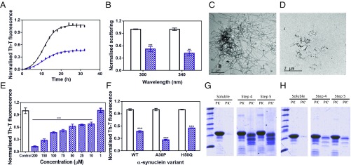
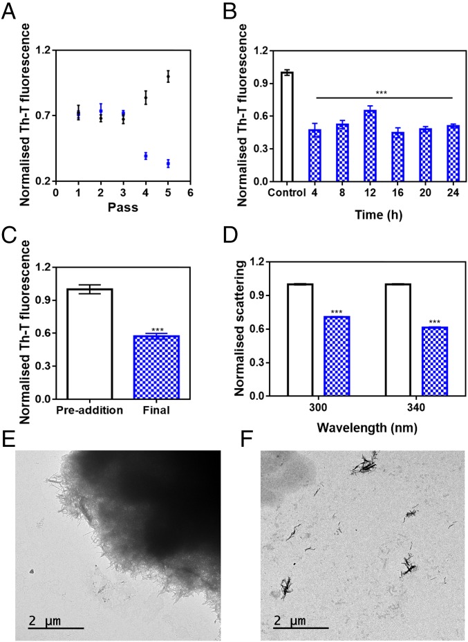
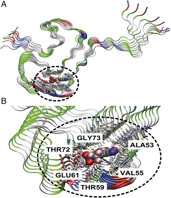
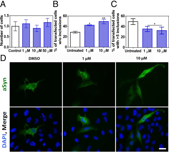
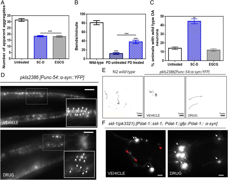
Parkinson's disease (PD) is characterized by a progressive loss of dopaminergic neurons, a process that current therapeutic approaches cannot prevent. In PD, the typical pathological hallmark is the accumulation of intracellular protein inclusions, known as Lewy bodies and Lewy neurites, which are mainly composed of α-synuclein. Here, we exploited a high-throughput screening methodology to identify a small molecule (SynuClean-D) able to inhibit α-synuclein aggregation. SynuClean-D significantly reduces the in vitro aggregation of wild-type α-synuclein and the familiar A30P and H50Q variants in a substoichiometric molar ratio. This compound prevents fibril propagation in protein-misfolding cyclic amplification assays and decreases the number of α-synuclein inclusions in human neuroglioma cells. Computational analysis suggests that SynuClean-D can bind to cavities in mature α-synuclein fibrils and, indeed, it displays a strong fibril disaggregation activity. The treatment with SynuClean-D of two PD Caenorhabditis elegans models, expressing α-synuclein either in muscle or in dopaminergic neurons, significantly reduces the toxicity exerted by α-synuclein. SynuClean-D-treated worms show decreased α-synuclein aggregation in muscle and a concomitant motility recovery. More importantly, this compound is able to rescue dopaminergic neurons from α-synuclein-induced degeneration. Overall, SynuClean-D appears to be a promising molecule for therapeutic intervention in Parkinson's disease.

Keywords: Parkinson’s disease; aggregation inhibition; dopaminergic degeneration; protein aggregation; α-synuclein.


Conflict of interest statement

Conflict of interest statement: J.P., S.P.-D., M.C.-G., J.S., E.D., and S.V. are inventors on a patent application (PCT/EP2018/054540) related to the compound in this study.

Figures

Fig. 1.
Fig. 1.
Effect of SynuClean-D on the aggregation of α-Syn in vitro. (A) α-Syn aggregation kinetics in the absence (black) and presence (blue) of SC-D followed by Th-T–derived fluorescence. (B) Light-scattering signal at 300 and 340 nm, both in the absence (white) and presence (blue) of SC-D. (C and D) Representative TEM images in the absence (C) and presence (D) of SC-D. (E) Inhibition of α-Syn aggregation in the presence of different concentrations of SC-D. (F) H50Q and A30P α-Syn variant aggregation in the absence (white) and presence (blue) of SC-D. (G and H) Bis/Tris gels of PMCA samples in the absence (G) and presence (H) of SC-D, both analyzed after PK digestion. Soluble α-Syn and PMCA steps 4 and 5 are shown. Th-T fluorescence is plotted as normalized means. Final points were obtained at 48 h. Error bars are represented as SE of mean values; **P < 0.01 and ***P < 0.001.
Fig. 2.
Fig. 2.
Disaggregational capacity of SynuClean-D. (A) Th-T fluorescence of the different PMCA passes of both treated (blue) and untreated (black) samples with SC-D. (B) Aggregation kinetics of α-Syn after the addition of SC-D at different time points. (C and D) Th-T–derived fluorescence (C) and light-scattering (D) assays before and after the addition of SC-D to preformed α-Syn fibrils. (E and F) Representative TEM images in the absence (E) and presence (F) of SC-D. Th-T fluorescence is plotted as normalized means. Error bars are represented as SE of mean values; ***P < 0.001.
Fig. 3.
Fig. 3.
Characterization of SynuClean-D–fibril interaction. General view (A) and zoom (B) of the most stable binding pose of SC-D on the α-Syn fibril model.
Fig. 4.
Fig. 4.
Inhibition of α-Syn aggregate formation in cultured cells. (A) Human neuroglioma cell (H4) survival when incubated with different concentrations of compound (blue) and without (white) the compound. (B and C) Reduction of α-Syn inclusion formation in human cultured cells in the presence of different concentrations of SC-D. (B) Percentage of transfected cells devoid of α-Syn aggregates. (C) Percentage of transfected cells bearing >5 α-Syn aggregates. (D) Representative epifluorescent images from cells treated with SC-D. n = 3. (Scale bar, 30 μm.) Error bars are represented as SE of means; *P < 0.05 and **P < 0.01.
Fig. 5.
Fig. 5.
Inhibition effect of the compound in the formation of α-Syn inclusions and protection from the α-Syn–induced dopaminergic cell death in C. elegans models of PD. (A) Quantification of α-Syn muscle inclusions per area of NL5901 worms in the absence (white) and presence of SC-D (blue) or EGCG (gray). (B) Worm-thrashing representation as the number of bends per minute of N2 wild-type and NL5901 worms treated without (white) and with SC-D (blue). (C) Percentage of UA196 worms that maintain a complete set of dopaminergic neurons (four pairs located in the head) after treatment without (white) and with SC-D (blue) or EGCG (gray) for 7 d after the L4 stage. (D) Representative images of α-Syn muscle aggregates obtained by epifluorescence microscopy of NL5901 worms treated without (Top, vehicle) and with SC-D (Bottom, drug). (Scale bars, 10 μm.) Between 40 and 50 animals were analyzed per condition. Aggregates are indicated by white arrows. (E) Path representation of the mobility of N2 wild-type (Left, vehicle) and NL5901 worms grown without (Middle, vehicle) and with SC-D (Right, drug). (Scale bars, 1 mm.) (F) Representative worms expressing GFP–α-Syn specifically in DA neurons without (Left, vehicle) and with SC-D (Right, drug) for 7 d after L4. Healthy neurons are labeled with white arrows, whereas neurodegenerated or missing neurons are labeled with red arrows. (Scale bars, 30 μm.) Between 40 and 50 animals were analyzed per condition in each experiment. Data are shown as means, and error bars are shown as the SE of means; **P < 0.01 and ***P < 0.001.

References

    1. Kalia LV, Lang AE. Parkinson’s disease. Lancet. 2015;386:896–912. - PubMed
    1. Fanciulli A, Wenning GK. Multiple-system atrophy. N Engl J Med. 2015;372:1375–1376. - PubMed
    1. Spillantini MG, Crowther RA, Jakes R, Hasegawa M, Goedert M. Alpha-synuclein in filamentous inclusions of Lewy bodies from Parkinson’s disease and dementia with Lewy bodies. Proc Natl Acad Sci USA. 1998;95:6469–6473. - PMC - PubMed
    1. Luk KC, et al. Pathological α-synuclein transmission initiates Parkinson-like neurodegeneration in nontransgenic mice. Science. 2012;338:949–953. - PMC - PubMed
    1. Winner B, et al. In vivo demonstration that alpha-synuclein oligomers are toxic. Proc Natl Acad Sci USA. 2011;108:4194–4199. - PMC - PubMed

Publication types

MeSH terms