. 2018 Dec 3;217(12):4184-4198.
doi: 10.1083/jcb.201805039. Epub 2018 Sep 24.

GTP hydrolysis promotes disassembly of the atlastin crossover dimer during ER fusion

Affiliations

GTP hydrolysis promotes disassembly of the atlastin crossover dimer during ER fusion

James Winsor et al. J Cell Biol. .

Abstract

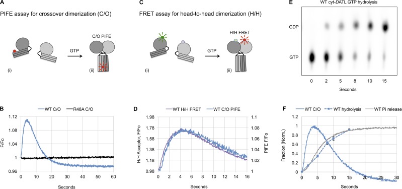
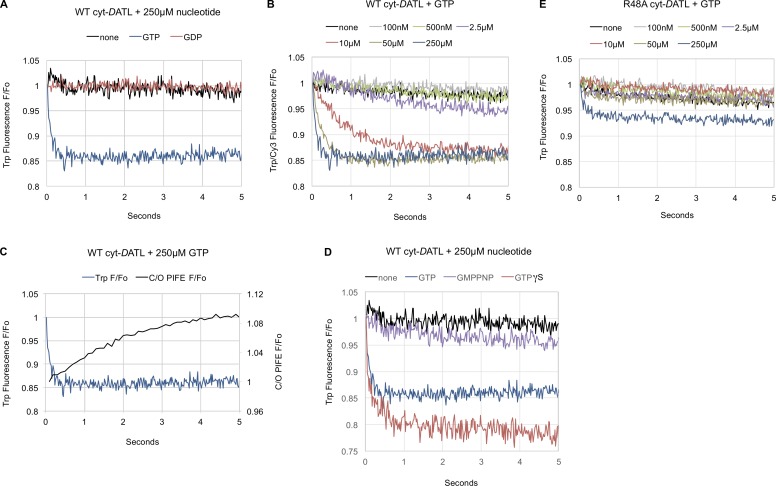
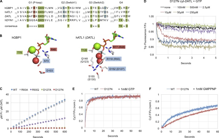
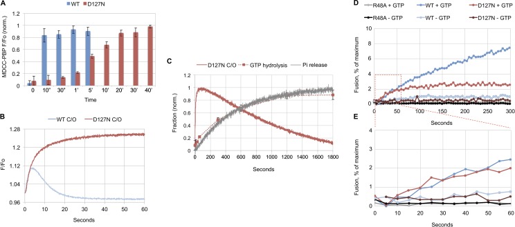
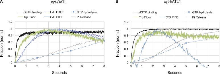
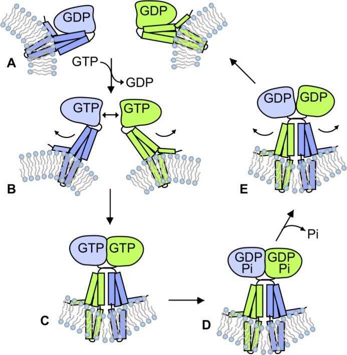
Membrane fusion of the ER is catalyzed when atlastin GTPases anchored in opposing membranes dimerize and undergo a crossed over conformational rearrangement that draws the bilayers together. Previous studies have suggested that GTP hydrolysis triggers crossover dimerization, thus directly driving fusion. In this study, we make the surprising observations that WT atlastin undergoes crossover dimerization before hydrolyzing GTP and that nucleotide hydrolysis and Pi release coincide more closely with dimer disassembly. These findings suggest that GTP binding, rather than its hydrolysis, triggers crossover dimerization for fusion. In support, a new hydrolysis-deficient atlastin variant undergoes rapid GTP-dependent crossover dimerization and catalyzes fusion at an initial rate similar to WT atlastin. However, the variant cannot sustain fusion activity over time, implying a defect in subunit recycling. We suggest that GTP binding induces an atlastin conformational change that favors crossover dimerization for fusion and that the input of energy from nucleotide hydrolysis promotes complex disassembly for subunit recycling.


Figures

Figure 1.
Figure 1.
Atlastin (DATL) dimerization and crossover precedes GTP hydrolysis and Pi release. (A–F) Single-turnover kinetics of cyt-DATL. (A) Schematic of Cy3 fluorescence enhancement (PIFE) as Cy3-labeled cyt-DATL (i) undergoes dimer formation and crossover (ii). (B) Stopped-flow PIFE of WT or R48A cyt-DATL after addition of GTP. (C) Schematic of FRET as cyt-DATL monomers labeled with a FRET donor and acceptor (i) undergo H/H dimerization (ii). (D) Comparison of WT H/H FRET with WT crossover PIFE (C/O) after GTP addition under stopped flow. (E) Hydrolysis of GTP (containing a-32P-GTP) by WT cyt-DATL at room temperature. Reactions were acid quenched at the indicated times (one of three replicates shown). (F) Comparison of WT crossover PIFE (C/O), GTP hydrolysis, and Pi release. PIFE (C/O) trace is from B except normalized (initial value = 0; maximum value = 1). GTP hydrolysis was the average of three replicates from E (±SEM). Pi release was measured by including 15 µM MDCC-PBP (final) in the stopped flow and normalized (initial value = 0; maximum value = 1). All reactions contained 15 µM cyt-DATL and 7.5 µM GTP (final concentrations) and were performed at 25°C except where indicated. All traces are the average of three to five individual traces and are representative of two independent protein preparations.
Figure 2.
Figure 2.
Neither GMPPNP nor the R48A mutation replicates a GTP-induced atlastin G domain conformational change. (A–E) Intrinsic tryptophan fluorescence of 125 nM WT, D127N, or R48A cyt-DATL after addition of the indicated concentrations of the indicated nucleotides under stopped flow at 25°C. (A) WT after mixing with 250 µM GTP, GDP, or buffer. (B) WT tryptophan fluorescence quenching with the indicated concentrations of GTP. (C) Crossover dimerization is slower than tryptophan fluorescence quenching under the same conditions. WT tryptophan fluorescence trace with 250 µM GTP (from A) replotted relative to WT crossover PIFE under the same conditions. (D) WT with 250 µM GTP, GMPPNP, or GTPγS. (E) R48A with the indicated concentrations of GTP. All concentrations are final after mixing. All traces are the average of three to five individual traces.
Figure 3.
Figure 3.
A novel D127N active site mutation in cyt-DATL inhibits GTP hydrolysis but not the GTP-induced G domain conformational change or crossover dimer formation. (A) Sequence alignment of hGBP1, hATL1, DATL, and human dynamin1 (hDYN1) showing the positions of D127 and R93 in DATL relative to other signature GTPase residues. Residues conserved across GTPases are in green, catalytic arginine is in red, and D127 and R93 are in blue. (B) Side chains of active site residues in hGBP1 (PDB ID 2B92) and hATL1 (PDB ID 4IDQ) bound to GDP ⋅ AlF4 rendered in PyMOL. Magnesium ion and waters are shown as green and red spheres, respectively. (C) Steady-state GTPase assay of Pi release by WT, D127A, D127N, R93A, and R93Q cyt-DATL upon addition of GTP (n = 3; ± SEM). (D) Stopped-flow intrinsic tryptophan fluorescence quenching by D127N cyt-DATL after mixing with the indicated concentrations of GTP (as described in Fig. 2). (E and F) Stopped-flow PIFE of 2 µM WT or D127N cyt-DATL after addition of 1 mM GTP (E) or 1 mM GMPPNP (F). Traces in E and F were normalized (minimum value = 0; maximum value = 1), are the average of three runs, and are representative of two independent protein preparations.
Figure 4.
Figure 4.
GTP hydrolysis is not required for crossover dimerization or initial fusion. (A–C) WT and D127N DATL single-turnover kinetics. All reactions contained 15 µM cyt-DATL and 7.5 µM GTP (final concentrations) and were performed at 25°C except where indicated. (A) GTP hydrolysis by WT or D127N cyt-DATL at room temperature. Reactions were quenched at the indicated times, and total Pi produced was measured after 10-fold dilution into 1.5 µM MDCC-PBP (n = 3; ±SD). (B) Stopped-flow PIFE of WT or D127N cyt-DATL after addition of GTP. The WT trace is the same as in Fig. 1 B. (C) Comparison of D127N crossover PIFE (C/O), GTP hydrolysis, and Pi release. D127N C/O PIFE was replotted from B after normalization. GTP hydrolysis was replotted from A. Single-turnover D127N Pi release was measured by including 15 µM MDCC-PBP (final) in the stopped flow. Traces were normalized (initial value = 0; maximum value = 1). (D and E) Fusion assay. (D) Full-length WT, R48A, or D127N DATL was reconstituted into donor and acceptor vesicles at a 1:1,000 protein/lipid ratio, and fusion was monitored as the dequenching of NBD-labeled lipid present in the donor vesicles over time at 28°C after addition of 1 mM GTP (average of three runs plotted). (E) Magnified view of the early time points of traces boxed in D. All stopped flow, GTP hydrolysis, and fusion kinetics are representative of two independent protein preparations.
Figure 5.
Figure 5.
Crossover dimerization of hATL1 also precedes GTP hydrolysis and Pi release. (A–C) Single-turnover kinetics of cyt-hATL1. (A) Stopped-flow PIFE of WT cyt-hATL1 after addition of GTP. (B) Hydrolysis of GTP (containing a-32P-GTP) by WT cyt-hATL1 at room temperature. Reactions were acid quenched at the indicated times (one of three replicates shown). (C) Comparison of WT crossover PIFE (C/O), GTP hydrolysis, and Pi release. Traces were normalized (initial value = 0; maximum value = 1). Normalized PIFE (C/O) trace is from A. GTP hydrolysis was the average of three replicates from B (±SEM). Pi release was measured by including 15 µM MDCC-PBP (final) in the stopped flow. All reactions contained 15 µM cyt-hATL1 and 7.5 µM GTP (final concentrations) and were performed at 25°C except where indicated. All traces are the average of three to five individual traces and are representative of two independent protein preparations.
Figure 6.
Figure 6.
DATL and hATL1 undergo a similar sequence of events during their GTPase cycle. (A and B) Timing of GTP-induced events for cyt-DATL (A) and cyt-hATL1 (B). dGTP binding measured using mant-dGTP and intrinsic tryptophan fluorescence quenching (Trp fluor) after GTP addition. All reactions were performed with stopped flow under single-turnover conditions with 15 µM cyt-DATL or cyt-hATL1 and 7.5 µM GTP or mant-dGTP (final concentrations). H/H FRET, C/O PIFE, GTP hydrolysis, and Pi release data for cyt-DATL (A) are replotted from Fig. 1 (D and F). C/O PIFE, GTP hydrolysis, and Pi release data for cyt-hATL1 (B) are replotted from Fig. 5 C. All traces were normalized (initial value = 0; maximum value = 1). Tryptophan fluorescence data are plotted as (1-normalized fluorescence) for ease of comparison. All traces are the average of three to five individual traces.
Figure 7.
Figure 7.
Model depicting the role of GTP hydrolysis in atlastin-catalyzed fusion. (A–E) GDP-bound monomers (A) undergo a conformational change upon GTP binding (B) that triggers dimerization and crossover for fusion (C). After fusion, hydrolysis of GTP (D) and release of Pi trigger dimer disassembly (E) to restart the cycle (A). Note that the order of dimer dissociation and GDP release is not necessarily being specified in this figure, and GDP could be released before dimer disassembly.

Similar articles

Cited by

References

    1. Anwar K., Klemm R.W., Condon A., Severin K.N., Zhang M., Ghirlando R., Hu J., Rapoport T.A., and Prinz W.A.. 2012. The dynamin-like GTPase Sey1p mediates homotypic ER fusion in S. cerevisiae. J. Cell Biol. 197:209–217. 10.1083/jcb.201111115 - DOI - PMC - PubMed
    1. Bian X., Klemm R.W., Liu T.Y., Zhang M., Sun S., Sui X., Liu X., Rapoport T.A., and Hu J.. 2011. Structures of the atlastin GTPase provide insight into homotypic fusion of endoplasmic reticulum membranes. Proc. Natl. Acad. Sci. USA. 108:3976–3981. 10.1073/pnas.1101643108 - DOI - PMC - PubMed
    1. Brune M., Hunter J.L., Corrie J.E., and Webb M.R.. 1994. Direct, real-time measurement of rapid inorganic phosphate release using a novel fluorescent probe and its application to actomyosin subfragment 1 ATPase. Biochemistry. 33:8262–8271. 10.1021/bi00193a013 - DOI - PubMed
    1. Byrnes L.J., and Sondermann H.. 2011. Structural basis for the nucleotide-dependent dimerization of the large G protein atlastin-1/SPG3A. Proc. Natl. Acad. Sci. USA. 108:2216–2221. 10.1073/pnas.1012792108 - DOI - PMC - PubMed
    1. Byrnes L.J., Singh A., Szeto K., Benvin N.M., O’Donnell J.P., Zipfel W.R., and Sondermann H.. 2013. Structural basis for conformational switching and GTP loading of the large G protein atlastin. EMBO J. 32:369–384. 10.1038/emboj.2012.353 - DOI - PMC - PubMed

Publication types

MeSH terms