Review
. 2018 Sep 24;373(1759):20170321.
doi: 10.1098/rstb.2017.0321.

Mechanics of cortical folding: stress, growth and stability

Affiliations
Review

Mechanics of cortical folding: stress, growth and stability

K E Garcia et al. Philos Trans R Soc Lond B Biol Sci. .

Abstract

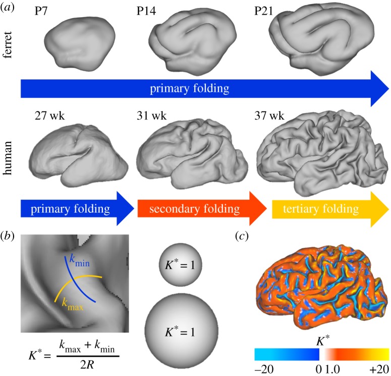
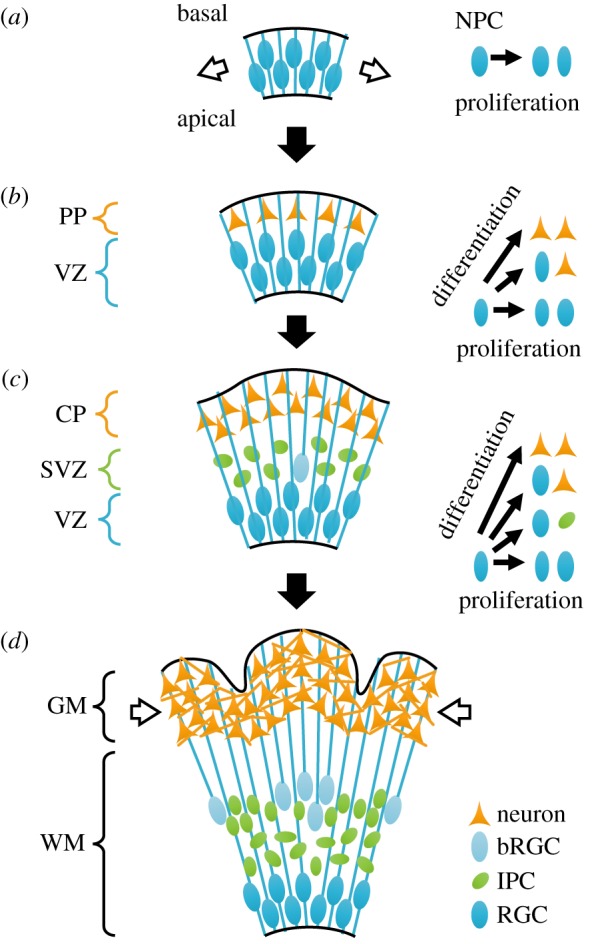
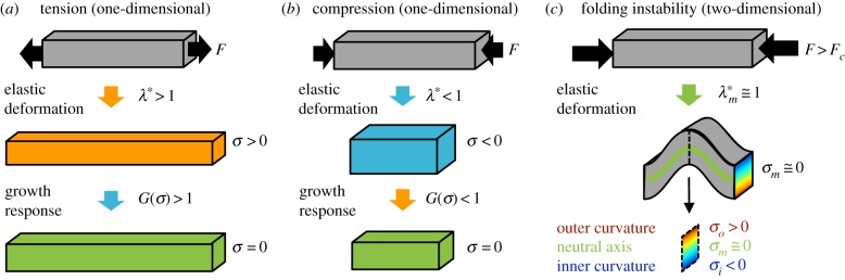
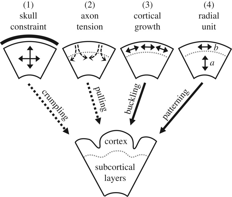
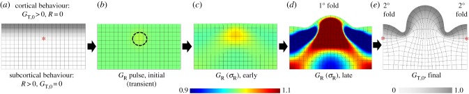
Cortical folding, or gyrification, coincides with several important developmental processes. The folded shape of the human brain allows the cerebral cortex, the thin outer layer of neurons and their associated projections, to attain a large surface area relative to brain volume. Abnormal cortical folding has been associated with severe neurological, cognitive and behavioural disorders, such as epilepsy, autism and schizophrenia. However, despite decades of study, the mechanical forces that lead to cortical folding remain incompletely understood. Leading hypotheses have focused on the roles of (i) tangential growth of the outer cortex, (ii) spatio-temporal patterns in the birth and migration of neurons, and (iii) internal tension in axons. Recent experimental studies have illuminated not only the fundamental cellular and molecular processes underlying cortical development, but also the stress state, mechanical properties and spatio-temporal patterns of growth in the developing brain. The combination of mathematical modelling and physical measurements has allowed researchers to evaluate hypothesized mechanisms of folding, to determine whether each is consistent with physical laws. This review summarizes what physical scientists have learned from models and recent experimental observations, in the context of recent neurobiological discoveries regarding cortical development. Here, we highlight evidence of a combined mechanism, in which spatio-temporal patterns bias the locations of primary folds (i), but tangential growth of the cortical plate induces mechanical instability (ii) to propagate primary and higher-order folds.This article is part of the Theo Murphy meeting issue 'Mechanics of development'.

Keywords: cortical folding; growth; gyrification; instability; modelling; stress.


Conflict of interest statement

We declare we have no competing interests.

Figures

Figure 1.
Figure 1.
Measuring cortical folding. (a) The progression of cortical folding is shown after birth in ferret (top) and preterm human (bottom). (b) Dimensionless mean curvature (K*), the average of maximum and minimum principal curvatures (kmax and kmin) multiplied by the surface's characteristic radius (R), offers a useful metric of local folding. As shown for two spheres of different radius, K* is unaffected by size (K* = 1 everywhere on a sphere, regardless of scale). (c) Map of dimensionless mean curvature displays the complexity of the 37 week human surface in (a). P = postnatal day, wk = weeks postmenstrual age. (Online version in colour.)
Figure 2.
Figure 2.
Development in the gyrencephalic brain. (a) During early embryonic stages, the brain wall consists of pseudostratified neuroepithelial progenitor cells (NPCs, blue ellipses). Each cell maintains contact with both apical (inner) and basal (outer) surfaces. During embryonic stages, these cells experience mechanical tension (white arrows) [11,12]. (b) As development continues, some NPCs divide asymmetrically to produce differentiated neurons (orange triangles), which do not maintain contact with the apical wall. Instead, neurons migrate toward the basal, outer surface along the radial scaffold (blue lines) provided by NPCs, the precursors to radial glial cells (RGCs). (c) In later stages, some NPCs divide to produce intermediate progenitor cells (IPCs, green ellipses), and some lose contact with the apical surface to form basal radial glial cells (bRGCs, light blue ellipses). These proliferative cell types populate the subventricular zone (SVZ), between the ventricular zone (VZ, filled with RGCs) and cortical plate (CP, filled with neurons). (d) As neurons morphologically mature in the cortical plate, cortical grey matter (GM) expands faster than subcortical white matter (WM). At late stages, constrained growth of the grey matter may result in mechanical compression (white arrows). PP = preplate. (Online version in colour.)
Figure 3.
Figure 3.
Elastic, viscoelastic and unstable behaviour in response to mechanical force. (a–c) Application of axial force (F) is shown for a hypothetical bar-shaped element of tissue. In all cases, the sum of forces on the bar is equal, denoted by black arrows in opposing directions. (a) Under pulling forces, the bar will stretch elastically (λ* > 1), resulting in tensile stress (σ > 0). In the case of either viscoelastic tissue or growing tissue, sustained tension may lead to permanent growth and relaxation of the original stress. (b) Conversely, under pushing forces, the bar will shorten elastically (λ* < 1), resulting in compressive stress (σ < 0) and—potentially—tissue shrinking to relieve stress. (c) Under sufficiently high compressive force, the same bar may buckle to a lower energy configuration. In the resulting fold, stress is tensile along the outer curvature region (red) and compressive along the inner curvature region (blue) due to bending. The neutral axis (green) represents the intermediate location where stresses due to bending are zero. (Online version in colour.)
Figure 4.
Figure 4.
Competing (or complementary) hypotheses for cortical folding. Dotted arrows (1 and 2) denote mechanisms not consistent with prior experiments. Additional experiments are needed to clarify the true mechanism(s) of cortical folding, which could include a combination of these hypotheses.
Figure 5.
Figure 5.
Evolution of folding based on initial radial patterning and growth-induced instability. (a) As described in Bayly et al. [33], cortex (grey) was defined to grow tangentially at a pre-determined rate (GT,0), while growth of subcortical layers evolves in response to stress GR(σR) and GT(σT), with rate constant R. (b) A small, transient perturbation in radial growth was applied (region circled by dotted line) to influence the location of the first fold. (c) After the perturbation, mechanical feedback, combined with tangential growth of the cortex, causes increased radial stress and growth in this region. (d) A primary (1°) gyrus later develops above the initial perturbation, and regions of higher radial growth develop to the right and left. (e) As the model continues, secondary (2°) gyri emerge from these new regions of higher radial growth. Red asterisks denote local growth concentrations beneath prospective gyri. (Online version in colour.)
Figure 6.
Figure 6.
Dynamic patterns of tangential growth in human brain development. (a) In preterm infants, patterns of cortical expansion (red) change from 28–30 weeks (top) to 30–34 weeks (middle) to 34–38 weeks (bottom). (b) In healthy infants, areas of highest cortical expansion (red) are consistent with the trajectory from preterm development. (c) Schematic illustrating the trajectory of the maximum growth region from primary motor, sensory and visual cortices (labelled ‘pre’) to frontal, parietal and temporal lobes (labelled ‘post’). Pre, prenatal/preterm; post, postnatal. Reproduced from [7] with permission. (Online version in colour.)

References

    1. Welker W. 1990. Why does the cortex fissure and fold: a review of determinants of gyri and sulci. In Cerebral cortex: comparative structure and evolution of cerebral cortex (eds Jones EG, Peters A), pp. 3–136. New York, NY: Plenum Press.
    1. Le Gros Clark WE. 1945. Deformation patterns in the cerebral cortex. In Essays on growth and form (eds Le Gros Clark WE, Medawar PB), pp. 1–22. London, UK: Oxford University Press.
    1. Smart IH, McSherry GM. 1986. Gyrus formation in the cerebral cortex of the ferret. II. Description of the internal histological changes. J. Anat. 147, 27–43. - PMC - PubMed
    1. Smart IH, McSherry GM. 1986. Gyrus formation in the cerebral cortex in the ferret. I. Description of the external changes. J. Anat. 146, 141–152. - PMC - PubMed
    1. Sun T, Hevner RF. 2014. Growth and folding of the mammalian cerebral cortex: from molecules to malformations. Nat. Rev. Neurosci. 15, 217–232. (10.1038/nrn3707) - DOI - PMC - PubMed

Publication types

LinkOut - more resources