. 2018 Nov 7;38(45):9768-9780.
doi: 10.1523/JNEUROSCI.0495-18.2018. Epub 2018 Sep 24.

Regulation of the Natriuretic Peptide Receptor 2 (Npr2) by Phosphorylation of Juxtamembrane Serine and Threonine Residues Is Essential for Bifurcation of Sensory Axons

Affiliations

Regulation of the Natriuretic Peptide Receptor 2 (Npr2) by Phosphorylation of Juxtamembrane Serine and Threonine Residues Is Essential for Bifurcation of Sensory Axons

Hannes Schmidt et al. J Neurosci. .

Abstract

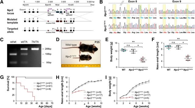
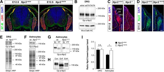
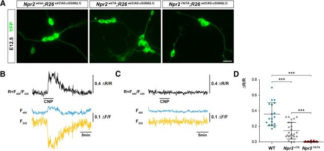
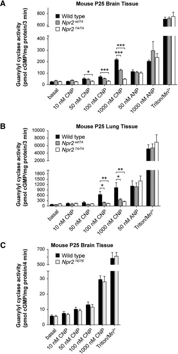
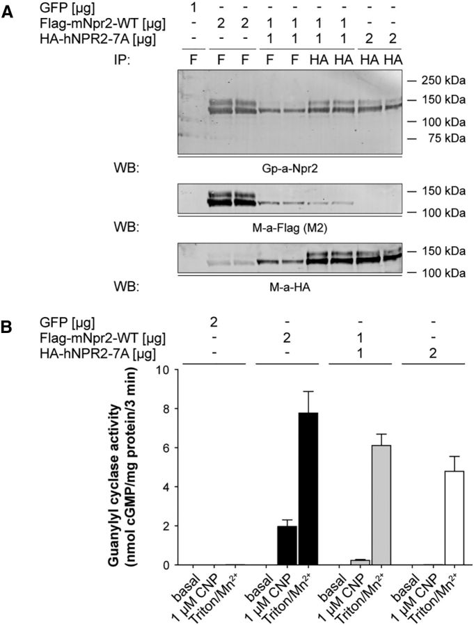
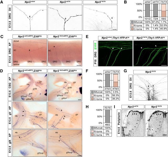
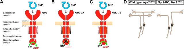
cGMP signaling elicited by activation of the transmembrane receptor guanylyl cyclase Npr2 (also known as guanylyl cyclase B) by the ligand CNP controls sensory axon bifurcation of DRG and cranial sensory ganglion (CSG) neurons entering the spinal cord or hindbrain, respectively. Previous studies have shown that Npr2 is phosphorylated on serine and threonine residues in its kinase homology domain (KHD). However, it is unknown whether phosphorylation of Npr2 is essential for axon bifurcation. Here, we generated a knock-in mouse line in which the seven regulatory serine and threonine residues in the KHD of Npr2 were substituted by alanine (Npr2-7A), resulting in a nonphosphorylatable enzyme. Real-time imaging of cGMP in DRG neurons with a genetically encoded fluorescent cGMP sensor or biochemical analysis of guanylyl cyclase activity in brain or lung tissue revealed the absence of CNP-induced cGMP generation in the Npr27A/7A mutant. Consequently, bifurcation of axons, but not collateral formation, from DRG or CSG in this mouse mutant was perturbed at embryonic and mature stages. In contrast, axon branching was normal in a mouse mutant in which constitutive phosphorylation of Npr2 is mimicked by a replacement of all of the seven serine and threonine sites by glutamic acid (Npr2-7E). Furthermore, we demonstrate that the Npr27A/7A mutation causes dwarfism as described for global Npr2 mutants. In conclusion, our in vivo studies provide strong evidence that phosphorylation of the seven serine and threonine residues in the KHD of Npr2 is an important regulatory element of Npr2-mediated cGMP signaling which affects physiological processes, such as axon bifurcation and bone growth.SIGNIFICANCE STATEMENT The branching of axons is a morphological hallmark of virtually all neurons. It allows an individual neuron to innervate different targets and to communicate with neurons located in different regions of the nervous system. The natriuretic peptide receptor 2 (Npr2), a transmembrane guanylyl cyclase, is essential for the initiation of bifurcation of sensory axons when entering the spinal cord or the hindbrain. By using two genetically engineered mouse lines, we show that phosphorylation of specific serine and threonine residues in juxtamembrane regions of Npr2 are required for its enzymatic activity and for axon bifurcation. These investigations might help to understand the regulation of Npr2 and its integration in intracellular signaling systems.

Keywords: Npr2; axonal branching; cGMP signaling; phosphorylation; sensory axons.


Figures

Figure 1.
Figure 1.
Generation and validation of the Npr2-7A mouse mutant. A, Targeting strategy for the replacement of seven serine and threonine phosphorylation sites that are encoded in exons 8 and 9 of the murine Npr2 gene by Crispr/Cas9. PAM, Protospacer adjacent motif; sgRNA, synthetic guide RNA. P1 and P2 indicate primer locations for genotyping. B, Sequencing of genomic DNA of the mutated locus in Npr27A/7A and in wild-type, demonstrating the replacement of specific nucleotides in the kinase homology domain. These replacements result in codons, which encode alanine residues instead of serine or threonine residues as indicated below the sequencing information. C, PCR genotyping using primer P1 and P2 followed by digestion with restriction endonuclease NheI, which specifically splits the wild-type sequence but not the mutated segment. D–F, Depiction and quantification of wild-type and Npr27A/7A littermates at postnatal day 25, demonstrating a severely reduced body length and body weight due to impaired endochondral ossification as described previously for other Npr2 loss-of-function mutants. G, Survival rate of Npr27A/7A, Npr2wt/7A, and Npr2wt/wt mice at different ages. Numbers of animals inspected are given in parentheses. H, Naso-anal length of Npr27A/7A, Npr2wt/7A, and Npr2wt/wt male mice at different ages. I, Body weight of Npr27A/7A, Npr2wt/7A, and Npr2wt/wt male mice at different ages. Females are not shown because they were underrepresented in these cohorts, but the same tendency was observed. *p < 0.05; **p < 0.01.
Figure 2.
Figure 2.
Npr2-7A protein is expressed on the cell surface of DRGs. A, Cross sections of the spinal cord from Npr27A/7A and Npr2wt/7A E12.5 embryos stained with anti-neurofilament, anti-cGKI, and DAPI reveal a normal overall structure of the spinal cord in Npr27A/7A mutants. Scale bar, 100 μm. B, Western blots of detergent extracts of DRGs from E13.5 Npr27A/7A, Npr2wt/7A, and Npr2wt/wt embryos, indicating similar amounts of Npr2 protein in all three genotypes. Npr2 and Npr2-7A are composed of two major bands. The bottom served as loading control using a monoclonal antibody to the heavy chain of clathrin. Right, Molecular mass markers. C, D, Transversal sections of the spinal cord from Npr27A/7A, Npr2LacZ/LacZ (representing Npr2−/−), and Npr2wt/wt stained with anti-Npr2, demonstrating expression of Npr2-7A-mutated protein in the somata of DRG neurons, in the dorsal root and dorsal root entry zone and the developing dorsal funiculus axons. Hyphenated line indicates the border of the spinal cord. Scale bar, 50 μm. D, Dorsal; DR, dorsal root; DREZ, dorsal root entry zone; V, ventral. E, F, Expression of Npr2 variants on the surface of DRGs and of cortical astrocyte cultures from wild-type or Npr27A/7A mutants. Cell surface proteins on freshly picked DRGs or cultivated astrocytes were labeled by sulfo-NHS-biotin followed by isolation with streptavidin agarose. Bottom, Equal protein loading was controlled by streptavidin-HRP. Right, Molecular mass markers. G, Western blot of Npr2 and Np2–7A from cortical astrocyte cultures that were cell surface biotinylated by sulfo-NHS-biotin followed by purification using streptavidin beads. The biotinylated fraction is composed of one major band in both genotypes, whereas the unbound fraction contains two bands (for DRGs, see also Fig. 2B). EL, Eluate of streptavidin beads (bound, biotinylated fraction of Npr2); FT, flow through of streptavidin beads (unbound fraction of Npr2). H, Western blot of Npr2 from astrocyte cultures treated with endoglycosidase F (EnF) or endoglycosidase H (EnH). EnF treatment resulted in one major band, whereas EnH treatment resulted in a significant shift of the lower Npr2 band. A weak stained band between both major bands might represent not fully endoglycosidase H-dependent deglycosylated Npr2. I, Quantification of Western blots of the ratio of the two major components from DRGs or astrocyte cultures from Npr27A/7A or Npr2wt/wt. Upper band is considered as mature Npr2 expressed at the cell surface. Lower band is considered as immature Npr2 expressed intracellularly. Number of blots analyzed: 9 for DRGs of each genotype, 12 for wild-type, and 9 for mutant astrocytes. Error bar indicates SEM. *p = 0.0197, for astrocytes (t test).
Figure 3.
Figure 3.
FRET-based cGMP imaging of individual DRG neurons from E12.5 Npr2wt/wt, Npr2wt/7A, and Npr27A/7A embryos expressing the genetically encoded cGi500 cGMP biosensor. A, Representative images of cGi500 biosensor fluorescence in dissociated E12.5 DRG neurons from Npr27A/7A, Npr2wt/7A, and Npr2wt/wt 24 h after plating. Scale bar, 10 μm. B, C, Changes in fluorescence of the genetically encoded cGi500 cGMP biosensor elicited by application of 200 nm CNP (horizontal bar) recorded from individual DRG neurons from wild-type (B) and Npr27A/7A (C) mice. Traces indicate CFP emission (F480, blue), YFP emission (F535, yellow), and the CFP/YFP emission ratio (r = F480/F535, black). Emission intensities and ratios were normalized to average baseline signals and given as ΔF/F and ΔR/R, respectively. Alterations of ΔR/R indicate changes of the intracellular cGMP concentration. Shown are representative results from five experiments with independent cell cultures. D, Scatter blot of the peak amplitudes of normalized CFP/YFP emission ratios recorded from DRG neurons perfused with 200 nm CNP from wild-type, Npr2wt/7A, and Npr27A/7A mice. Data are mean ± SD (wild-type, n = 18; Npr2wt/7A, n = 25; Npr27A/7A, n = 32). ***p < 0.0001 (Mann–Whitney test).
Figure 4.
Figure 4.
Guanylyl cyclase measurements of tissues from Npr2-7A and Npr-7E mutants. A, Changes in ANP- and CNP-dependent guanylyl cyclase activities in brains from Npr2wt/wt, Npr2wt/7A, and Npr27A/7A mice. Crude membranes were prepared from flash-frozen brains, and 60–75 μg was assayed in duplicate for each condition; n = 5 or 6. Error bars indicate mean ± SEM. *p ≤ 0.05, statistical significance from wild-type samples. **p ≤ 0.01, statistical significance from wild-type samples. ***p ≤ 0.001. B, Changes in ANP- and CNP-dependent guanylyl cyclase activities in lungs from Npr2wt/wt, Npr2wt/7A, and Npr27A/7A mice. Crude membranes were prepared from flash-frozen lungs, and 10–17 μg was assayed in duplicate for each condition; n = 5 or 6. Error bars indicate mean ± SEM. *p ≤ 0.05, statistical significance from WT samples. **p ≤ 0.01, statistical significance from WT samples. C, CNP-stimulated guanylyl cyclase activity measured in brains from Npr2wt/wt and Npr27E/7E mice does not differ. Guanylyl cyclase activity was measured in crude membranes from postnatal day 25 male and female Npr2wt/wt and Npr27E/7E mice in the presence of excess MgCl2, 0.1 mm GTP, 1 mm ATP, and the indicated concentrations of CNP or with 0.1 mm GTP and excess Mn2+ and 1% Triton X-100 (Mn/Triton). n = 6 mice for each genotype with 3 males and 3 females for each treatment. A, B, cGMP measurements were done by a highly sensitive radioimmunoassay without removal of cross-reacting GTP. C, cGMP was determined by an ELISA-based method after removal of GTP by sodium carbonate precipitation and purification of cGMP over an alumina column.
Figure 5.
Figure 5.
Npr2-7A inactivates Npr2. A, Npr2 and Npr2-7A coimmunoprecipitate. The 293T cells were transfected with 1 μg GFP plasmid, 2 μg FLAG-mouse-Npr2-WT, 1 μg FLAG-mouse-Npr2-WT, and 1 μg HA-human-Npr2-7A or 2 μg HA-human-Npr2-7A. After 48 h, crude membranes were prepared and assayed for guanylyl cyclase activity. The remaining membranes were lysed, and then the Npr2 proteins were immunoprecipitated with the indicated antibodies. The proteins were separated by SDS-PAGE before transfer to PVDF. The blot was probed for Npr2 and FLAG and then stripped and reprobed for HA. B, Npr2-7A has a dominant-negative effect on Npr2. The . The 293T cells were transfected with 1 μg GFP plasmid, 2 μg FLAG-mouse-Npr2-WT, 1 μg FLAG-mouse-Npr2, and 1 μg HA-human-Npr2-7A or 2 μg HA-human-Npr2-7A. After 48 h, crude membranes were prepared and assayed for guanylyl cyclase activity. Values represent mean ± SEM from three experiments assayed in duplicate for n = 6.
Figure 6.
Figure 6.
Axon bifurcation in Npr27A/7A mutants is impaired, whereas axonal branching in Npr27E/7E mutants is normal. A, DiI tracing of DRG axons from Npr27A/7A mutants reveals the absence of bifurcation in the dorsal root entry zone at E13.5. Arrows indicate bifurcation. Arrowhead indicates turn without bifurcation. Scale bar, 25 μm. B, Quantification of branching errors by DiI tracing. Numbers indicate number of axons counted and number of embryos analyzed. C, Alkaline phosphatase stained DRG axons in whole-mount spinal cord preparations from tamoxifen-stimulated Npr2wt/CreERT2;Z/APtg and Npr27A/CreERT2;Z/APtg mice demonstrate loss of axon bifurcation in the spinal cord (SC). Scale bar, 100 μm. D, Lack of bifurcation of axons from cranial sensory ganglion neurons in Npr27A/7A mutant at E13.5 using a genetic approach for sparse labeling of Npr2-positive axons as described previously (Ter-Avetisyan et al., 2014). gV, Ganglion trigeminale; gVII, ganglion geniculi; gVIII, ganglion vestibulare; gIX, ganglion glossopharyngeal superior or inferior; Hb, hindbrain. Scale bar, 200 μm. E, Lack of DRG axon bifurcation in P15 Thy1-YFP-H reporter crossed into Npr27A/7A mutant mice, indicating that compensation does not occur at postnatal stages. Scale bar, 100 μm. F, Quantification of bifurcation errors in the spinal cord using Thy-1-YFP-H reporter mice. G, H, DiI tracing of DRG axons from Npr27E/7E mutants reveals normal sensory axon bifurcation in the dorsal root entry zone at E13.5. Ectopic or repeated bifurcation was not observed, suggesting that permanent responsive Npr2 does not impair bifurcation. Scale bar, 50 μm. I, Collateral formation is not impaired in E14.5 Npr27A/7A mutant mice. Spinal cord cross sections were stained with anti-peripherin. Arrowheads indicate collaterals. MN, Motoneurons. Scale bar, 100 μm.
Figure 7.
Figure 7.
Scheme depicting CNP-induced generation of cGMP by Npr2 in the wild-type and the effect of the Npr2-7A and Npr2–7E mutants (A–C) and the corresponding axon branching phenotype (D) of sensory neurons. Although phosphorylation is mimicked by glutamic acid substitutions, Npr2–7E does not generate cGMP in the absence of the ligand CNP. Npr2 dimers display only one catalytic site (Robinson and Potter, 2012).

References

    1. Abbey SE, Potter LR (2002) Vasopressin-dependent inhibition of the C-type natriuretic peptide receptor, NPR-B/GC-B, requires elevated intracellular calcium concentrations. J Biol Chem 277:42423–42430. 10.1074/jbc.M206686200 - DOI - PubMed
    1. Abbey-Hosch SE, Smirnov D, Potter LR (2005) Differential regulation of NPR-B/GC-B by protein kinase c and calcium. Biochem Pharmacol 70:686–694. 10.1016/j.bcp.2005.04.034 - DOI - PubMed
    1. Akiyama H, Fukuda T, Tojima T, Nikolaev VO, Kamiguchi H (2016) Cyclic nucleotide control of microtubule dynamics for axon guidance. J Neurosci 36:5636–5649. 10.1523/JNEUROSCI.3596-15.2016 - DOI - PMC - PubMed
    1. Amano N, Mukai T, Ito Y, Narumi S, Tanaka T, Yokoya S, Ogata T, Hasegawa T (2014) Identification and functional characterization of two novel NPR2 mutations in japanese patients with short stature. J Clin Endocrinol Metab 99:E713–E718. 10.1210/jc.2013-3525 - DOI - PubMed
    1. Armijo-Weingart L, Gallo G (2017) It takes a village to raise a branch: cellular mechanisms of the initiation of axon collateral branches. Mol Cell Neurosci 84:36–47. 10.1016/j.mcn.2017.03.007 - DOI - PMC - PubMed

Publication types

LinkOut - more resources